Future Directions in Coronary CT Angiography: CT-Fractional Flow Reserve, Plaque Vulnerability, and Quantitative Plaque Assessment
- Affiliations
-
- 1Department of Radiology, UT Southwestern Medical Center, Dallas, TX, USA. fernando.kay@utsouthwestern.edu
- KMID: 2470904
- DOI: http://doi.org/10.4070/kcj.2019.0315
Abstract
- Coronary computed tomography angiography (CCTA) is a well-validated and noninvasive imaging modality for the assessment of coronary artery disease (CAD) in patients with stable ischemic heart disease and acute coronary syndromes (ACSs). CCTA not only delineates the anatomy of the heart and coronary arteries in detail, but also allows for intra- and extraluminal imaging of coronary arteries. Emerging technologies have promoted new CCTA applications, resulting in a comprehensive assessment of coronary plaques and their clinical significance. The application of computational fluid dynamics to CCTA resulted in a robust tool for noninvasive assessment of coronary blood flow hemodynamics and determination of hemodynamically significant stenosis. Detailed evaluation of plaque morphology and identification of high-risk plaque features by CCTA have been confirmed as predictors of future outcomes, identifying patients at risk for ACSs. With quantitative coronary plaque assessment, the progression of the CAD or the response to therapy could be monitored by CCTA. The aim of this article is to review the future directions of emerging applications in CCTA, such as computed tomography (CT)-fractional flow reserve, imaging of vulnerable plaque features, and quantitative plaque imaging. We will also briefly discuss novel methods appearing in the coronary imaging scenario, such as machine learning, radiomics, and spectral CT.
Keyword
MeSH Terms
Figure
-
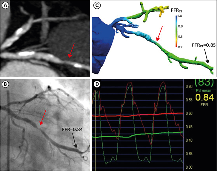
Figure 1 Stenosis without ischemia. (A) Coronary computed tomography angiography with stenosis graded >50% (arrow) in the obtuse marginal branch of the left circumflex artery. Multiple calcified plaques with stenosis between 40% and 69% are noted proximally and distally to the lesion. (B) Severe stenosis is confirmed on ICA (arrow). (C) FFRCT shows a value of 0.85. (D) A reference value of 0.84 was confirmed on ICA-fractional flow reserve. Reused with permission from J Am Coll Cardiol 2011;58:1989-97.10) FFR = fractional flow reserve; FFRCT = fractional flow reserve derived from coronary computed tomographic angiography data; ICA = invasive coronary angiography.
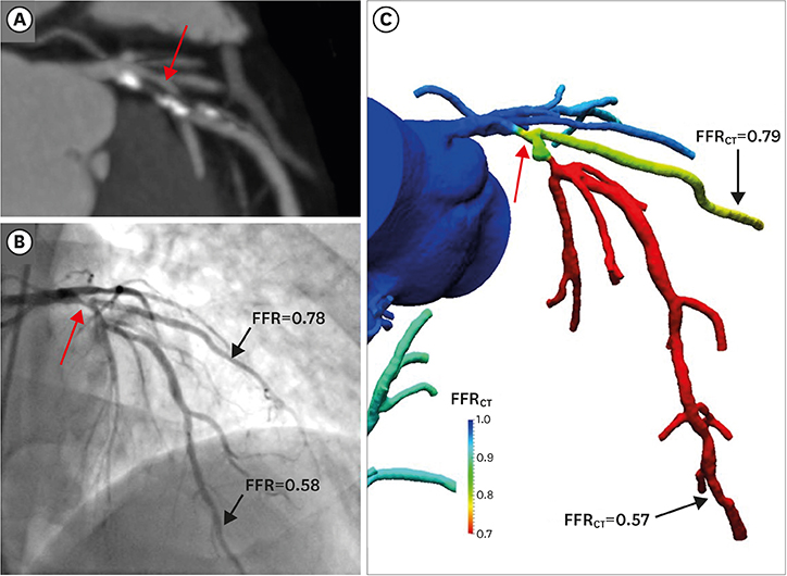
Figure 2 Stenosis with ischemia. (A) Lesion graded with >50% stenosis in the proximal LAD on multiplanar reformat of coronary computed tomography angiography (arrow). (B) Invasive coronary angiography showing the stenosis (arrow) with the corresponding reductions in coronary FFR in the first diagonal branch (FFR=0.78) and distal LAD (FFR=0.58). (C) FFRCT of the first diagonal branch (0.79) and distal LAD (0.57) were determined to be 0.78 and 0.58 by computational fluid dynamics. Reused with permission from J Am Coll Cardiol 2011;58:1989-97.10) FFR = fractional flow reserve; FFRCT = fractional flow reserve derived from coronary computed tomographic angiography data; LAD = left anterior descending artery.
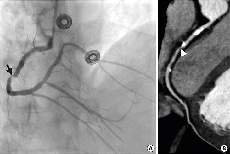
Figure 3 Direct plaque assessment. (A) Invasive coronary angiography, catheterization of the RCA. Severe stenosis of the mid RCA (arrow). (B) Coronary CT angiography, curved multiplanar reconstruction of the RCA. CT reveals a predominantly noncalcified component (arrowhead) in the obstructive mid RCA plaque. The additional calcified plaques do not cause significantly stenosis. CT = computed tomography; RCA = right coronary artery.
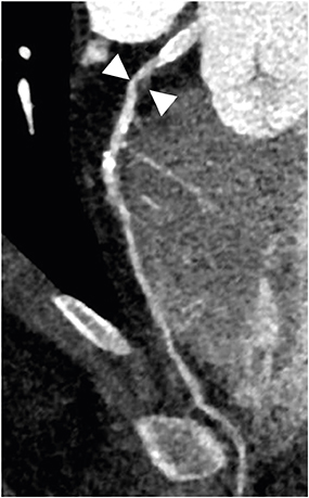
Figure 4 Positive remodeling. Coronary computed tomography angiography, curved multiplanar reconstruction of the left anterior descending artery showing a predominantly noncalcified plaque in the proximal segment causing stenosis between 50% and 69%. Note the increased diameter of the vessel at the level of the stenosis (arrowheads) when compared to the segments immediately before and after the stenosis (i.e., remodeling index >1.1).
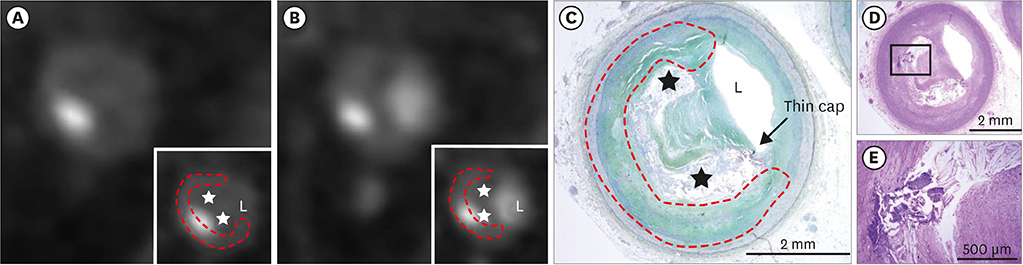
Figure 5 Napkin ring sign and spotty calcifications. Cross-sectional imaging of coronary atherosclerotic plaque showing higher computed tomography attenuation numbers within the circumferential outer rim (red dashed line) in both the noncontrast (A) and contrast-enhanced (B) images as compared to the attenuation within the central plaque (stars). Correlation with histopathology showed a thin cap fibroatheroma (C, D) with spotty calcification (E), and a necrotic core representing the low attenuation plaque core (stars in C). Fibrous plaque tissue corresponded to the hyperattenuating component within the outer rim (red dashed line). Reused with permission from JACC Cardiovasc Imaging 2010;3:440-4.44)
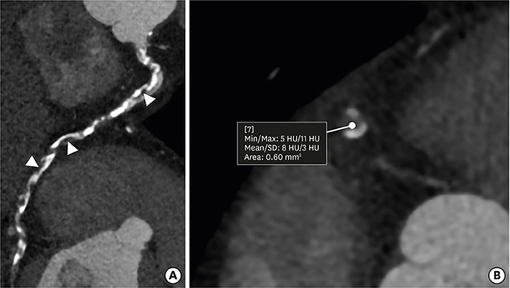
Figure 6 Low-attenuation plaque core. (A) Curved multiplanar reconstruction showing extensive atherosclerosis in the RCA, with plaques showing low-attenuation core (arrowheads). (B) Region of interest drawn in the core of the proximal RCA plaque shows a mean attenuation value of 8 HU. HU = Hounsfield units; RCA = right coronary artery.
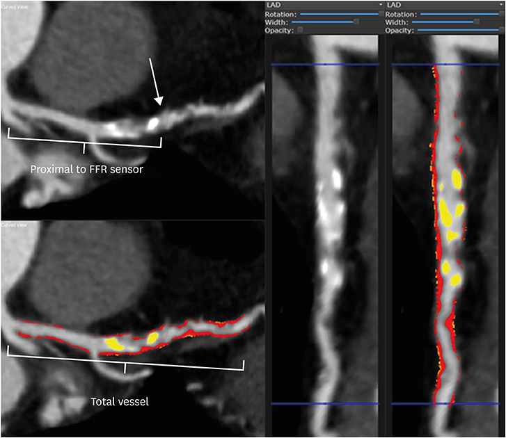
Figure 7 Quantitative plaque analysis. Left anterior descending artery. A region of interest was placed in the ascending aorta at the level of the left main coronary artery, and scan-specific thresholds for calcified plaque (yellow) and noncalcified plaque (in red) were automatically generated. Reused with permission from J Cardiovasc Comput Tomogr 2018;12:344-9.66) FFR = fractional flow reserve; LAD = left anterior descending artery.
Cited by 4 articles
-
Non-Invasive Physiological Assessment after Coronary Stent Implantation
Joseph C. Lee, Kylie M. Burns
Korean Circ J. 2021;51(6):547-548. doi: 10.4070/kcj.2021.0156.Clinical Utility of Coronary Computed Tomography Angiography, Beyond the Gatekeeper for Invasive Coronary Angiography
Yeonyee Elizabeth Yoon
Korean Circ J. 2022;52(11):826-828. doi: 10.4070/kcj.2022.0265.Coronary Physiology-Based Approaches for Plaque Vulnerability: Implications for Risk Prediction and Treatment Strategies
Seokhun Yang, Bon-Kwon Koo
Korean Circ J. 2023;53(9):581-593. doi: 10.4070/kcj.2023.0117.While Awaiting CT-FFR Era: A Novel Approach to Assess Functionally Significant Stenosis With On-Site CT-FFR
Jin-Sin Koh
Korean Circ J. 2024;54(7):395-397. doi: 10.4070/kcj.2024.0139.
Reference
-
1. Task Force Members, Montalescot G, Sechtem U, et al. 2013 ESC guidelines on the management of stable coronary artery disease: the Task Force on the management of stable coronary artery disease of the European Society of Cardiology. Eur Heart J. 2013; 34:2949–3003.2. Wolk MJ, Bailey SR, Doherty JU, et al. ACCF/AHA/ASE/ASNC/HFSA/HRS/SCAI/SCCT/SCMR/STS 2013 multimodality appropriate use criteria for the detection and risk assessment of stable ischemic heart disease: a report of the American College of Cardiology Foundation Appropriate Use Criteria Task Force, American Heart Association, American Society of Echocardiography, American Society of Nuclear Cardiology, Heart Failure Society of America, Heart Rhythm Society, Society for Cardiovascular Angiography and Interventions, Society of Cardiovascular Computed Tomography, Society for Cardiovascular Magnetic Resonance, and Society of Thoracic Surgeons. J Am Coll Cardiol. 2014; 63:380–406.3. Fordyce CB, Newby DE, Douglas PS. Diagnostic strategies for the evaluation of chest pain: clinical implications from SCOT-HEART and PROMISE. J Am Coll Cardiol. 2016; 67:843–852.4. SCOT-HEART investigators. CT coronary angiography in patients with suspected angina due to coronary heart disease (SCOT-HEART): an open-label, parallel-group, multicentre trial. Lancet. 2015; 385:2383–2391.5. Douglas PS, Hoffmann U, Patel MR, et al. Outcomes of anatomical versus functional testing for coronary artery disease. N Engl J Med. 2015; 372:1291–1300.6. Kelion AD, Nicol ED. The rationale for the primacy of coronary CT angiography in the National Institute for Health and Care Excellence (NICE) guideline (CG95) for the investigation of chest pain of recent onset. J Cardiovasc Comput Tomogr. 2018; 12:516–522.
Article7. Hoffmann U, Truong QA, Schoenfeld DA, et al. Coronary CT angiography versus standard evaluation in acute chest pain. N Engl J Med. 2012; 367:299–308.8. Litt HI, Gatsonis C, Snyder B, et al. CT angiography for safe discharge of patients with possible acute coronary syndromes. N Engl J Med. 2012; 366:1393–1403.
Article9. Linde JJ, Kofoed KF, Sørgaard M, et al. Cardiac computed tomography guided treatment strategy in patients with recent acute-onset chest pain: results from the randomised, controlled trial: CArdiac cT in the treatment of acute CHest pain (CATCH). Int J Cardiol. 2013; 168:5257–5262.10. Koo BK, Erglis A, Doh JH, et al. Diagnosis of ischemia-causing coronary stenoses by noninvasive fractional flow reserve computed from coronary computed tomographic angiograms. Results from the prospective multicenter DISCOVER-FLOW (Diagnosis of Ischemia-Causing Stenoses Obtained Via Noninvasive Fractional Flow Reserve) study. J Am Coll Cardiol. 2011; 58:1989–1997.11. Singh G, Al'Aref SJ, Van Assen M, et al. Machine learning in cardiac CT: basic concepts and contemporary data. J Cardiovasc Comput Tomogr. 2018; 12:192–201.
Article12. Pijls NH, De Bruyne B, Peels K, et al. Measurement of fractional flow reserve to assess the functional severity of coronary-artery stenoses. N Engl J Med. 1996; 334:1703–1708.
Article13. Fearon WF, Tonino PA, De Bruyne B, Siebert U, Pijls NH. FAME Study Investigators. Rationale and design of the Fractional Flow Reserve versus Angiography for Multivessel Evaluation (FAME) study. Am Heart J. 2007; 154:632–636.
Article14. Tonino PA, De Bruyne B, Pijls NH, et al. Fractional flow reserve versus angiography for guiding percutaneous coronary intervention. N Engl J Med. 2009; 360:213–224.
Article15. Pijls NH, Fearon WF, Tonino PA, et al. Fractional flow reserve versus angiography for guiding percutaneous coronary intervention in patients with multivessel coronary artery disease: 2-year follow-up of the FAME (Fractional Flow Reserve Versus Angiography for Multivessel Evaluation) study. J Am Coll Cardiol. 2010; 56:177–184.16. Tonino PA, Fearon WF, De Bruyne B, et al. Angiographic versus functional severity of coronary artery stenoses in the FAME study fractional flow reserve versus angiography in multivessel evaluation. J Am Coll Cardiol. 2010; 55:2816–2821.17. Fearon WF, Bornschein B, Tonino PA, et al. Economic evaluation of fractional flow reserve-guided percutaneous coronary intervention in patients with multivessel disease. Circulation. 2010; 122:2545–2550.
Article18. Budoff MJ, Dowe D, Jollis JG, et al. Diagnostic performance of 64-multidetector row coronary computed tomographic angiography for evaluation of coronary artery stenosis in individuals without known coronary artery disease: results from the prospective multicenter ACCURACY (Assessment by Coronary Computed Tomographic Angiography of Individuals Undergoing Invasive Coronary Angiography) trial. J Am Coll Cardiol. 2008; 52:1724–1732.19. Miller JM, Rochitte CE, Dewey M, et al. Diagnostic performance of coronary angiography by 64-row CT. N Engl J Med. 2008; 359:2324–2336.
Article20. Meijboom WB, Meijs MF, Schuijf JD, et al. Diagnostic accuracy of 64-slice computed tomography coronary angiography: a prospective, multicenter, multivendor study. J Am Coll Cardiol. 2008; 52:2135–2144.21. Meijboom WB, Van Mieghem CA, van Pelt N, et al. Comprehensive assessment of coronary artery stenoses: computed tomography coronary angiography versus conventional coronary angiography and correlation with fractional flow reserve in patients with stable angina. J Am Coll Cardiol. 2008; 52:636–643.22. Taylor CA, Fonte TA, Min JK. Computational fluid dynamics applied to cardiac computed tomography for noninvasive quantification of fractional flow reserve: scientific basis. J Am Coll Cardiol. 2013; 61:2233–2241.23. Kim HJ, Vignon-Clementel IE, Coogan JS, Figueroa CA, Jansen KE, Taylor CA. Patient-specific modeling of blood flow and pressure in human coronary arteries. Ann Biomed Eng. 2010; 38:3195–3209.
Article24. Kim HJ, Jansen KE, Taylor CA. Incorporating autoregulatory mechanisms of the cardiovascular system in three-dimensional finite element models of arterial blood flow. Ann Biomed Eng. 2010; 38:2314–2330.
Article25. Kim HJ, Vignon-Clementel IE, Figueroa CA, et al. On coupling a lumped parameter heart model and a three-dimensional finite element aorta model. Ann Biomed Eng. 2009; 37:2153–2169.
Article26. Min JK, Leipsic J, Pencina MJ, et al. Diagnostic accuracy of fractional flow reserve from anatomic CT angiography. JAMA. 2012; 308:1237–1245.
Article27. Nørgaard BL, Leipsic J, Gaur S, et al. Diagnostic performance of noninvasive fractional flow reserve derived from coronary computed tomography angiography in suspected coronary artery disease: the NXT trial (Analysis of Coronary Blood Flow Using CT Angiography: Next Steps). J Am Coll Cardiol. 2014; 63:1145–1155.28. Coenen A, Lubbers MM, Kurata A, et al. Fractional flow reserve computed from noninvasive CT angiography data: diagnostic performance of an on-site clinician-operated computational fluid dynamics algorithm. Radiology. 2015; 274:674–683.
Article29. Zhang JM, Zhong L, Luo T, et al. Simplified models of non-invasive fractional flow reserve based on CT images. PLoS One. 2016; 11:e0153070.
Article30. Itu L, Rapaka S, Passerini T, et al. A machine-learning approach for computation of fractional flow reserve from coronary computed tomography. J Appl Physiol (1985). 2016; 121:42–52.
Article31. Ko BS, Cameron JD, Munnur RK, et al. Noninvasive CT-derived FFR based on structural and fluid analysis: a comparison with invasive FFR for detection of functionally significant stenosis. JACC Cardiovasc Imaging. 2017; 10:663–673.32. Chung JH, Lee KE, Nam CW, et al. Diagnostic performance of a Novel Method for Fractional Flow Reserve Computed from Noninvasive Computed Tomography Angiography (NOVEL-FLOW Study). Am J Cardiol. 2017; 120:362–368.
Article33. Pontone G, Baggiano A, Andreini D, et al. Dynamic stress computed tomography perfusion with a whole-heart coverage scanner in addition to coronary computed tomography angiography and fractional flow reserve computed tomography derived. JACC Cardiovasc Imaging. 2019; S1936-878X(19)30250-5.34. Douglas PS, Pontone G, Hlatky MA, et al. Clinical outcomes of fractional flow reserve by computed tomographic angiography-guided diagnostic strategies vs. usual care in patients with suspected coronary artery disease: the prospective longitudinal trial of FFR(CT): outcome and resource impacts study. Eur Heart J. 2015; 36:3359–3367.
Article35. Hlatky MA, De Bruyne B, Pontone G, et al. Quality-of-life and economic outcomes of assessing fractional flow reserve with computed tomography angiography: PLATFORM. J Am Coll Cardiol. 2015; 66:2315–2323.36. Fairbairn TA, Nieman K, Akasaka T, et al. Real-world clinical utility and impact on clinical decision-making of coronary computed tomography angiography-derived fractional flow reserve: lessons from the ADVANCE registry. Eur Heart J. 2018; 39:3701–3711.
Article37. Glagov S, Weisenberg E, Zarins CK, Stankunavicius R, Kolettis GJ. Compensatory enlargement of human atherosclerotic coronary arteries. N Engl J Med. 1987; 316:1371–1375.
Article38. Schaar JA, Muller JE, Falk E, et al. Terminology for high-risk and vulnerable coronary artery plaques. Report of a meeting on the vulnerable plaque, June 17 and 18, 2003, Santorini, Greece. Eur Heart J. 2004; 25:1077–1082.39. Chang HJ, Lin FY, Lee SE, et al. Coronary atherosclerotic precursors of acute coronary syndromes. J Am Coll Cardiol. 2018; 71:2511–2522.40. Falk E, Nakano M, Bentzon JF, Finn AV, Virmani R. Update on acute coronary syndromes: the pathologists' view. Eur Heart J. 2013; 34:719–728.
Article41. Hoffmann U, Moselewski F, Nieman K, et al. Noninvasive assessment of plaque morphology and composition in culprit and stable lesions in acute coronary syndrome and stable lesions in stable angina by multidetector computed tomography. J Am Coll Cardiol. 2006; 47:1655–1662.
Article42. Narula J, Nakano M, Virmani R, et al. Histopathologic characteristics of atherosclerotic coronary disease and implications of the findings for the invasive and noninvasive detection of vulnerable plaques. J Am Coll Cardiol. 2013; 61:1041–1051.
Article43. Kashiwagi M, Tanaka A, Kitabata H, et al. Feasibility of noninvasive assessment of thin-cap fibroatheroma by multidetector computed tomography. JACC Cardiovasc Imaging. 2009; 2:1412–1419.
Article44. Maurovich-Horvat P, Hoffmann U, Vorpahl M, Nakano M, Virmani R, Alkadhi H. The napkin-ring sign: CT signature of high-risk coronary plaques? JACC Cardiovasc Imaging. 2010; 3:440–444.
Article45. Otsuka K, Fukuda S, Tanaka A, et al. Napkin-ring sign on coronary CT angiography for the prediction of acute coronary syndrome. JACC Cardiovasc Imaging. 2013; 6:448–457.
Article46. Saremi F, Achenbach S. Coronary plaque characterization using CT. AJR Am J Roentgenol. 2015; 204:W249–60.
Article47. Wykrzykowska J, Lehman S, Williams G, et al. Imaging of inflamed and vulnerable plaque in coronary arteries with 18F-FDG PET/CT in patients with suppression of myocardial uptake using a low-carbohydrate, high-fat preparation. J Nucl Med. 2009; 50:563–568.
Article48. Alexanderson E, Slomka P, Cheng V, et al. Fusion of positron emission tomography and coronary computed tomographic angiography identifies fluorine 18 fluorodeoxyglucose uptake in the left main coronary artery soft plaque. J Nucl Cardiol. 2008; 15:841–843.
Article49. Rogers IS, Nasir K, Figueroa AL, et al. Feasibility of FDG imaging of the coronary arteries: comparison between acute coronary syndrome and stable angina. JACC Cardiovasc Imaging. 2010; 3:388–397.50. Dweck MR, Chow MW, Joshi NV, et al. Coronary arterial 18F-sodium fluoride uptake: a novel marker of plaque biology. J Am Coll Cardiol. 2012; 59:1539–1548.51. Oikonomou EK, Marwan M, Desai MY, et al. Non-invasive detection of coronary inflammation using computed tomography and prediction of residual cardiovascular risk (the CRISP CT study): a post-hoc analysis of prospective outcome data. Lancet. 2018; 392:929–939.
Article52. Motoyama S, Kondo T, Anno H, et al. Atherosclerotic plaque characterization by 0.5-mm-slice multislice computed tomographic imaging. Circ J. 2007; 71:363–366.53. Motoyama S, Kondo T, Sarai M, et al. Multislice computed tomographic characteristics of coronary lesions in acute coronary syndromes. J Am Coll Cardiol. 2007; 50:319–326.
Article54. Motoyama S, Sarai M, Harigaya H, et al. Computed tomographic angiography characteristics of atherosclerotic plaques subsequently resulting in acute coronary syndrome. J Am Coll Cardiol. 2009; 54:49–57.
Article55. Motoyama S, Ito H, Sarai M, et al. Plaque characterization by coronary computed tomography angiography and the likelihood of acute coronary events in mid-term follow-up. J Am Coll Cardiol. 2015; 66:337–346.56. Puchner SB, Liu T, Mayrhofer T, et al. High-risk plaque detected on coronary CT angiography predicts acute coronary syndromes independent of significant stenosis in acute chest pain: results from the ROMICAT-II trial. J Am Coll Cardiol. 2014; 64:684–692.57. Ferencik M, Mayrhofer T, Bittner DO, et al. Use of high-risk coronary atherosclerotic plaque detection for risk stratification of patients with stable chest pain: a secondary analysis of the PROMISE randomized clinical trial. JAMA Cardiol. 2018; 3:144–152.58. Williams MC, Moss AJ, Dweck M, et al. Coronary artery plaque characteristics associated with adverse outcomes in the SCOT-HEART Study. J Am Coll Cardiol. 2019; 73:291–301.59. Cury RC, Abbara S, Achenbach S, et al. Coronary Artery Disease - Reporting and Data System (CAD-RADS): an expert consensus document of SCCT, ACR and NASCI: endorsed by the ACC. JACC Cardiovasc Imaging. 2016; 9:1099–1113.60. Maroules CD, Hamilton-Craig C, Branch K, et al. Coronary artery disease reporting and data system (CAD-RADSTM): Inter-observer agreement for assessment categories and modifiers. J Cardiovasc Comput Tomogr. 2018; 12:125–130.61. Sullivan DC, Obuchowski NA, Kessler LG, et al. Metrology standards for quantitative imaging biomarkers. Radiology. 2015; 277:813–825.
Article62. Agatston AS, Janowitz WR, Hildner FJ, Zusmer NR, Viamonte M Jr, Detrano R. Quantification of coronary artery calcium using ultrafast computed tomography. J Am Coll Cardiol. 1990; 15:827–832.
Article63. de Vos BD, Wolterink JM, Leiner T, de Jong PA, Lessmann N, Isgum I. Direct automatic coronary calcium scoring in cardiac and chest CT. IEEE Trans Med Imaging. 2019; 38:2127–2138.
Article64. Boogers MJ, Schuijf JD, Kitslaar PH, et al. Automated quantification of stenosis severity on 64-slice CT: a comparison with quantitative coronary angiography. JACC Cardiovasc Imaging. 2010; 3:699–709.65. Dey D, Cheng VY, Slomka PJ, et al. Automated 3-dimensional quantification of noncalcified and calcified coronary plaque from coronary CT angiography. J Cardiovasc Comput Tomogr. 2009; 3:372–382.
Article66. Øvrehus KA, Gaur S, Leipsic J, et al. CT-based total vessel plaque analyses improves prediction of hemodynamic significance lesions as assessed by fractional flow reserve in patients with stable angina pectoris. J Cardiovasc Comput Tomogr. 2018; 12:344–349.
Article67. Nadjiri J, Hausleiter J, Jähnichen C, et al. Incremental prognostic value of quantitative plaque assessment in coronary CT angiography during 5 years of follow up. J Cardiovasc Comput Tomogr. 2016; 10:97–104.
Article68. Hell MM, Motwani M, Otaki Y, et al. Quantitative global plaque characteristics from coronary computed tomography angiography for the prediction of future cardiac mortality during long-term follow-up. Eur Heart J Cardiovasc Imaging. 2017; 18:1331–1339.
Article69. Bittner DO, Mayrhofer T, Puchner SB, et al. Coronary computed tomography angiography-specific definitions of high-risk plaque features improve detection of acute coronary syndrome. Circ Cardiovasc Imaging. 2018; 11:e007657.
Article70. Lee SE, Chang HJ, Rizvi A, et al. Rationale and design of the Progression of AtheRosclerotic PlAque DetermIned by Computed TomoGraphic Angiography IMaging (PARADIGM) registry: a comprehensive exploration of plaque progression and its impact on clinical outcomes from a multicenter serial coronary computed tomographic angiography study. Am Heart J. 2016; 182:72–79.
Article71. Shin S, Park HB, Chang HJ, et al. Impact of intensive LDL cholesterol lowering on coronary artery atherosclerosis progression: a serial CT angiography study. JACC Cardiovasc Imaging. 2017; 10:437–446.72. Kalisz K, Halliburton S, Abbara S, et al. Update on cardiovascular applications of multienergy CT. Radiographics. 2017; 37:1955–1974.
Article73. Scheske JA, O'Brien JM, Earls JP, et al. Coronary artery imaging with single-source rapid kilovolt peak-switching dual-energy CT. Radiology. 2013; 268:702–709.
Article74. Bamberg F, Dierks A, Nikolaou K, Reiser MF, Becker CR, Johnson TR. Metal artifact reduction by dual energy computed tomography using monoenergetic extrapolation. Eur Radiol. 2011; 21:1424–1429.
Article75. Barreto M, Schoenhagen P, Nair A, et al. Potential of dual-energy computed tomography to characterize atherosclerotic plaque: ex vivo assessment of human coronary arteries in comparison to histology. J Cardiovasc Comput Tomogr. 2008; 2:234–242.
Article76. Symons R, Sandfort V, Mallek M, Ulzheimer S, Pourmorteza A. Coronary artery calcium scoring with photon-counting CT: first in vivo human experience. Int J Cardiovasc Imaging. 2019; 35:733–739.
Article77. Symons R, De Bruecker Y, Roosen J, et al. Quarter-millimeter spectral coronary stent imaging with photon-counting CT: initial experience. J Cardiovasc Comput Tomogr. 2018; 12:509–515.
Article78. Thondapu V, Bourantas CV, Foin N, Jang IK, Serruys PW, Barlis P. Biomechanical stress in coronary atherosclerosis: emerging insights from computational modelling. Eur Heart J. 2017; 38:81–92.
Article79. Choi G, Lee JM, Kim HJ, et al. Coronary artery axial plaque stress and its relationship with lesion geometry: application of computational fluid dynamics to coronary CT angiography. JACC Cardiovasc Imaging. 2015; 8:1156–1166.80. Han D, Starikov A, Ó Hartaigh B, et al. Relationship between endothelial wall shear stress and high-risk atherosclerotic plaque characteristics for identification of coronary lesions that cause ischemia: a direct comparison with fractional flow reserve. J Am Heart Assoc. 2016; 5:e004186.
Article81. Lee JM, Choi G, Koo BK, et al. Identification of high-risk plaques destined to cause acute coronary syndrome using coronary computed tomographic angiography and computational fluid dynamics. JACC Cardiovasc Imaging. 2019; 12:1032–1043.82. Tuncay V, Vliegenthart R, den Dekker MA, et al. Non-invasive assessment of coronary artery geometry using coronary CTA. J Cardiovasc Comput Tomogr. 2018; 12:257–260.
Article83. Kolossváry M, Kellermayer M, Merkely B, Maurovich-Horvat P. Cardiac computed tomography radiomics: a comprehensive review on radiomic techniques. J Thorac Imaging. 2018; 33:26–34.84. Kolossváry M, Park J, Bang JI, et al. Identification of invasive and radionuclide imaging markers of coronary plaque vulnerability using radiomic analysis of coronary computed tomography angiography. Eur Heart J Cardiovasc Imaging. 2019; 20:1250–1258.
Article85. Kolossváry M, Karády J, Szilveszter B, et al. Radiomic features are superior to conventional quantitative computed tomographic metrics to identify coronary plaques with napkin-ring sign. Circ Cardiovasc Imaging. 2017; 10:e006843.
Article86. Dey D, Gaur S, Ovrehus KA, et al. Integrated prediction of lesion-specific ischaemia from quantitative coronary CT angiography using machine learning: a multicentre study. Eur Radiol. 2018; 28:2655–2664.
Article87. Goeller M, Achenbach S, Marwan M, et al. Epicardial adipose tissue density and volume are related to subclinical atherosclerosis, inflammation and major adverse cardiac events in asymptomatic subjects. J Cardiovasc Comput Tomogr. 2018; 12:67–73.
Article88. Zhou J, Chen Y, Zhang Y, et al. Epicardial fat volume improves the prediction of obstructive coronary artery disease above traditional risk factors and coronary calcium score. Circ Cardiovasc Imaging. 2019; 12:e008002.
Article89. Hedgire S, Baliyan V, Zucker EJ, et al. Perivascular epicardial fat stranding at coronary CT angiography: a marker of acute plaque rupture and spontaneous coronary artery dissection. Radiology. 2018; 287:808–815.
- Full Text Links
-
- Actions
-
Cited
- CITED
-
- Close
- Share
-
- Similar articles
-
- Assessment of Coronary Stenosis Using Coronary CT Angiography in Patients with High Calcium Scores: Current Limitations and Future Perspectives
- Beyond Coronary CT Angiography: CT Fractional Flow Reserve and Perfusion
- CT Assessment of Myocardial Perfusion and Fractional Flow Reserve in Coronary Artery Disease: A Review of Current Clinical Evidence and Recent Developments
- Coronary Physiology-Based Approaches for Plaque Vulnerability: Implications for Risk Prediction and Treatment Strategies
- Multimodality Imaging in Coronary Artery Disease: Focus on Computed Tomography
