Malignant Melanoma Presenting as Superior Mediastinal Mass without Extrathoracic Primary Melanoma
- Affiliations
-
- 1Department of Diagnostic Radiology, Kyung Hee University Hospital, Seoul, Korea. sungdw@paran.com
- KMID: 1748453
- DOI: http://doi.org/10.3348/jksr.2013.68.3.213
Abstract
- Malignant melanoma most commonly occurs in the skin, and other organs are secondarily involved. Malignant melanoma presenting in the mediastinum without an extrathoracic primary is very rare. Authors report a case of malignant melanoma of the superior mediastinum without clinical history of extrathoracic malignant melanoma primarily and discuss its radiologic findings. CT shows lobulated heterogenous enhanced mass. Magnetic resonance shows mild hyperintense mass on T1 and T2-weighted images contained focal hemorrhage and necrosis, and invasion to neural foramen. In addition, positron emission tomography/computed tomography shows high standard uptake values uptake of the mass.
MeSH Terms
Figure
-
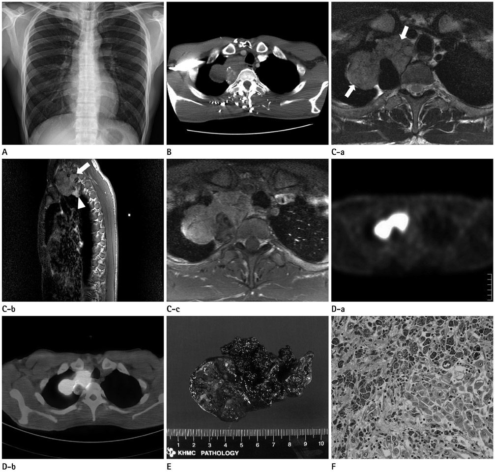
Fig. 1 Imaging findings of superior mediastinal mass in a 32-year-old man. A. Chest radiography shows lobulated contour of mass on the right lung apex with left side deviation of trachea. The lesion shows homogenous opacity, obtuse angle with adjacent pleura and smooth interface with right upper lung. Cervicothoracic sign is positive (mass opacity above the clavicle), so the lesion would be located in the posterior portion of superior mediastinum. B. Chest CT shows well margined lobulated and homogenously enhanced mass on right superior mediastinum. Right tracheoesophageal groove is obliterated, left side tracheal deviation and compression or invasion of great vessels (right common carotid, subclavian arteries and internal jugular vein). Some calcifications are visible on the mass. C. Axial T2-weighted MR image at the upper T2 level (a), shows heterogenous mass (arrows) with intermediate to high signal intensity (compared with adjacent muscles). (b) The mass lesion has also slightly hyperintense area on sagittal T1-weighted image. Invasion of T1-2 and T2-3 neural foramina is noted (arrow). Higher signal intensity area at the lower portion of the mass (arrowhead) correlated with the calcifications on the CT scan. (c) Contrast enhanced T1-weighted MR image with fat suppression shows heterogeneous enhancement of mass. D. (a, b) Positron emission tomography/computed tomography shows high intense signal (standard uptake values 8.25) at right apex, and not uptake at the portion of necrotic area. E. The gross pathologic examination reveals a dark brownish lobulating mass. Multiple tan-brown areas of soft to firm tissues with areas of hemorrhage and necrosis are noted at the cut-section of the mass. F. The histologic specimen shows large pleomorphic cells with prominent nucleoli with some of which contain melanin pigments (H&E, × 200).
Reference
-
1. Karuppiah SV, Buchan KG. Primary malignant melanoma: a rare cause of mediastinal mass. Jpn J Thorac Cardiovasc Surg. 2006. 54:396–398.2. Loewenthal B, Shiau MC, Garcia R. Metastatic melanoma: an unusual diagnosis for a large anterior mediastinal mass. Radiographics. 2004. 24:1714–1718.3. Lau CL, Bentley RC, Gockerman JP, Que LG, D'Amico TA. Malignant melanoma presenting as a mediastinal mass. Ann Thorac Surg. 1999. 67:851–852.4. Takao H, Shimizu S, Doi I, Watanabe T. Primary malignant melanoma of the anterior mediastinum: CT and MR findings. Clin Imaging. 2008. 32:58–60.5. Fishman EK, Kuhlman JE, Schuchter LM, Miller JA 3rd, Magid D. CT of malignant melanoma in the chest, abdomen, and musculoskeletal system. Radiographics. 1990. 10:603–620.6. Kalra A, Kalra A, Palaniswamy C, Gajera M, Rajput V. Primary malignant melanoma presenting as superior mediastinal mass. Int J Surg Case Rep. 2011. 2:239–240.7. Clerico R, Bottoni U, Paolino G, Ambrifi M, Corsetti P, Devirgiliis V, et al. Melanoma with unknown primary: report and analysis of 24 patients. Med Oncol. 2012. 29:2978–2984.8. Webb WR. Hilar and mediastinal lymph node metastases in malignant melanoma. AJR Am J Roentgenol. 1979. 133:805–810.
- Full Text Links
-
- Actions
-
Cited
- CITED
-
- Close
- Share
-
- Similar articles
-
- Primary Malignant Melanoma of the Mediastinum: Radiologic and Pathologic Correlation in Two Case
- Primary malignant melanoma arising in a cystic teratoma
- A Case of Primary Malignant Melanoma of the Vagina: Trial of a Wide Local Excision of Vagina and Rectum
- Primary Pulmonary Malignant Melanoma Presenting as Bilateral Multiple Subsolid Nodules: A Case Report
- Primary Malignant Melanoma Presenting as an Anterior Mediastinal Mass
