Tumor Treated by Endoscopy
- Affiliations
-
- 1Department of Orthopaedic Surgery, Kosin University Gospel Hospital, Busan, Korea. jdkim@ns.kosinmed.or.kr
- KMID: 1737634
- DOI: http://doi.org/10.4055/cios.2014.6.1.72
Abstract
- BACKGROUND
This study was conducted to examine the clinical usefulness and efficacy of endoscopic curettage on benign bone tumor.
METHODS
Thirty-two patients (20 men and 12 women) with benign bone tumor were included in the study. The patients were aged between five and 76 years; the mean follow-up period was 27.05 months (range, 9.6 to 39.9 months). The primary sites include simple bone cyst (9 cases), fibrous dysplasia (6 cases), enchondroma (5 cases), non-ossifying fibroma (4 cases), bone infarct (3 cases), aneurysmal bone cyst (1 case), chondroblastoma (1 case), osteoblastoma (1 case), intraosseous lipoma (1 case), and Brodie abscess (1 case). A plain radiography was performed to assess the radiological recovery. Radiological outcomes, including local recurrence and bone union, were evaluated as excellent, good, poor, and recurred.
RESULTS
In our series, there were 27 cases (84.4%) of good or better outcomes, six cases (18.8%) of complications (4 local recurrence, 1 wound infection, and 1 pathologic fracture).
CONCLUSIONS
Our results showed that endoscopic curettage and bone graft had a lower rate of recurrence and a higher cure rate in cases of benign bone tumor. It can, therefore, be concluded that endoscopic curettage and bone graft might be good treatment modalities for benign bone tumors.
Keyword
MeSH Terms
Figure
-

Fig. 1 (A) Intraoperative radiograph of left calcaneus shows osteolytic bony leision and guide K-wire. (B) Intraoperative radiograph of left calcaneus shows curettage of simple bone cyst under endoscopic guidance.
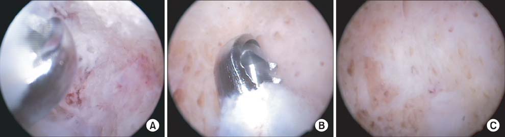
Fig. 2 (A) Endoscopic image of simple bone cyst which is seen as a soft white tissue. (B) Endoscopic image shows curettage of simple bone cyst under endoscopic guidance. (C) Endoscopic image after thorough crettage of the tumor: only normal bone is seen in the cavity.

Fig. 3 Simple bone cyst of the calcaneus in a 12-year-old boy. (A) Preoperative lateral radiograph shows bone loss in calcaneus. (B) Immediate postoperative radiograph taken after curettage and defect filling with allobone graft. (C) At postoperative 4 weeks, bone graft was partially resorbed. (D) At postoperative 3 months, bony trabeculation filled the preoperative bone defect. (E) At postoperative 6 months, simple bone cyst was completely resorbed.
Reference
-
1. Damien CJ, Parsons JR. Bone graft and bone graft substitutes: a review of current technology and applications. J Appl Biomater. 1991; 2(3):187–208.2. Csizy M, Buckley RE, Fennell C. Benign calcaneal bone cyst and pathologic fracture--surgical treatment with injectable calcium-phosphate bone cement (Norian): a case report. Foot Ankle Int. 2001; 22(6):507–510.3. Mainard D, Galois L. Treatment of a solitary calcaneal cyst with endoscopic curettage and percutaneous injection of calcium phosphate cement. J Foot Ankle Surg. 2006; 45(6):436–440.4. Wulle C. On the treatment of enchondroma. J Hand Surg Br. 1990; 15(3):320–330.5. Stricker SJ. Extraarticular endoscopic excision of femoral head chondroblastoma. J Pediatr Orthop. 1995; 15(5):578–581.6. Bonnel F, Canovas F, Faure P. Treatment of a simple bone cyst of the calcaneus by endoscopic curettage with cancellous bone injection. Acta Orthop Belg. 1999; 65(4):528–531.7. Dietz JF, Kachar SM, Nagle DJ. Endoscopically assisted excision of digital enchondroma. Arthroscopy. 2007; 23(6):678.e1–678.e4.8. Rockwood CA, Wilkins KE, Beaty JH, editors. Fractures in children. 4th ed. Philadelphia: Lippincott Williams and Wilkins;1996. p. 167–320.9. Wilber MC, Hyatt GW. Bone cysts: results of surgical treatment in 200 cases. J Bone Joint Surg Am. 1960; 42(5):879.10. Spence KF Jr, Bright RW, Fitzgerald SP, Sell KW. Solitary unicameral bone cyst: treatment with freeze-dried crushed cortical-bone allograft: a review of one hundred and forty-four cases. J Bone Joint Surg Am. 1976; 58(5):636–641.11. Jaffe KA, Dunham WK. Treatment of benign lesions of the femoral head and neck. Clin Orthop Relat Res. 1990; (257):134–137.12. Catier P, Bracq H, Canciani JP, Allouis M, Babut JM. The treatment of upper femoral unicameral bone cysts in children by Ender's nailing technique. Rev Chir Orthop Reparatrice Appar Mot. 1981; 67(2):147–149.13. Baek SG, Oh CW, Jeon IH, Min WK, Park IH. Comparison of results by its location and treatment modality of the simple bone cyst. J Korean Musculoskelet Transplant Soc. 2006; 6(2):88–97.14. Yandow SM, Lundeen GA, Scott SM, Coffin C. Autogenic bone marrow injections as a treatment for simple bone cyst. J Pediatr Orthop. 1998; 18(5):616–620.15. Neer CS, Francis KC, Johnston AD, Kiernan HA Jr. Current concepts on the treatment of solitary unicameral bone cyst. Clin Orthop Relat Res. 1973; (97):40–51.16. Nakashima Y, Kotoura Y, Nagashima T, Yamamuro T, Hamashima Y. Monostotic fibrous dysplasia in the femoral neck: a clinicopathologic study. Clin Orthop Relat Res. 1984; (191):242–248.17. Harris WH, Dudley HR Jr, Barry RJ. The natural history of fibrous dysplasia: an orthopaedic, pathological, and roentgenographic study. J Bone Joint Surg Am. 1962; 44(2):207–233.18. Guille JT, Kumar SJ, MacEwen GD. Fibrous dysplasia of the proximal part of the femur: long-term results of curettage and bone-grafting and mechanical realignment. J Bone Joint Surg Am. 1998; 80(5):648–658.19. Bae DK, Rhee YG, Kim SK, Son YL. Clinical analysis of fibrous dysplasia. J Korean Orthop Assoc. 1992; 27(5):1418–1425.20. Kang ES, Roh KJ, Yoo JD. Comparative study of the simple curettage and the curettage with bonegraft in enchondroma of the hand. J Korean Orthop Assoc. 1997; 32(1):156–162.21. Kuur E, Hansen SL, Lindequist S. Treatment of solitary enchondromas in fingers. J Hand Surg Br. 1989; 14(1):109–112.22. Tordai P, Hoglund M, Lugnegard H. Is the treatment of enchondroma in the hand by simple curettage a rewarding method? J Hand Surg Br. 1990; 15(3):331–334.23. Jaffe HL, Lichtenstein L. Non-osteogenic fibroma of bone. Am J Pathol. 1942; 18(2):205–221.24. Compere CL, Coleman SS. Nonosteogenic fibroma of bone. Surg Gynecol Obstet. 1957; 105(5):588–598.
- Full Text Links
-
- Actions
-
Cited
- CITED
-
- Close
- Share
-
- Similar articles
-
- Comparisons of Gastric Endoscopy and Upper Gastrointestinal Series in The Submucosal Tumor
- Upper Endoscopy in International Digestive Endoscopy Network 2012: Towards Upper End of Quality
- Estimation by Gross Findings in Early Gastric Cancer
- Hemorrhagic Small Bowel Tumor Diagnosed with Using Capsule Endoscopy and It was Treated with Laparoscopic Surgery: Report of a Case
- Image-Enhanced Endoscopy for Diagnosis and Treatment of Gastrointestinal Tumor
