Assessment of metal artifacts in three-dimensional dental surface models derived by cone-beam computed tomography
- Affiliations
-
- 1Department of Orthodontics, School of Dentistry, Chonnam National University, Gwangju, Korea.
- 2Department of Orthodontics, School of Dentistry, Dental Science Research Institute, Chonnam National University, Gwangju, Korea. hhwang@chonnam.ac.kr
- KMID: 1726416
- DOI: http://doi.org/10.4041/kjod.2014.44.5.229
Abstract
OBJECTIVE
The aim of this study was to assess artifacts induced by metallic restorations in three-dimensional (3D) dental surface models derived by cone-beam computed tomography (CBCT).
METHODS
Fifteen specimens, each with four extracted human premolars and molars embedded in a plaster block, were scanned by CBCT before and after the cavitated second premolars were restored with dental amalgam. Five consecutive surface models of each specimen were created according to increasing restoration size: no restoration (control) and small occlusal, large occlusal, disto-occlusal, and mesio-occluso-distal restorations. After registering each restored model with the control model, maximum linear discrepancy, area, and intensity of the artifacts were measured and compared.
RESULTS
Artifacts developed mostly on the buccal and lingual surfaces. They occurred not only on the second premolar but also on the first premolar and first molar. The parametric values increased significantly with increasing restoration size.
CONCLUSIONS
Metallic restorations induce considerable artifacts in 3D dental surface models. Artifact reduction should be taken into consideration for a proper diagnosis and treatment planning when using 3D surface model derived by CBCT in dentofacial deformity patients.
Keyword
MeSH Terms
Figure
-

Figure 1 Illustration of the restorations. A, Small occlusal restoration; B, large occlusal restoration; C, disto-occlusal restoration; D, mesio-occluso-distal restoration.
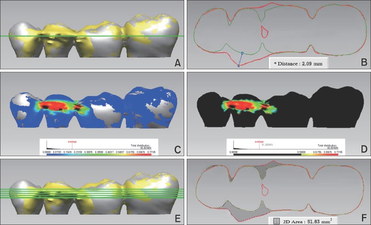
Figure 2 Quantitative assessment of artifacts on the basis of discrepancies of two models, each restoration model and no restoration model as the control. The present figures show the discrepancies between a mesio-occluso-distal restoration and the control. A and B, Maximum linear discrepancy was measured as the distance between the shells at the most protruded point of the graphic. C and D, Artifact area was defined as an area of discrepancy over 0.5 mm. E and F, Artifact intensity was defined as the sum of five discrepancy areas measured in cross-sectional graphics captured at the level of the maximum linear discrepancy and 0.5 and 1.0 mm above and below.
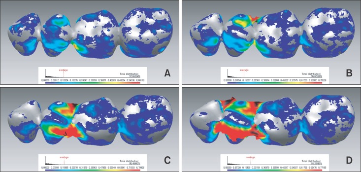
Figure 3 Color-mapped graphics obtained by registering three-dimensional surface models of the restorations with the control (no restoration). Discrepancies between the shells indicate artifacts due to amalgam restoration. Blue and red represent the minimum and maximum discrepancies, respectively. A, Small occlusal restoration; B, large occlusal restoration; C, disto-occlusal restoration; D, mesio-occluso-distal restoration.
Reference
-
1. Hwang HS, Hwang CH, Lee KH, Kang BC. Maxillofacial 3-dimensional image analysis for the diagnosis of facial asymmetry. Am J Orthod Dentofacial Orthop. 2006; 130:779–785. PMID: 17169741.
Article2. Terajima M, Nakasima A, Aoki Y, Goto TK, Tokumori K, Mori N, et al. A 3-dimensional method for analyzing the morphology of patients with maxillofacial deformities. Am J Orthod Dentofacial Orthop. 2009; 136:857–867. PMID: 19962610.
Article3. Girod S, Keeve E, Girod B. Advances in interactive craniofacial surgery planning by 3D simulation and visualization. Int J Oral Maxillofac Surg. 1995; 24:120–125. PMID: 7782646.
Article4. Xia J, Ip HH, Samman N, Wang D, Kot CS, Yeung RW, et al. Computer-assisted three-dimensional surgical planning and simulation: 3D virtual osteotomy. Int J Oral Maxillofac Surg. 2000; 29:11–17. PMID: 10691136.
Article5. Xia JJ, Gateno J, Teichgraeber JF, Christensen AM, Lasky RE, Lemoine JJ, et al. Accuracy of the computer-aided surgical simulation (CASS) system in the treatment of patients with complex craniomaxillofacial deformity: A pilot study. J Oral Maxillofac Surg. 2007; 65:248–254. PMID: 17236929.
Article6. Gateno J, Xia JJ, Teichgraeber JF, Christensen AM, Lemoine JJ, Liebschner MA, et al. Clinical feasibility of computer-aided surgical simulation (CASS) in the treatment of complex cranio-maxillofacial deformities. J Oral Maxillofac Surg. 2007; 65:728–734. PMID: 17368370.
Article7. Schulze R, Heil U, Gross D, Bruellmann DD, Dranischnikow E, Schwanecke U, et al. Artefacts in CBCT: a review. Dentomaxillofac Radiol. 2011; 40:265–273. PMID: 21697151.
Article8. Endo M, Tsunoo T, Nakamori N, Yoshida K. Effect of scattered radiation on image noise in cone beam CT. Med Phys. 2001; 28:469–474. PMID: 11339743.
Article9. Mozzo P, Procacci C, Tacconi A, Martini PT, Andreis IA. A new volumetric CT machine for dental imaging based on the cone-beam technique: preliminary results. Eur Radiol. 1998; 8:1558–1564. PMID: 9866761.
Article10. Hsieh J, Molthen RC, Dawson CA, Johnson RH. An iterative approach to the beam hardening correction in cone beam CT. Med Phys. 2000; 27:23–29. PMID: 10659734.
Article11. De Man B, Nuyts J, Dupont P, Marchal G, Suetens P. Metal streak artifacts in x-ray computed tomography: a simulation study. IEEE Trans Nucl Sci. 1999; 46:691–696.
Article12. De Man B, Nuyts J, Dupont P, Marchal G, Suetens P. Reduction of metal streak artefacts in X-ray computed tomography using a transmission maximum a posteriori algorithm. IEEE Trans Nucl Sci. 2000; 47:977–981.13. Gateno J, Xia J, Teichgraeber JF, Rosen A. A new technique for the creation of a computerized composite skull model. J Oral Maxillofac Surg. 2003; 61:222–227. PMID: 12619001.
Article14. Nkenke E, Zachow S, Benz M, Maier T, Veit K, Kramer M, et al. Fusion of computed tomography data and optical 3D images of the dentition for streak artefact correction in the simulation of orthognathic surgery. Dentomaxillofac Radiol. 2004; 33:226–232. PMID: 15533975.
Article15. Uechi J, Okayama M, Shibata T, Muguruma T, Hayashi K, Endo K, et al. A novel method for the 3-dimensional simulation of orthognathic surgery by using a multimodal image-fusion technique. Am J Orthod Dentofacial Orthop. 2006; 130:786–798. PMID: 17169742.
Article16. Kim BC, Lee CE, Park W, Kang SH, Zhengguo P, Yi CK, et al. Integration accuracy of digital dental models and 3-dimensional computerized tomography images by sequential point- and surface-based markerless registration. Oral Surg Oral Med Oral Pathol Oral Radiol Endod. 2010; 110:370–378. PMID: 20591700.
Article17. Noh H, Nabha W, Cho JH, Hwang HS. Registration accuracy in the integration of laser-scanned dental images into maxillofacial cone-beam computed tomography images. Am J Orthod Dentofacial Orthop. 2011; 140:585–591. PMID: 21967948.
Article18. Lin HH, Chiang WC, Lo LJ, Sheng-Pin Hsu S, Wang CH, Wan SY. Artifact-resistant superimposition of digital dental models and cone-beam computed tomography images. J Oral Maxillofac Surg. 2013; 71:1933–1947. PMID: 23911142.
Article19. Lee WB, Theodore MR, Aldridge DW. Class I, II, and VI amalgam restorations. In : Herald OH, Edward JS, Andre VR, editors. Sturdevant's art and science of operative dentistry. 6th ed. St Louis: Mosby;2012.20. Besl PJ, Mckay ND. A method for registration for 3-D shapes. IEEE Trans Patt Anal Machine Intell. 1992; 14:239–256.21. Hassan B, Couto Souza P, Jacobs R, de Azambuja Berti S, van der Stelt P. Influence of scanning and reconstruction parameters on quality of three-dimensional surface models of the dental arches from cone beam computed tomography. Clin Oral Investig. 2010; 14:303–310.
Article22. Coward TJ, Scott BJ, Watson RM, Richards R. A comparison between computerized tomography, magnetic resonance imaging, and laser scanning for capturing 3-dimensional data from an object of standard form. Int J Prosthodont. 2005; 18:405–413. PMID: 16220806.23. Liang X, Lambrichts I, Sun Y, Denis K, Hassan B, Li L, et al. A comparative evaluation of cone beam computed tomography (CBCT) and multi-slice CT (MSCT). Part II: On 3D model accuracy. Eur J Radiol. 2010; 75:270–274. PMID: 19423257.
Article24. Ye N, Jian F, Xue J, Wang S, Liao L, Huang W, et al. Accuracy of in-vitro tooth volumetric measurements from cone-beam computed tomography. Am J Orthod Dentofacial Orthop. 2012; 142:879–887. PMID: 23195374.
Article25. Schulze RK, Berndt D, d'Hoedt B. On cone-beam computed tomography artifacts induced by titanium implants. Clin Oral Implants Res. 2010; 21:100–107. PMID: 19845706.
Article26. Zhang Y, Zhang L, Zhu XR, Lee AK, Chambers M, Dong L. Reducing metal artifacts in cone-beam CT images by preprocessing projection data. Int J Radiat Oncol Biol Phys. 2007; 67:924–932. PMID: 17161556.
Article27. Swennen GR, Mommaerts MY, Abeloos J, De Clercq C, Lamoral P, Neyt N, et al. A cone-beam CT based technique to augment the 3D virtual skull model with a detailed dental surface. Int J Oral Maxillofac Surg. 2009; 38:48–57. PMID: 19118978.
Article28. Biao Y. Registration accuracy according to the removal of artifact area in the integration of laser-scanned dental images into maxillofacial cone-beam CT images [Master's thesis]. Gwangju: Chonnam National University;2012.
- Full Text Links
-
- Actions
-
Cited
- CITED
-
- Close
- Share
-
- Similar articles
-
- Three-dimensional imaging modalities in endodontics
- Does the metal artifact reduction algorithm activation mode influence the magnitude of artifacts in CBCT images?
- A rare case of dilated invaginated odontome with talon cusp in a permanent maxillary central incisor diagnosed by cone beam computed tomography
- Metal artifact production and reduction in CBCT with different numbers of basis images
- Magnitude of beam-hardening artifacts produced by gutta-percha and metal posts on cone-beam computed tomography with varying tube current
