Magnitude of beam-hardening artifacts produced by gutta-percha and metal posts on cone-beam computed tomography with varying tube current
- Affiliations
-
- 1Department of Oral Diagnosis, Division of Oral Radiology, Piracicaba Dental School, University of Campinas, Piracicaba, Sao Paulo, Brazil.
- 2Department of Dental Materials and Prosthodontics, School of Dentistry of Ribeirao Preto, University of Sao Paulo, Ribeirao Preto, Sao Paulo, Brazil.
- 3Department of Stomatology, Public Oral Health and Forensic Dentistry, Division of Oral Radiology, School of Dentistry of Ribeirao Preto, University of Sao Paulo, Ribeirao Preto, Sao Paulo, Brazil. oliveirach@usp.br
- KMID: 2471839
- DOI: http://doi.org/10.5624/isd.2020.50.1.1
Abstract
- PURPOSE
This study was performed to evaluate the magnitude of artifacts produced by gutta-percha and metal posts on cone-beam computed tomography (CBCT) scans obtained with different tube currents and with or without metal artifact reduction (MAR).
MATERIALS AND METHODS
A tooth was inserted in a dry human mandible socket, and CBCT scans were acquired after root canal instrumentation, root canal filling, and metal post placement with various tube currents with and without MAR activation. The artifact magnitude was assessed by the standard deviation (SD) of gray values and the contrast-to-noise ratio (CNR) at the various distances from the tooth. Data were compared using multi-way analysis of variance.
RESULTS
At all distances, a current of 4 mA was associated with a higher SD and a lower CNR than 8 mA or 10 mA (P<0.05). For the metal posts without MAR, the artifact magnitude as assessed by SD was greatest at 1.5 cm or less (P<0.05). When MAR was applied, SD values for distances 1.5 cm or closer to the tooth were reduced (P<0.05). MAR usage did not influence the magnitude of artifacts in the control and gutta-percha groups (P>0.05).
CONCLUSION
Increasing the tube current from 4 mA to 8 mA may reduce the magnitude of artifacts from metal posts. The magnitude of artifacts arising from metal posts was significantly higher at distances of 1.5 cm or less than at greater distances. MAR usage improved image quality near the metal post, but had no significant influence farther than 1.5 cm from the tooth.
Keyword
MeSH Terms
Figure
-
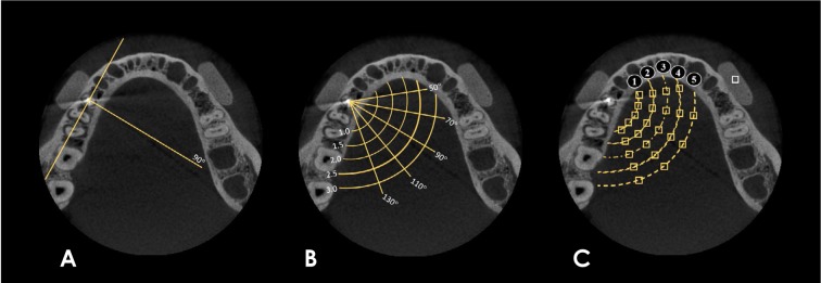
Fig. 1 A. A line was drawn perpendicular to the mandibular body and through the center of the artifact-generating object. B. 20°-step lines were drawn above and below the perpendicular line, and 5 concentric semicircles were created with different radii (0.5-cm intervals). C. In the regions of intersection between the lines and the semicircles, 24 square regions of interest (ROIs) of the same size were established. A square ROI of the same size as the others was established in the silicone block, serving as the control area.
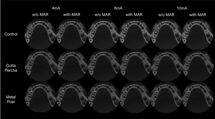
Fig. 2 Axial images represent intracanal materials (control in the first row, gutta-percha in the second row, and metal post in the third row) with different tube currents (4 mA, 8 mA, and 10 mA) and without and with the application of a metal artifact reduction tool.
Reference
-
1. Fontenele RC, Nascimento EH, Vasconcelos TV, Noujeim M, Freitas DQ. Magnitude of cone beam CT image artifacts related to zirconium and titanium implants: impact on image quality. Dentomaxillofac Radiol. 2018; 47:20180021. PMID: 29668300.
Article2. Freitas DQ, Fontenele RC, Nascimento EH, Vasconcelos TV, Noujeim M. Influence of acquisition parameters on the magnitude of cone beam computed tomography artifacts. Dentomaxillofac Radiol. 2018; 47:20180151. PMID: 29916722.
Article3. Candemil AP, Salmon B, Freitas DQ, Ambrosano GM, Haiter-Neto F, Oliveira ML. Metallic materials in the exomass impair cone beam CT voxel values. Dentomaxillofac Radiol. 2018; 47:20180011. PMID: 29582689.4. Cebe F, Aktan AM, Ozsevik AS, Ciftci ME, Surmelioglu HD. The effects of different restorative materials on the detection of approximal caries in cone-beam computed tomography scans with and without metal artifact reduction mode. Oral Surg Oral Med Oral Pathol Oral Radiol. 2017; 123:392–400. PMID: 28111155.
Article5. Neves FS, Freitas DQ, Campos PS, Ekestubbe A, Lofthag-Hansen S. Evaluation of cone-beam computed tomography in the diagnosis of vertical root fractures: the influence of imaging modes and root canal materials. J Endod. 2014; 40:1530–1536. PMID: 25127934.
Article6. Freitas DQ, Vasconcelos TV, Noujeim M. Diagnosis of vertical root fracture in teeth close and distant to implant: an in vitro study to assess the influence of artifacts produced in cone beam computed tomography. Clin Oral Investig. 2019; 23:1263–1270.
Article7. Freitas DQ, Nascimento EH, Vasconcelos TV, Noujeim M. Diagnosis of external root resorption in teeth close and distant to zirconium implants: influence of acquisition parameters and artefacts produced during cone beam computed tomography. Int Endod J. 2019; 52:866–873. PMID: 30585641.
Article8. de-Azevedo-Vaz SL, Peyneau PD, Ramirez-Sotelo LR, Vasconcelos Kde F, Campos PS, Haiter-Neto F. Efficacy of a cone beam computed tomography metal artifact reduction algorithm for the detection of peri-implant fenestrations and dehiscences. Oral Surg Oral Med Oral Pathol Oral Radiol. 2016; 121:550–556. PMID: 27068312.
Article9. Sancho-Puchades M, Hämmerle CH, Benic GI. In vitro assessment of artifacts induced by titanium, titanium-zirconium and zirconium dioxide implants in cone-beam computed tomography. Clin Oral Implants Res. 2015; 26:1222–1228. PMID: 25040484.10. Queiroz PM, Oliveira ML, Groppo FC, Haiter-Neto F, Freitas DQ. Evaluation of metal artefact reduction in cone-beam computed tomography images of different dental materials. Clin Oral Investig. 2018; 22:419–423.
Article11. Lira de Farias Freitas AP, Cavalcanti YW, Costa FC, Peixoto LR, Maia AM, Rovaris K, et al. Assessment of artefacts produced by metal posts on CBCT images. Int Endod J. 2018; 52:223–236. PMID: 30107037.
Article12. Pauwels R, Silkosessak O, Jacobs R, Bogaerts R, Bosmans H, Panmekiate S. A pragmatic approach to determine the optimal kVp in cone beam CT: balancing contrast-to-noise ratio and radiation dose. Dentomaxillofac Radiol. 2014; 43:20140059. PMID: 24708447.
Article13. Jaju PP, Jaju SP. Cone-beam computed tomography: time to move from ALARA to ALADA. Imaging Sci Dent. 2015; 45:263–265. PMID: 26730375.
Article14. Pauwels R, Seynaeve L, Henriques JC, de Oliveira-Santos C, Souza PC, Westphalen FH, et al. Optimization of dental CBCT exposures through mAs reduction. Dentomaxillofac Radiol. 2015; 44:20150108. PMID: 26090934.
Article15. Schulze R, Heil U, Gross D, Bruellmann DD, Dranischnikow E, Schwanecke U, et al. Artefacts in CBCT: a review. Dentomaxillofac Radiol. 2011; 40:265–273. PMID: 21697151.
Article16. Pauwels R, Stamatakis H, Bosmans H, Bogaerts R, Jacobs R, Horner K, et al. Quantification of metal artifacts on cone beam computed tomography images. Clin Oral Implants Res. 2013; 24 Suppl A100:94–99. PMID: 22168574.
Article17. Bechara B, McMahan CA, Geha H, Noujeim M. Evaluation of a cone beam CT artefact reduction algorithm. Dentomaxillofac Radiol. 2012; 41:422–428. PMID: 22362221.
Article18. Nascimento EH, Fontenele RC, Santaella GM, Freitas DQ. Difference in the artefacts production and the performance of the metal artefact reduction (MAR) tool between the buccal and lingual cortical plates adjacent to zirconium dental implant. Dentomaxillofac Radiol. 2019; 48:20190058. PMID: 31276425.
Article19. Parsa A, Ibrahim N, Hassan B, Syriopoulos K, van der Stelt P. Assessment of metal artefact reduction around dental titanium implants in cone beam CT. Dentomaxillofac Radiol. 2014; 43:20140019. PMID: 25135316.
Article20. Kamburoǧlu K, Yilmaz F, Yeta EN, Özen D. Assessment of furcal perforations in the vicinity of different root canal sealers using a cone beam computed tomography system with and without the application of artifact reduction mode: an ex vivo investigation on extracted human teeth. Oral Surg Oral Med Oral Pathol Oral Radiol. 2016; 121:657–665. PMID: 27039007.21. Nikbin A, Dalili Kajan Z, Taramsari M, Khosravifard N. Effect of object position in the field of view and application of a metal artifact reduction algorithm on the detection of vertical root fractures on cone-beam computed tomography scans: an in vitro study. Imaging Sci Dent. 2018; 48:245–254. PMID: 30607348.
Article22. Vasconcelos KF, Codari M, Queiroz PM, Nicolielo LF, Freitas DQ, Sforza C, et al. The performance of metal artifact reduction algorithms in cone beam computed tomography images considering the effects of materials, metal positions, and fields of view. Oral Surg Oral Med Oral Pathol Oral Radiol. 2019; 127:71–76. PMID: 30528550.
Article
- Full Text Links
-
- Actions
-
Cited
- CITED
-
- Close
- Share
-
- Similar articles
-
- Assessment of vertical root fracture using cone-beam computed tomography
- Expression of beam hardening artifacts on horizontally stitched cone-beam computed tomography images
- Cone-beam computed tomography artifacts in the presence of dental implants and associated factors: An integrative review
- Influence of CBCT metal artifact reduction on vertical radicular fracture detection
- Comparative evaluation of computed tomography for dental implants on the mandibular edentulous area
