Change in Effective Leg Length after Angular Deformity Correction by Hemiepiphyseal Stapling
- Affiliations
-
- 1Department of Orthopaedic Surgery, Chung-Ang University College of Medicine, Seoul, Korea.
- 2Department of Orthopaedic Surgery, Seoul National University College of Medicine, Seoul, Korea. tjcho@snu.ac.kr
- KMID: 1719334
- DOI: http://doi.org/10.4055/cios.2010.2.2.85
Abstract
- BACKGROUND
The hemiepiphyseal stapling has both positive and negative effects on effective leg length. The purpose of this study was to analyze change in effective leg length after angular correction by hemiepiphyseal stapling, and to validate in clinical cases.
METHODS
Mathematical analysis of a hemiepiphyseal stapling model was conducted. The induced formula was validated in 6 cases fulfilling the assumptions of the model. Anatomical parameters involved in this formula were measured in additional 21 cases undergoing hemiepiphyseal stapling or hemiepiphysiodesis.
RESULTS
Effective leg length increased or decreased according to three parameters in this model: 1) limb length distal to the operated physis (L), 2) width of the operated physis (d), and 3) the amount of angular deformity to be corrected (theta). Actual change in effective leg length of 6 cases similar to this model coincided with the predicted change at least in its direction. L/d ratio was 4.82 +/- 0.51.
CONCLUSIONS
Considering the narrow range of the L/d ratio, hemiepiphyseal stapling is likely to decrease effective leg length if the amount of angular correction is less than 10degrees, whereas to increase it if the amount of angular correction is larger than 16degrees. This should be taken into consideration when selecting the surgical method for angular deformity correction in skeletally immature patients.
MeSH Terms
Figure
-
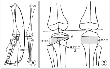
Fig. 1 Schematic diagram of the mathematical analysis. (A) The amount of angular correction and the length of the operated limb was designated as θ and L, respectively. Gain of effective leg length, when angular correction has been achieved, will be L · (1 - cos θ). "A" is the effective leg length. (B) The width of the operated physis was designated as d · When angular correction is achieved by hemiepiphyseal stapling, the contralateral limb will grow at this physis by d · tan θ; linear growth of the operated limb will be a half of this.
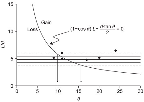
Fig. 2 Equation 2 is plotted on plane of L/d ratio and the amount of angular correction (θ). Dots depict six cases in Tables 1 and 2. If a dot is above and right to the curve, gain of effective leg length is expected. Vertical arrows indicate range of the evalue corresponding to the range of L/d ratio with 2 standard deviations. A thick transverse line denotes the mean value, thin lines range of 1 standard deviation, and dotted line that of 2 standard deviations.

Fig. 3 Teleradiographs, preoperative (A) and post-correction (B) of case 3 in Tables 1 and 2. Note the improvement in preoperative pelvic obliquity after angular correction using hemiepiphyseal stapling of the distal femur.
Reference
-
1. Stevens PM, Maguire M, Dales MD, Robins AJ. Physeal stapling for idiopathic genu valgum. J Pediatr Orthop. 1999. 19(5):645–649.
Article2. Zuege RC, Kempken TG, Blount WP. Epiphyseal stapling for angular deformity at the knee. J Bone Joint Surg Am. 1979. 61(3):320–329.
Article3. Arkin AM, Katz JF. The effects of pressure on epiphyseal growth: the mechanism of plasticity of growing bone. J Bone Joint Surg Am. 1956. 38(5):1056–1076.4. Goff CW. Histologic arrangements from biopsies of epiphyseal plates of children before and after stapling: correlated with roentgenographic studies. Am J Orthop. 1967. 9(5):87–89.5. Mielke CH, Stevens PM. Hemiepiphyseal stapling for knee deformities in children younger than 10 years: a preliminary report. J Pediatr Orthop. 1996. 16(4):423–429.
Article6. Canale ST, Harper MC. Biotrigonometric analysis and practical applications of osteotomies of tibia in children. Instr Course Lect. 1981. 30:85–101.7. Aykut US, Yazici M, Kandemir U, et al. The effect of temporary hemiepiphyseal stapling on the growth plate: a radiologic and immunohistochemical study in rabbits. J Pediatr Orthop. 2005. 25(3):336–341.8. Blount WP. A mature look at epiphyseal stapling. Clin Orthop Relat Res. 1971. (77):158–163.9. Blount WP, Clarke GR. Control of bone growth by epiphyseal stapling: a preliminary report. J Bone Joint Surg Am. 1949. 31(3):464–478.10. Frantz CH. Epiphyseal stapling: a comprehensive review. Clin Orthop Relat Res. 1971. (77):149–157.11. Harper MC, Canale ST. Angulation osteotomy: a trigonometric analysis. Clin Orthop Relat Res. 1982. (166):173–181.12. Harper MC, Canale ST, Cobb RM. Proximal femoral osteotomy: a trigonometric analysis of effect on leg length. J Pediatr Orthop. 1983. 3(4):431–434.13. Hernigou P, Jaafar A, Hamdadou A. Leg length changes after upper tibial osteotomy: analysis of different preoperative planning methods. Rev Chir Orthop Reparatrice Appar Mot. 2002. 88(1):68–73.14. Hernigou P, Ovadia H, Goutallier D. Mathematical modelization of open-wedge tibial osteotomy and correction tables. Rev Chir Orthop Reparatrice Appar Mot. 1992. 78(4):258–263.15. Paley D. Principles of deformity correction. 2002. New York: Springer-Verlag;276–278.16. Farnum CE, Nixon A, Lee AO, Kwan DT, Belanger L, Wilsman NJ. Quantitative three-dimensional analysis of chondrocytic kinetic responses to short-term stapling of the rat proximal tibial growth plate. Cells Tissues Organs. 2000. 167(4):247–258.
Article
- Full Text Links
-
- Actions
-
Cited
- CITED
-
- Close
- Share
-
- Similar articles
-
- Remodelling of Angular DeforMity after Femoral Shaft Fractures in Children
- Clinical Experience with Epiphyseal Stapling
- Limb Angular Deformity Correction Using Dyna-ATC: Surgical Technique, Calculation Method, and Clinical Outcome
- Clinical Studies of Corrective Osteotomy for Various Angular Deformities of Tibia
- Surgical Correction and Osteosynthesis for Cranial Displaced Pelvic Nonunion: Technical Note and Two Cases Report Regarding Anterior Correction and Osteosynthesis Following Posterior Release
