Clin Orthop Surg.
2011 Jun;3(2):121-127. 10.4055/cios.2011.3.2.121.
The Correlations of the Radiological Parameters of Hip Dysplasia and Proximal Femoral Deformity in Clinically Normal Hips of a Korean Population
- Affiliations
-
- 1Department of Orthopaedics, Dongguk University Ilsan Hospital, Goyang, Korea. gunil@duih.org
- KMID: 1719308
- DOI: http://doi.org/10.4055/cios.2011.3.2.121
Abstract
- BACKGROUND
The aim of this study was to answer the following two questions: 1) Do the radiological parameters of dysplasia have significant correlations between themselves or with the parameters of the proximal femoral deformity and vice versa? 2) Do the physical parameters have a significant correlation with the radiological parameters of hip dysplasia and proximal femoral deformity?
METHODS
Four hundred and twenty eight consecutive patients with no clinical evidence of hip osteoarthritis and who underwent pelvic radiography in the supine position for hip contusion or a routine health check were analyzed for the relationships between the center-edge (CE) angle, acetabular depth, acetabular angle, the head-neck ratio and the neck-shaft angle as well as the relationships of the above-mentioned variables with age, gender, body height and the body mass index.
RESULTS
The CE angle, acetabular depth and acetabular angle showed a strong correlation with each other. The neck-shaft angle and the head-neck ratio showed no correlation with each other or with the CE angle, acetabular depth and acetabular angle. Age was positively associated with the CE angle, and inversely associated with the acetabular depth or acetabular angle. Male gender was significantly associated with the increased neck-shaft angle, and inversely associated with the head-neck ratio.
CONCLUSIONS
The radiological parameters of hip dysplasia are all strongly, if not perfectly, inter-correlated. Age was associated with the radiological parameters of hip dysplasia whereas gender was associated with the radiological parameters of a proximal femoral deformity.
Keyword
MeSH Terms
-
Acetabulum/*abnormalities/radiography
Adolescent
Adult
Age Factors
Aged
Aged, 80 and over
Biomechanics
Body Height
Body Mass Index
Female
Femur Head/*abnormalities/radiography
Femur Neck/*abnormalities/radiography
Hip Dislocation, Congenital/radiography
Hip Joint/*abnormalities/radiography
Humans
Linear Models
Male
Middle Aged
Republic of Korea
Sex Factors
Young Adult
Figure
-
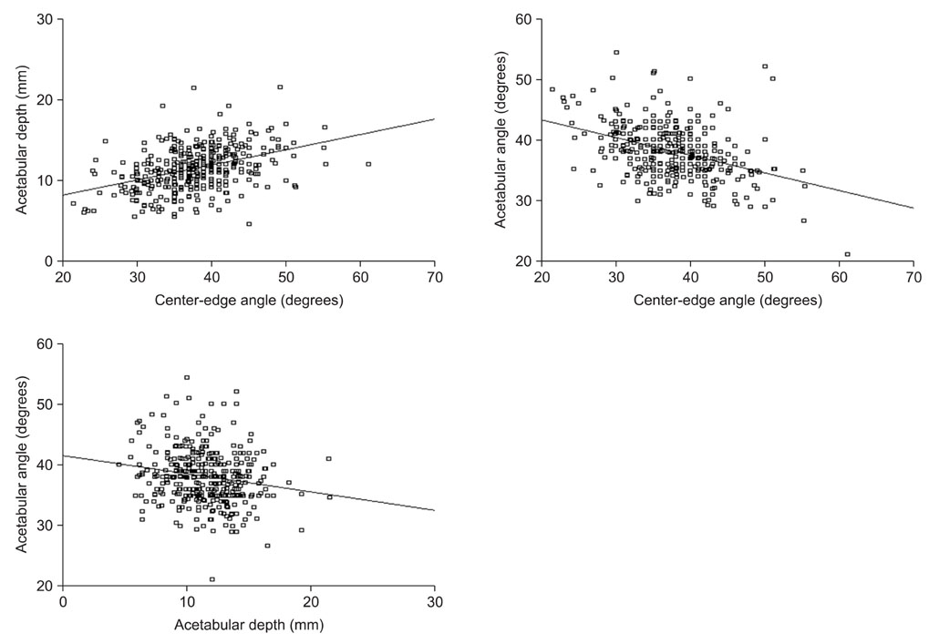
Fig. 1 The correlations between the radiological parameters of hip dysplasia.
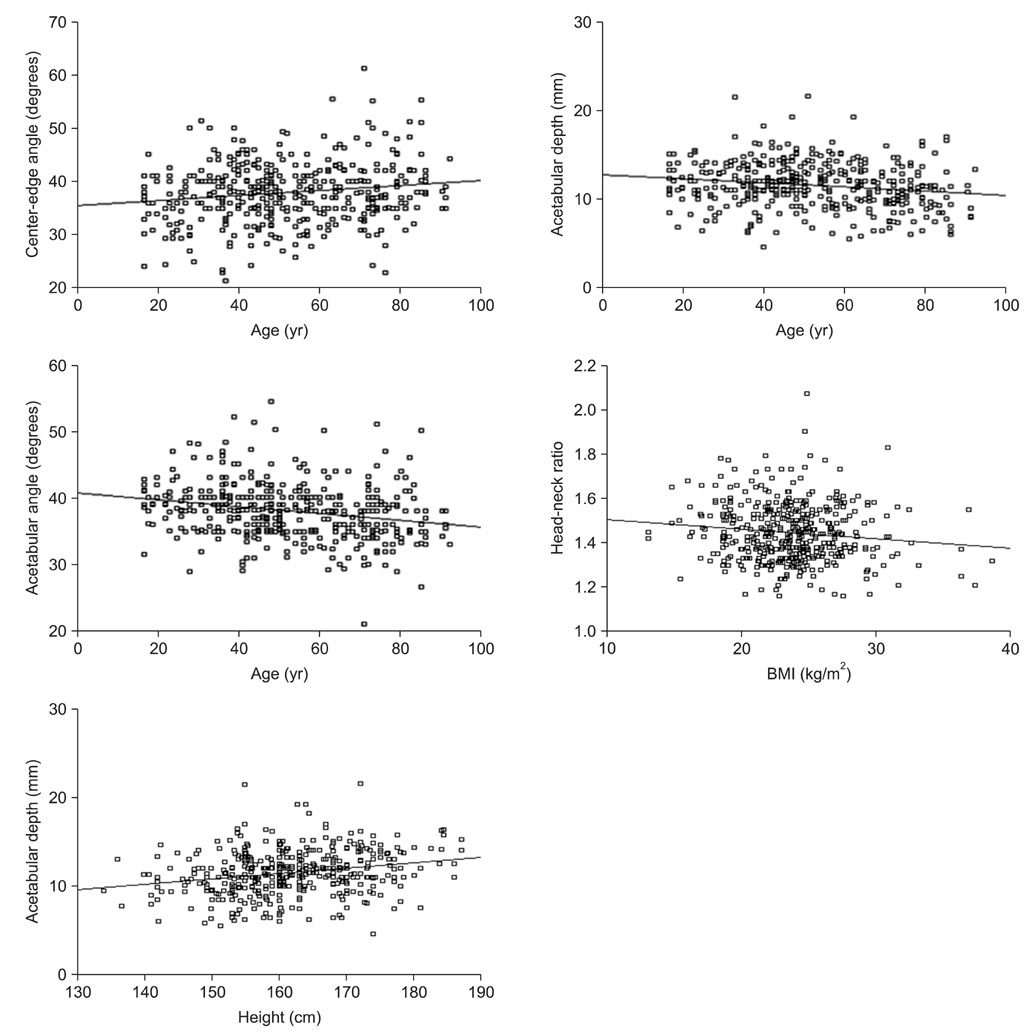
Fig. 2 The correlation between age and the radiological parameters of hip dysplasia.
Reference
-
1. Murphy SB, Ganz R, Muller ME. The prognosis in untreated dysplasia of the hip: a study of radiographic factors that predict the outcome. J Bone Joint Surg Am. 1995. 77(7):985–989.2. Reijman M, Hazes JM, Pols HA, Koes BW, Bierma-Zeinstra SM. Acetabular dysplasia predicts incident osteoarthritis of the hip: the Rotterdam study. Arthritis Rheum. 2005. 52(3):787–793.
Article3. Doherty M, Courtney P, Doherty S, et al. Nonspherical femoral head shape (pistol grip deformity), neck shaft angle, and risk of hip osteoarthritis: a case-control study. Arthritis Rheum. 2008. 58(10):3172–3182.
Article4. Jessel RH, Zurakowski D, Zilkens C, Burstein D, Gray ML, Kim YJ. Radiographic and patient factors associated with pre-radiographic osteoarthritis in hip dysplasia. J Bone Joint Surg Am. 2009. 91(5):1120–1129.
Article5. Daysal GA, Goker B, Gonen E, et al. The relationship between hip joint space width, center edge angle and acetabular depth. Osteoarthritis Cartilage. 2007. 15(12):1446–1451.
Article6. Im GI, Kim JY. Radiological joint space width in the clinically normal hips of a Korean population. Osteoarthritis Cartilage. 2010. 18(1):61–64.
Article7. Kellgren JH, Lawrence JS. Radiological assessment of osteoarthrosis. Ann Rheum Dis. 1957. 16(4):494–502.
Article8. Bland JM, Altman DG. Statistical methods for assessing agreement between two methods of clinical measurement. Lancet. 1986. 1(8476):307–310.
Article9. Wiberg G. Studies on dysplastic acetabula and congenital subluxation of the hip joint with special reference to the complication of osteo-arthritis. Acta Chir Scand. 1939. 83:Suppl 58. 1–135.10. Murray RO. The aetiology of primary osteoarthritis of the hip. Br J Radiol. 1965. 38(455):810–824.
Article11. Fredensborg N. The CE angle of normal hips. Acta Orthop Scand. 1976. 47(4):403–405.
Article12. Sharp IK. Acetabular dysplasia: the acetabular angle. J Bone Joint Surg Br. 1961. 43(2):268–272.13. Han CD, Yoo JH, Lee WS, Choe WS. Radiographic parameters of acetabulum for dysplasia in Korean adults. Yonsei Med J. 1998. 39(5):404–408.
Article14. Goker B, Sancak A, Arac M, Shott S, Block JA. The radiographic joint space width in clinically normal hips: effects of age, gender and physical parameters. Osteoarthritis Cartilage. 2003. 11(5):328–334.
Article
- Full Text Links
-
- Actions
-
Cited
- CITED
-
- Close
- Share
-
- Similar articles
-
- Femoral Revisional Arthroplasty using Proximal Modular Femoral Stem: Short Term Follow Up
- Dynamic ultrasonography in developmental dysplasia of the hip treated with Pavlik harness
- Total Hip Arthroplasty with Use of Proximal Modular Femoral Stem in Secondary Coxarthrosis of Hip Associated with Deformed Femur
- Varus Positined Femoral Stem in Cementless Total Hip Arthroplasty
- Total Hip Revision with Proximal Modular Femoral Stem
