Effect of Imaging Time in the Magnetic Resonance Detection of Intracerebral Metastases Using Single Dose Gadobutrol
- Affiliations
-
- 1Department of Radiology, Konkuk University Medical Center, Konkuk University School of Medicine, Seoul 143-914, Korea. mdmoonwj@naver.com
- KMID: 1711490
- DOI: http://doi.org/10.3348/kjr.2014.15.1.145
Abstract
OBJECTIVE
To compare the effect of imaging time delay on the MR detection of intracerebral metastases using single dose gadobutrol.
MATERIALS AND METHODS
Twenty-one patients with intracerebral metastases underwent contrast-enhanced MR with three-dimensional T1-weighted sequence at 1 minute, 5 minutes and 10 minutes after a single dose injection of gadobutrol. One hundred index metastatic lesions (1 to 30 mm; median, 7 mm) were chosen for the analysis. For the qualitative analysis, lesion conspicuity were assessed on a 1 (worst) to 5 (best) scale of the index lesions by an expert reader. For the quantitative analysis, signal intensity (SI) of enhancing lesions and normal parenchyma was measured to determine the contrast rate (CR, %) ([postcontrast SI lesion - postcontrast SI white matter] x 100 / postcontrast SI white matter) and the enhancement rate (ER, %) ([postcontrast SI lesion - baseline SI gray matter] x 100 / baseline SI gray matter). Statistical comparisons were made between three different time delays.
RESULTS
Lesion conspicuity did not differ significantly among the three time delays (p = 0.097). Although the SI, CR and ER of lesions did not reveal any significant difference between 1 minute and 5 minutes delayed images, both the 1 minute and 5 minutes delayed images showed significantly higher CRs of lesions compared with the 10 minutes delayed images (p = 0.004 and p = 0.001, respectively).
CONCLUSION
With single dose gadobutrol, imaging time delay did not have an effect on lesion conspicuity. Both 1-minute and 5-minute-delayed imaging after gadobutrol injection appears to be effective for the detection of intracerebral metastases.
MeSH Terms
-
Adult
Aged
Aged, 80 and over
Brain Neoplasms/*diagnosis/*secondary
Contrast Media/administration & dosage/*diagnostic use
Female
Humans
Image Enhancement/methods
Magnetic Resonance Imaging/*methods
Male
Middle Aged
Observer Variation
Organometallic Compounds/administration & dosage/*diagnostic use
Time Factors
Contrast Media
Organometallic Compounds
Figure
-
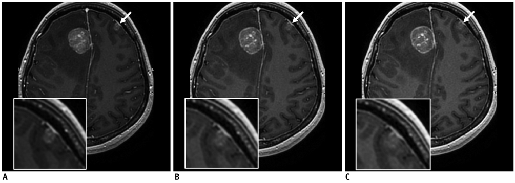
Fig. 1 28-year-old woman displaying brain metastases from breast cancer. Axial contrast-enhanced 3D-FSPGR images at 1 minute (A), 5 minutes (B), and 10 minutes (C) after injection of 1.0-mol/L gadobutrol show enhancing brain metastasis, which shows higher contrast enhancement in 1 minute and 5 minutes delayed images than in 10 minutes delayed images (arrows). This lesion in 10 minutes delayed image was missed in initial reading session. Larger enhancing mass shows good contrast enhancement at any time-point. 3D = three-dimensional, FSPGR = fast spoiled gradient echo
Cited by 2 articles
-
Application of Synthetic MRI for Direct Measurement of Magnetic Resonance Relaxation Time and Tumor Volume at Multiple Time Points after Contrast Administration: Preliminary Results in Patients with Brain Metastasis
Koung Mi Kang, Seung Hong Choi, Moonjung Hwang, Roh-Eul Yoo, Tae Jin Yun, Ji-hoon Kim, Chul-Ho Sohn
Korean J Radiol. 2018;19(4):783-791. doi: 10.3348/kjr.2018.19.4.783.Does Multiphasic Contrast Enhanced Fluid Attenuated Inversion Recovery Magnetic Resonance Imaging Enhance the Detectability of Small Intracerebral Metastases?
Jung Hwan Kim, Kyung Sik Yi, Chi-Hoon Choi, Seung Tae Woo, Sang-Hoon Cha
J Korean Soc Radiol. 2018;78(3):179-189. doi: 10.3348/jksr.2018.78.3.179.
Reference
-
1. Attenberger UI, Runge VM, Jackson CB, Baumann S, Birkemeier K, Michaely HJ, et al. Comparative evaluation of lesion enhancement using 1 M gadobutrol vs. 2 conventional gadolinium chelates, all at a dose of 0.1 mmol/kg, in a rat brain tumor model at 3 T. Invest Radiol. 2009; 44:251–256.2. Kim ES, Chang JH, Choi HS, Kim J, Lee SK. Diagnostic yield of double-dose gadobutrol in the detection of brain metastasis: intraindividual comparison with double-dose gadopentetate dimeglumine. AJNR Am J Neuroradiol. 2010; 31:1055–1058.3. Healy ME, Hesselink JR, Press GA, Middleton MS. Increased detection of intracranial metastases with intravenous Gd-DTPA. Radiology. 1987; 165:619–624.4. Byrne TN. Imaging of gliomas. Semin Oncol. 1994; 21:162–171.5. Uysal E, Erturk SM, Yildirim H, Seleker F, Basak M. Sensitivity of immediate and delayed gadolinium-enhanced MRI after injection of 0.5 M and 1.0 M gadolinium chelates for detecting multiple sclerosis lesions. AJR Am J Roentgenol. 2007; 188:697–702.6. Maravilla KR, Maldjian JA, Schmalfuss IM, Kuhn MJ, Bowen BC, Wippold FJ 2nd, et al. Contrast enhancement of central nervous system lesions: multicenter intraindividual crossover comparative study of two MR contrast agents. Radiology. 2006; 240:389–400.7. Jacobs AH, Kracht LW, Gossmann A, Rüger MA, Thomas AV, Thiel A, et al. Imaging in neurooncology. NeuroRx. 2005; 2:333–347.8. Suh CH, Kim HS, Choi YJ, Kim N, Kim SJ. Prediction of Pseudoprogression in Patients with Glioblastomas Using the Initial and Final Area Under the Curves Ratio Derived from Dynamic Contrast-Enhanced T1-Weighted Perfusion MR Imaging. AJNR Am J Neuroradiol. 2013; 34:2278–2286.9. Furutani K, Harada M, Mawlan M, Nishitani H. Difference in enhancement between spin echo and 3-dimensional fast spoiled gradient recalled acquisition in steady state magnetic resonance imaging of brain metastasis at 3-T magnetic resonance imaging. J Comput Assist Tomogr. 2008; 32:313–319.10. Moon WJ. Measurement of signal-to-noise ratio in MR imaging with sensitivity encoding. Radiology. 2007; 243:908–909.11. Anzalone N, Gerevini S, Scotti R, Vezzulli P, Picozzi P. Detection of cerebral metastases on magnetic resonance imaging: intraindividual comparison of gadobutrol with gadopentetate dimeglumine. Acta Radiol. 2009; 50:933–940.12. Benner T, Reimer P, Erb G, Schuierer G, Heiland S, Fischer C, et al. Cerebral MR perfusion imaging: first clinical application of a 1 M gadolinium chelate (Gadovist 1.0) in a double-blinded randomized dose-finding study. J Magn Reson Imaging. 2000; 12:371–380.13. Knopp MV, Runge VM, Essig M, Hartman M, Jansen O, Kirchin MA, et al. Primary and secondary brain tumors at MR imaging: bicentric intraindividual crossover comparison of gadobenate dimeglumine and gadopentetate dimeglumine. Radiology. 2004; 230:55–64.14. Yuh WT, Tali ET, Nguyen HD, Simonson TM, Mayr NA, Fisher DJ. The effect of contrast dose, imaging time, and lesion size in the MR detection of intracerebral metastasis. AJNR Am J Neuroradiol. 1995; 16:373–380.15. Barrett T, Brechbiel M, Bernardo M, Choyke PL. MRI of tumor angiogenesis. J Magn Reson Imaging. 2007; 26:235–249.16. Ocak I, Baluk P, Barrett T, McDonald DM, Choyke P. The biologic basis of in vivo angiogenesis imaging. Front Biosci. 2007; 12:3601–3616.17. Kuhl CK, Mielcareck P, Klaschik S, Leutner C, Wardelmann E, Gieseke J, et al. Dynamic breast MR imaging: are signal intensity time course data useful for differential diagnosis of enhancing lesions? Radiology. 1999; 211:101–110.18. Kramer H, Runge VM, Naul LG, Loynachan AT, Reiser MF, Wintersperger BJ. Brain MRI with single-dose (0.1 mmol/kg) Gadobutrol at 1.5 T and 3 T: comparison with 0.15 mmol/kg Gadoterate meglumine. AJR Am J Roentgenol. 2010; 194:1337–1342.
- Full Text Links
-
- Actions
-
Cited
- CITED
-
- Close
- Share
-
- Similar articles
-
- A Comparison of Low-Dose and Normal-Dose Gadobutrol in MR Renography and Renal Angiography
- Comparison of Contrast-Enhanced T2 FLAIR and 3D T1 Black-Blood Fast Spin-Echo for Detection of Leptomeningeal Metastases
- Does Multiphasic Contrast Enhanced Fluid Attenuated Inversion Recovery Magnetic Resonance Imaging Enhance the Detectability of Small Intracerebral Metastases?
- Contrast-enhanced Magnetic Resonance Imaging of Brain Metastases at 7.0T versus 1.5T: A Preliminary Result
- Diffusion-Weighted Magnetic Resonance Imaging of Spine
