Development of (1)H-(31)P Animal RF Coil for pH Measurement Using a Clinical MR Scanner
- Affiliations
-
- 1Department of Radiology, Severance Hospital, YUHS-KRIBB Medical Convergence Research Institute, Yonsei University College of Medicine, Seoul, Korea. jss@yuhs.ac
- 2Molecular Imaging & Therapy Branch, National Cancer Center, Gyeonggi-do, Korea.
- 3GE Healthcare. Global Applied Science Laboratory, Korea.
- 4Department of Radiology, Severance Hospital, Yonsei University College of Medicine, Seoul, Korea.
- 5Nanomedical National Core Research Center, Yonsei University, Seoul, Korea.
- KMID: 1683780
- DOI: http://doi.org/10.13104/jksmrm.2014.18.1.52
Abstract
- PURPOSE
To establish a pH measurement system for a mouse tumor study using a clinical scanner, to develop the (1)H and (31)P radio frequency (RF) coil system and to test pH accuracy with phantoms.
MATERIALS AND METHODS
The (1)H and the (31)P surface coils were designed to acquire signals from mouse tumors. Two coils were positioned orthogonally for geometric decoupling. The pH values of various pH phantoms were calculated using the (1)H decoupled (31)P MR spectrum with the Henderson-Hasselbalch equation. The calculated pH value was compared to that of a pH meter.
RESULTS
The mutual coil coupling was shown in a standard S12. Coil coupling (S12) were -73.0 and -62.3 dB respectively. The signal-to-noise ratio (SNR) obtained from the homogeneous phantom (1)H image was greater than 300. The high resolution in vivo mice images were acquired using a (31)P-decoupled (1)H coil. The pH values calculated from the (1)H-decoupled (31)P spectrum correlated well with the values measured by pH meter (R(2)=0.97).
CONCLUSION
Accurate pH values can be acquired using a (1)H-decoupled (31)P RF coil with a clinical scanner. This two-surface coil system could be applied to other nuclear MRS or MRI.
Keyword
Figure
-
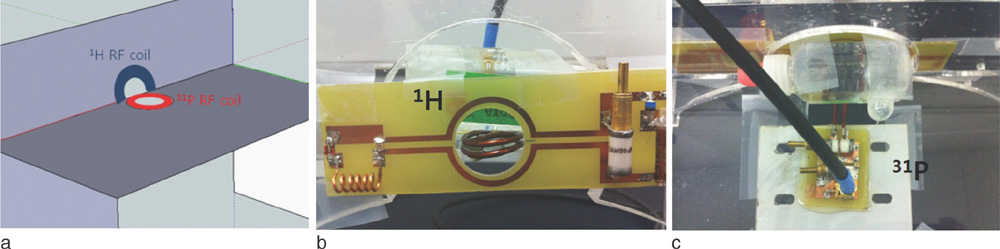
Fig. 1 The set-up of 1H and 31P RF coils. The two RF coil planes were located orthogonally for geometric decoupling (a). Photo of a 1H receive-only coil (b) and a 31P transmit-and-receive coil (c).
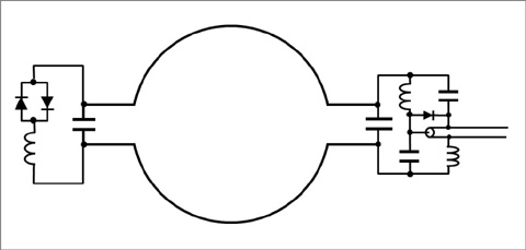
Fig. 2 The 1H receive-only coil scheme. Active decoupling consists of a PIN diode switching design and passive decoupling with crossed high-speed switching diodes and a detuning inductor.
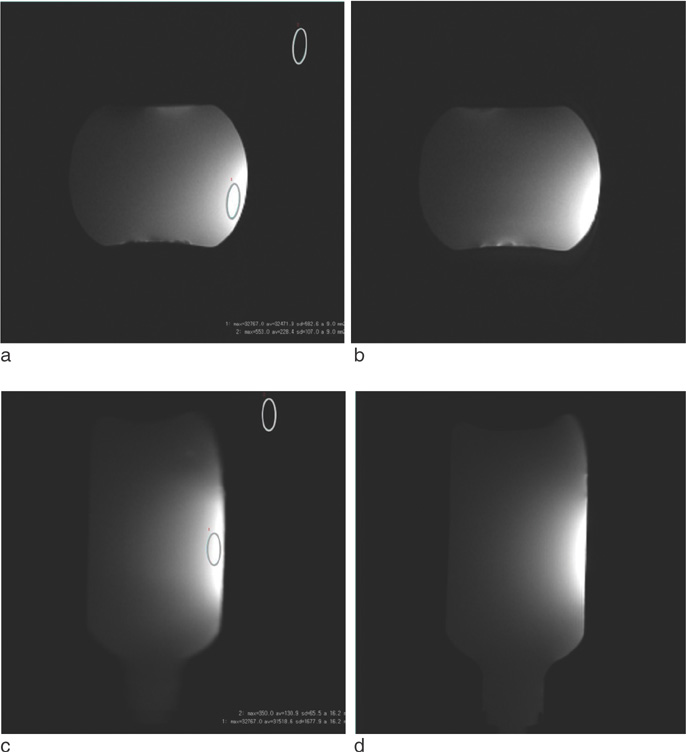
Fig. 3 The 1H spin-echo images of a homogeneous phantom: (a, b) axial images (TR/TE =167/15 ms, FOV 6 × 6 cm2, 256 × 256), (c, d) coronal images (TR/TE = 150/15 ms, FOV 8 × 8 cm2, 256 × 256). The SNR was measured at ROI (gray ellipse) in (a) and (c).
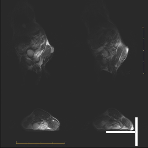
Fig. 4 The T2 weighted in vivo mouse images acquired from a 1H receive-only coil with a Fast Recovery Fast Spin-Echo pulse sequence. The parameters of these images were TR=3500 ms, TE=89.3 (coronal)/88.4 (axial) ms, thickness 4 mm, FOV 12 × 12 cm2, matrix 256 × 256 (coronal), 256 × 224 (axial) 2 NEX. One segment of the scale bar denotes 10 mm. The two white bars show the positions of the 1H and 31P RF coils.

Fig. 5 (a) 31P MR spectra of the phosphorus phantom. The pH determined by 31P MRS was 7.6 for the upper spectrum and 7.03 for the lower spectrum. (b) Comparison of pH values determined by 31P MRS and pH meter.
Reference
-
1. Glunde K, Bhujwalla ZM. Metabolic tumor imaging using magnetic resonance spectroscopy. Semin Oncol. 2011; 38:26–41.2. Stubbs M, Bhujwalla ZM, Tozer GM, et al. An assessment of 31P MRS as a method of measuring pH in rat tumours. NMR Biomed. 1992; 5:351–359.3. Zhou R, Bansal N, Leeper DB, Glickson JD. Intracellular acidification of human melanoma xenografts by the respiratory inhibitor m-iodobenzylguanidine plus hyperglycemia: a 31P magnetic resonance spectroscopy study. Cancer Res. 2000; 60:3532–3236.4. Gallagher FA, Kettunen MI, Brindle KM. Imaging pH with hyperpolarized 13C. NMR Biomed. 2011; 24:1006–1015.5. Mason RP, Antich PP, Babcock EE, Gerberich JL, Nunnally RL. Perfluorocarbon imaging in vivo: a 19F MRI study in tumor-bearing mice. Magn Reson Imaging. 1989; 7:475–485.6. McSheehy PM, Seymour MT, Ojugo AS, et al. A pharmacokinetic and pharmacodynamic study in vivo of human HT29 tumours using 19F and 31P magnetic resonance spectroscopy. Eur J Cancer. 1997; 33:2418–2427.7. Gillies RJ, Raghunand N, Garcia-Martin ML, Gatenby RA. pH imaging. A review of pH measurement methods and applications in cancers. IEEE Eng Med Biol Mag. 2004; 23:57–56.8. Iessi E, Marino ML, Lozupone F, Fais S, Milito AD. Tumor acidity and malignancy: novel aspects in the design of antitumor therapy. Cancer Ther. 2008; 6:55–66.9. Hashim AI, Zhang X, Wojtkowiak JW, Martinez GV, Gillies RJ. Imaging pH and metastasis. NMR Biomed. 2011; 24:582–591.10. Gillies RJ, Raghunand N, Karczmar GS, Bhujwalla ZM. MRI of the tumor microenvironment. J Magn Reson Imaging. 2002; 16:430–450.11. Negendank W. Studies of human tumors by MRS: a review. NMR Biomed. 1992; 5:303–324.12. Bottomley PA, Hardy CJ, Roemer PB, Mueller OM. Proton-decoupled, Overhauser-enhanced, spatially localized carbon-13 spectroscopy in humans. Magn Reson Med. 1989; 12:348–363.13. Merkle H, Wei HR, Garwood M, Ugurbil K. B1-insensitive heteronuclear adiabatic polarization transfer for signal enhancement. J Magn Reson. 1992; 99:480–494.14. Adriany G, Gruetter R. A half-volume coil for efficient proton decoupling in humans at 4 Tesla. J Magn Reson. 1997; 125:178–184.15. WMIC 2013 P266 New Design of dual Tuned RF coil for Fluorine MR Molecular Imaging.16. Barberi EA, Gati JS, Rutt BK, Menon RS. A transmit-only/receive-only (TORO) RF system for high-field MRI/MRS applications. Magn Reson Med. 2000; 43:284–289.17. Cassidy PJ, Clarke K, Edwards DJ. Determining the tuning and matching requirements of RF coils using electromagnetic simulation and electric circuit analysis. Concepts Magn Reson Part B Magn Reson Eng. 2005; 25:27–41.18. De Graaf RA. In vivo NMR Spectroscopy - Principles and Techniques. 2nd ed. John Wiley & Sons Ltd;2007. p. 80–82.19. Larcombe-McDouall J, Buttell N, Harrison N, Wray S. In vivo pH and metabolite changes during a single contraction in rat uterine smooth muscle. J Physiol. 1999; 518:783–790.20. Babic SI, Akyel C. Calculating mutual inductance between circular coils with inclined axes in air. IEEE Trans Magn. 2008; 44:1743–1750.21. Grover FW. Inductance Calculations. New York: Dover;1964. p. 193–208.
- Full Text Links
-
- Actions
-
Cited
- CITED
-
- Close
- Share
-
- Similar articles
-
- Dual Birdcage RF Coil for Leg MR Angiography
- Analysis of Endcap Effect for MRI Birdcage RF Coil by FDTD Method
- Development of Solenoid RF Coil for Animal Imaging in 3T High Magnetic Field MRI
- Improvement of a 4-Channel Spiral-Loop RF Coil Array for TMJ MR Imaging at 7T
- The Effect of Coating Material of Copper-wire RF Coil on the Signal-to-Noise Ratio in MR Images
