Thoracic Pedicle Screw Insertion in Scoliosis Using Posteroanterior C-arm rotation Method
- Affiliations
-
- 1Department of Orthopedic Surgery, Asan Medical Center, College of Medicine, Ulsan University, Seoul, Korea. cslee@amc.seoul.kr
- 2Department of Orthopedic Surgery, National Police Hospital, Seoul, Korea.
- KMID: 1583455
- DOI: http://doi.org/10.4184/jkss.2005.12.2.123
Abstract
- STUDY DESIGN: A prospective study of the accuracy of thoracic pedicle screws inserted in scoliotic patients.
OBJECTIVES
To evaluate and present a practical, safe and accurate method for thoracic pedicle screw insertion in the surgical treatment of scoliosis using the posteroanterior c-arm fluoroscopy rotating method. SUMMARY OF LITERATURE REVIEW: Previous studies have emphasized the clinical importance, yet difficulty, of accurate thoracic pedicle screw insertion in scoliotic patients. Three-dimensional alterations in the pedicle orientation of scoliotic patients makes the accurate insertion challenging. No reports exist on the accuracy and benefits of posteroanterior c-arm fluoroscopy, which is rotated to allow visualization from en face, in real patients.
MATERIALS AND METHODS
A total of 350 thoracic pedicle screws were inserted in 29 patients, including 24 with idiopathic scoliosis, using the posteroanterior (PA) c-arm rotation method. The smallest patient weighed 14 kg, and the next smallest 17 kg. The average preoperative curve was 60.9 degrees(range, 45 degrees~101 degrees). CT scans were taken, postoperatively, in the transverse and sagittal sections to evaluate the pedicle screw placement.
RESULTS
The mean preoperative curve of 60.9 degrees was corrected to 15.4 degrees(range, 3 degrees~45 degrees) in the coronal plane, a correction of 74.7%. A mean of 12.1 thoracic screws were inserted per patient. On analysis of the postoperative CT scans, 39(11.1%) of the 350 screws penetrated the medial or lateral pedicle cortices, 8(2.3%) into the medial cortex and 31(8.9%) into the lateral cortex, by mean distances of 3.3 and 3.6 mm, respectively. No screws penetrated the inferior or superior cortices in the sagittal plane, but 16(4.6%) penetrated the anterior cortex. No neurological or vascular complications were encountered, and none of the screws required subsequent replacement.
CONCLUSIONS
Thoracic pedicle screw insertion in scoliotic patients, using a posteroanterior c-arm rotation method, allows the en face visualization of both pedicles by rotating the c-arm to compensate for rotational deformity, which makes it a practical, simple and safe method.
MeSH Terms
Figure
-
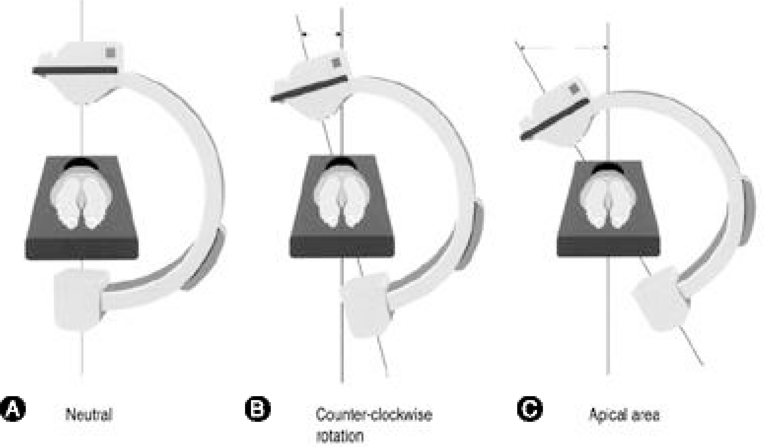
Fig. 1. Positioning of the c-arm. (A) Place in the posteroanterior position. (B) Rotate the c-arm according to the rotation of the pedicles until they are seen symmetrically on both sides. Most patients require counter-clockwise rotation (when viewed from the feet). (C) More counter-clockwise rotation is usually required at the apical vertebra.
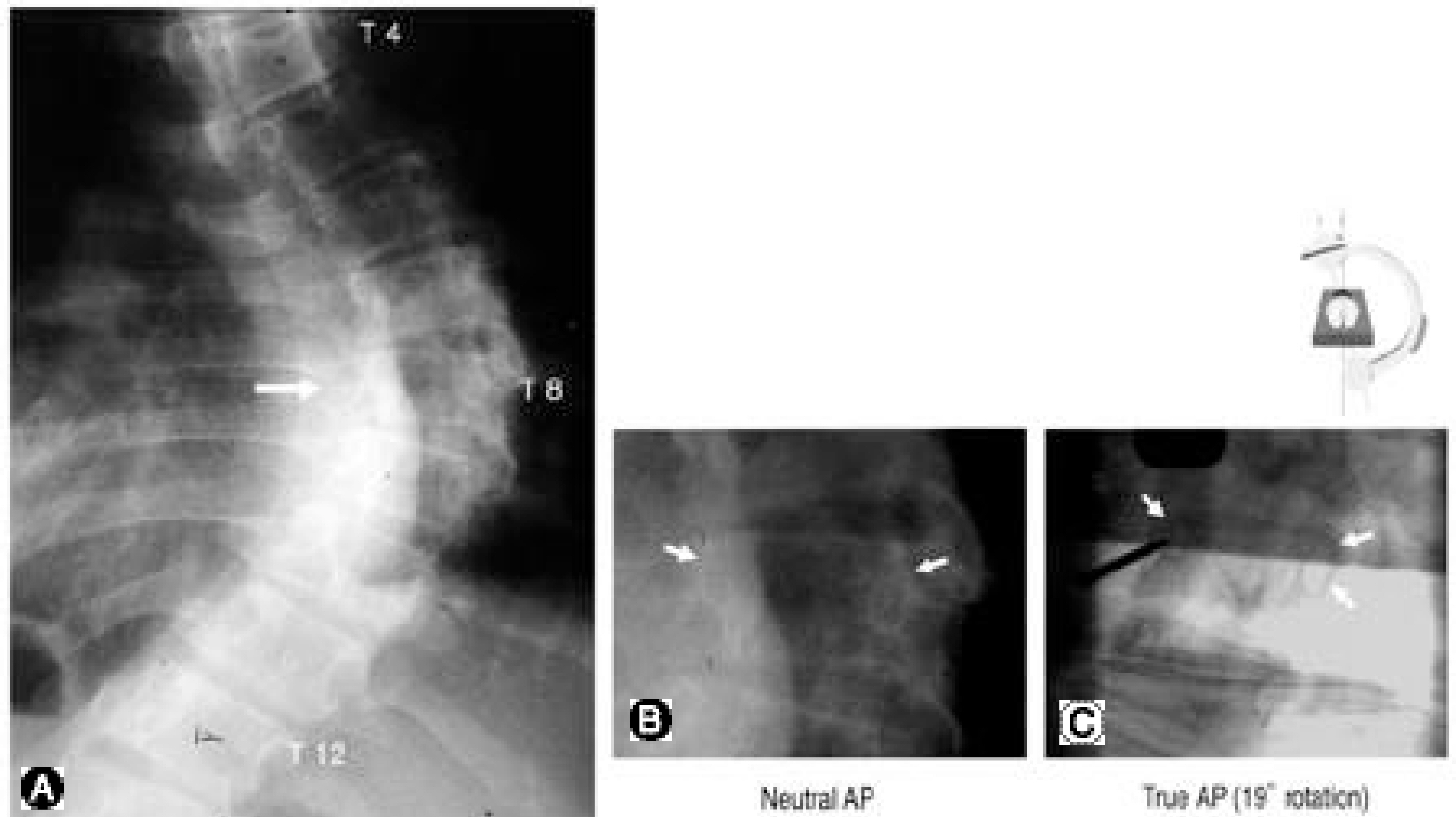
Fig. 2. (A) Case with a 69 degrees Cobb’s angle. (B) Without c-arm rotation, posteroanterior fluoroscopy reveals asymmetrical visualization of both pedicles with a Nash-Moe grade II deformity. (C) 19 degrees counter-clockwise rotation of the c-arm pro-vides en face visualization of both pedicles. This image is familiar to spine surgeons and entry sites for pedicle screw insertion is now easily determined. Entry of the right pedicle at 10 o’ clock and the left pedicle at 2 o’ clock is initiated.
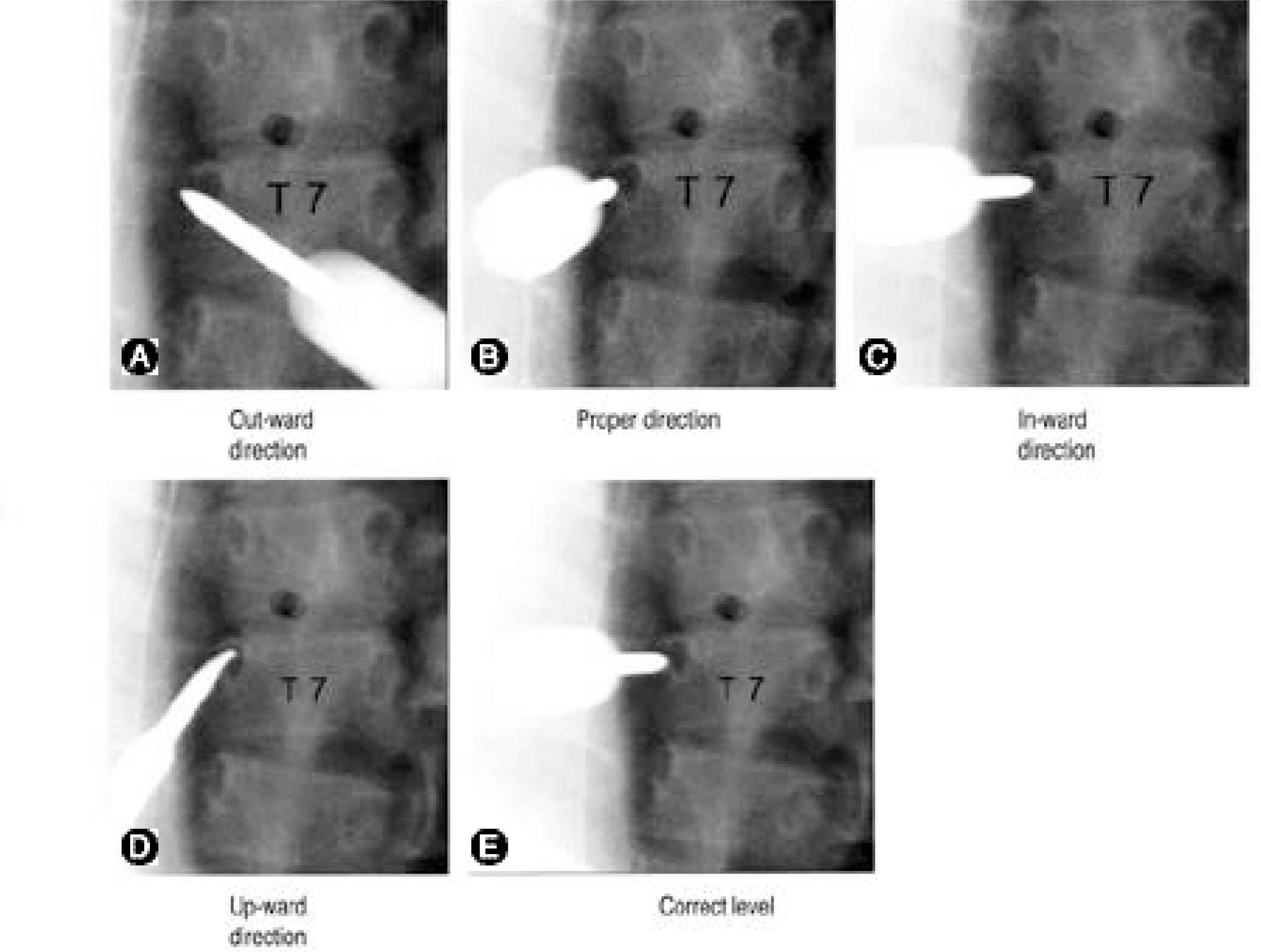
Fig. 3. Pedicle screw trajectory can be evaluated with the PA imagery because insertion is performed with both pedicles en face. (A) Axial image revealing: lateral deviation of screw trajectory. (B) Proper trajectory. (C) Medial deviation. (D) Up-ward direction. (E) Correct trajectory.
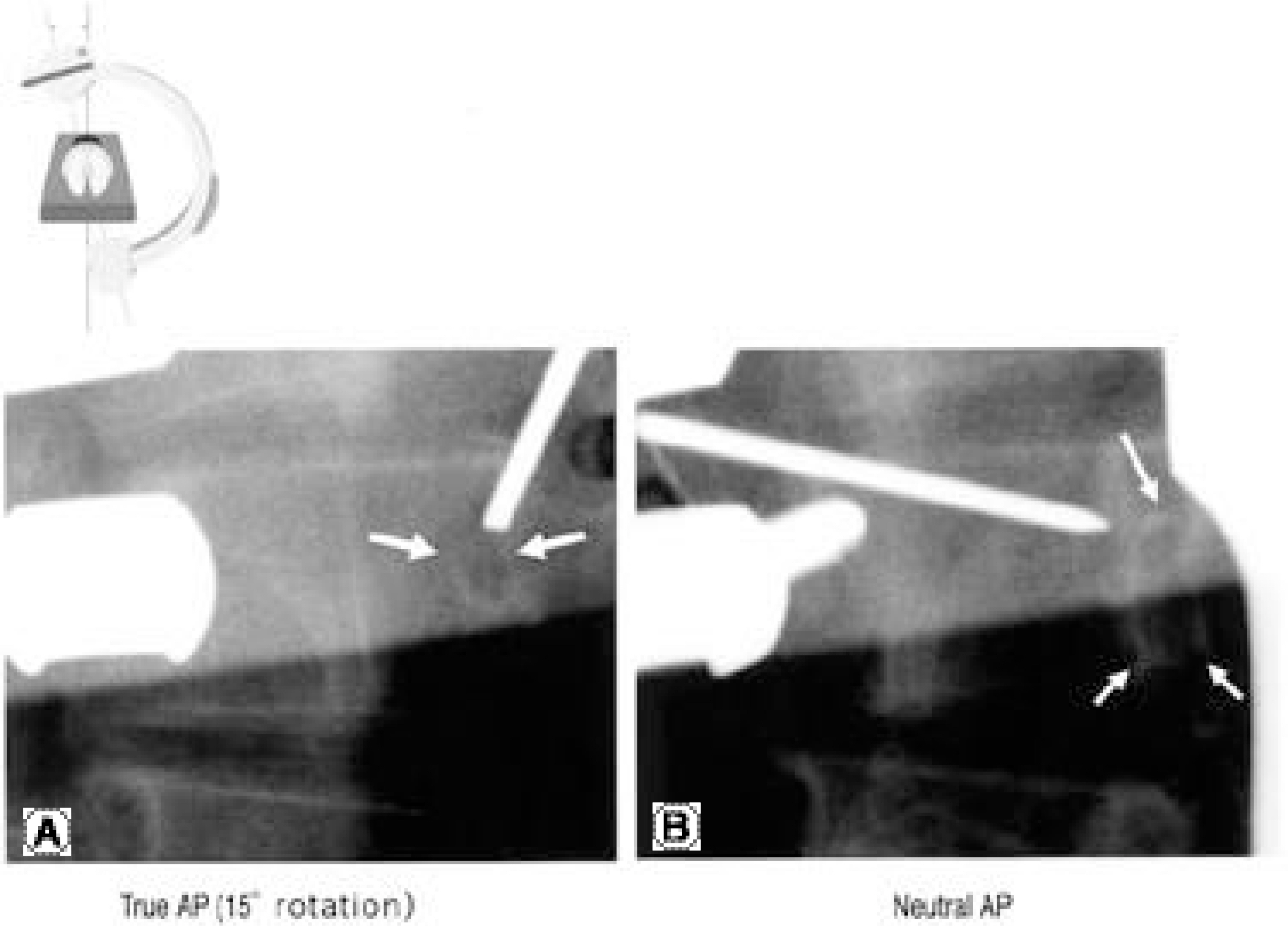
Fig. 4. Comparison of screw insertion using biplanar radiographs and c-arm rotation. (A) With the c-arm rotated 15 degrees, both pedicles are symmetrical and a K-wire is inserted into the right pedicle at 2 o’ clock. The entry point and trajectory can be accurately evaluated. (B) Same K-wire without no c-arm rotation. The entry point is 3 mm medial to the pedicle and the trajectory can not be easily evaluated.
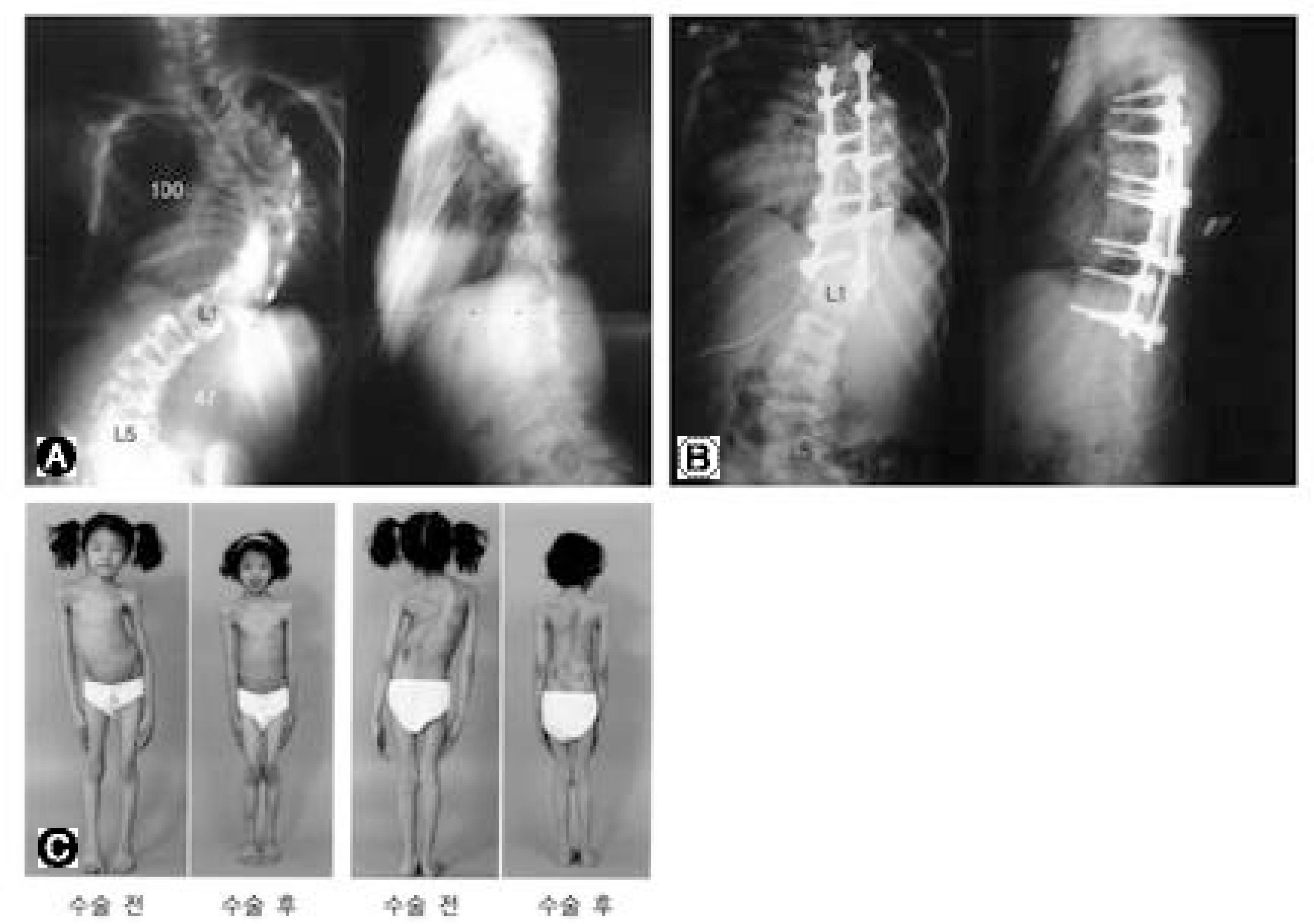
Fig. 5. A 14 kg scoliosis patient using the rotating posteroanterior c-arm fluoroscopic guidance technique. (A) The patient with severe imbalance and a Cobb's angle of 100 degrees. (B) Pedicle screws used to correct the thoracic curve. (C) Improvement of truncal imbalance post-operatively.
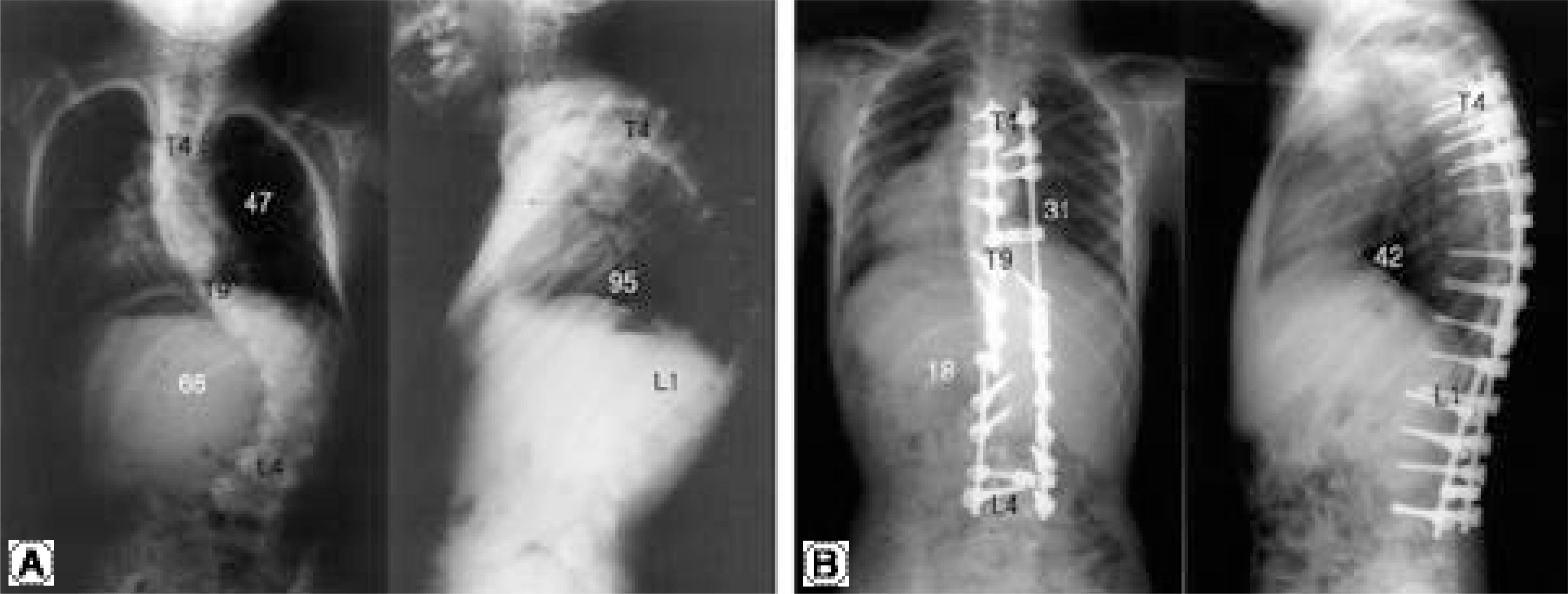
Fig. 6. A 17 kg kyphosis patient using the rotating posteroanterior c-arm fluoroscopic guidance technique. (A) The patient with 95 degrees kyphosis and 47 degrees scoliosis of the thoracic spine. (B) An almost normal curvature after pedicle screw insertion and correction of T4-L4.
Reference
-
1). Vaccaro AR, Rizzolo SJ, Balderston RA, et al. Placement of pedicle screws in the thoracic spine. Part II: An anatomical and radiographic assessment. J Bone Joint Surg Am. 1995; 77(8):1200–1206.2). Lenke LG, Rinella A, YJ K. Freehand thoracic pedicle screw placement. Seminars in Spine Surgery. 2002; 14:48–57.3). Liljenqvist U, Hackenberg L, Link T, Halm H. Pullout strength of pedicle screws versus pedicle and laminar hooks in the thoracic spine. Acta Orthop Belg. 2001; 67(2):157–163.4). Liljenqvist U, Lepsien U, Hackenberg L, Niemeyer T, Halm H. Comparative analysis of pedicle screw and hook instrumentation in posterior correction and fusion of idiopathic thoracic scoliosis. Eur Spine J. 2002; 11(4):336–343.
Article5). Suk SI, Kim WJ, Lee SM, Kim JH, Chung ER. Thoracic pedicle screw fixation in spinal deformities: are they really safe? Spine. 2001; 26(18):2049–2057.6). Belmont PJ Jr, Klemme WR, Dhawan A, Polly DW Jr. In vivo accuracy of thoracic pedicle screws. Spine. 2001; 26(21):2340–2346.
Article7). Ebraheim NA, Jabaly G, Xu R, Yeasting RA. Anatomic relations of the thoracic pedicle to the adjacent neural structures. Spine. 1997; 22(14):1553–1556.
Article8). Cinotti G, Gumina S, Ripani M, Postacchini F. Pedicle instrumentation in the thoracic spine. A morphometric and cadaveric study for placement of screws. Spine. 1999; 24(2):114–119.9). Gertzbein SD, Robbins SE. Accuracy of pedicular screw placement in vivo. Spine. 1990; 15(1):11–14.
Article10). Mirza SK, Wiggins GC, Kuntz CT, et al. Accuracy of thoracic vertebral body screw placement using standard fluoroscopy, fluoroscopic image guidance, and computed tomographic image guidance: a cadaver study. Spine. 2003; 28(4):402–413.11). Zindrick MR, Wiltse LL, Widell EH, et al. A biomechanical study of intrapeduncular screw fixation in the lumbosacral spine. Clin Orthop. 1986; 203:99–112.
Article12). O’ Brien MF, Lenke LG, Mardjetko S, et al. Pedic le morphology in thoracic adolescent idiopathic scoliosis: is pedicle fixation an anatomically viable technique? Spine. 2000; 25(18):2285–93.13). Liljenqvist U, Link T, Halm H. Morphometric analysis of thoracic and lumbar vertebrae in idiopathic scoliosis. Spine. 2000; 25(10):1247–1253.
Article14). Liljenqvist U, Allkemper T, Hackenberg L. Analysis of vertebral morphology in idiopathic scoliosis with use of magnetic resonance imaging and multiplanar reconstruction. J Bone Joint Surg Am. 2002; 84-A(3):359–368.
Article15). Kim YJ, Lenke LG, Bridwell KH, Cho YS, Riew KD. Free hand pedicle screw placement in the thoracic spine: Is it safe? Spine. 2004; 29(3):333–342.
Article16). Liljenqvist U, Halm H, Link T. Pedicle screw instrumentation of the thoracic spine in idiopathic scoliosis. Spine. 1997; 22(19):2239–2245.
Article17). Xu R, Ebraheim NA, Ou Y, Yeasting RA. Anat omi c considerations of pedicle screw placement in the thoracic spine. Roy-Camille technique versus open-lamina tech nique. Spine. 1998; 23(9):1065–1068.18). Kim KD, Patrick J, Bloch BO, Masciopinto JE. Com -puter-assisted thoracic pedicle screw placement: an in vitro feasibility study. Spine. 2001; 26(4):360–364.19). Youkilis AS, Quint DJ, McGillicuddy JE, Papadopoulos SM. Stereotactic navigation for placement of pedicle screws in the thoracic spine. Neurosurgery. 2001; 48(4):771–778.
Article20). Foley KT, Simon DA, Rampersaud YR. Virtual fluoroscopy: computer- assisted fluoroscopic navigation. Spine. 2001; 26(4):347–351.21). Rampersaud YR. Advanced image guidance for the placement of thoracic pedicle screws. Seminars in Spine Surgery. 2002; 14:58–65.
- Full Text Links
-
- Actions
-
Cited
- CITED
-
- Close
- Share
-
- Similar articles
-
- Scoliosis Correction with Thoracic Pedicle Screws: Posteroanterior C-arm Rotation Method
- Pedicle Screw Instrumentation for Adolescent Idiopathic Scoliosis: The Insertion Technique, the Fusion Levels and Direct Vertebral Rotation
- Safety of Pedicle Screws in Adolescent Idiopathic Scoliosis Surgery
- Clinical Results of Selective Thoracic Fusion by Segmental Pedicle Screws in King type II Adolescent Idiopathic Scoliosis ( AIS )
- Changes in Vertebral Rotation Following Segmental Pedicle Screw Instrumentation and Rod Derotation in Idiopathic Thoracic Scoliosis : Part I - CT Evaluation
