Usefulness of Additional Delayed Regional F-18 Fluorodeoxyglucose Positron Emission Tomography in the Lymph Node Staging of Non-Small Cell Lung Cancer Patients
- Affiliations
-
- 1Department of Nuclear Medicine and Thoracic Surgery, Seoul National University College of Medicine, Seoul, Korea. jkchung@plaza.snu.ac.kr
Abstract
- PURPOSE
In this study, we examined whether additional, delayed regional FDG PET scans could increase the accuracy of the lymph node staging of NSCLC patients. MATERIALS AND METHODS: Among 87 patients who underwent open thoracotomy or mediastinoscopic biopsy under the suspicion of NSCLC, 35 (32 NSCLC and 3 infectious diseases) who had visible lymph nodes on both preoperative whole body scan and regional FDG PET scan were included. The following 3 calculations were made for each biopsy-proven, visible lymph node: maximum SUV of whole body scan (WB SUV), maximum SUV of delayed chest regional scan (Reg SUV), and the percent change of SUV between WB and regional scans (% SUV Change). ROC curve analyses were performed for WB SUVs, Reg SUVs and % SUV Changes. RESULTS: Seventy lymph nodes (29 benign, 41 malignant) were visible on both preoperative whole bodyscan and regional scan. The means of WB SUVs, Reg SUVs and % SUV Changes of the 41 malignant nodes, 3.71+/-1.08, 5.18+/-1.60, and 42.59+/-33.41%, respectively, were all significantly higher than those of the 29 benign nodes, 2.45+/-0.73, 3.00+/-0.89, and 22.71+/-20.17%, respectively. ROC curve analysis gave sensitivity and specificity values of 80.5% and 82.8% at a cutoff of 2.89 (AUC 0.839) for WB SUVs, 87.8% and 82.8% at a cutoff of 3.61 (AUC 0.891) for Reg SUVs, and 87.8% and 41.4% at a cutoff of 12.3% (AUC 0.671) for % SUV Changes. CONCLUSION: Additional, delayed regional FDG PET scans may improve the accuracy of lymph node staging of whole body FDG PET scan by providing additional criteria of Reg SUV and % SUV Change.
MeSH Terms
Figure
-
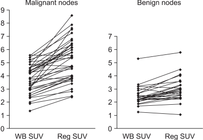
Fig. 1 Comparison of whole body maximum SUVs and regional maximum SUVs of 41 dissected malignant nodes and 29 dissected benign nodes which were visible on preoperative F-18 FDG PET.
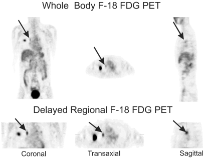
Fig. 2 Whole body F-18 FDG PET image (upper row) and delayed regional F-18 FDG PET image (lower row) of a 56-year-old man with adenocarcinoma in the upper lobe of the right lung. On the whole body PET image, only weak FDG accumulation was noted at 10R (right hilar) lymph node station; WB SUV was 1.33. On the regional PET image, however, FDG uptake was increased compared with the whole body PET image; Reg SUV was 2.39. Open thoracotomy biopsy revealed cancer cell invasion at this lymph node station.
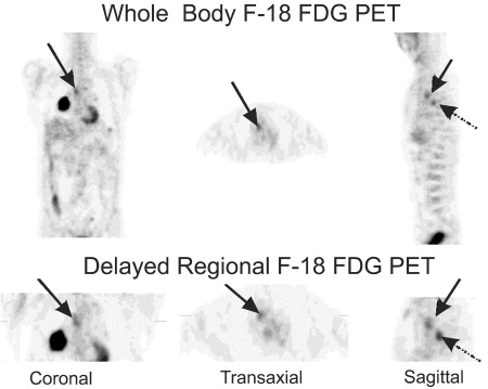
Fig. 3 Whole body F-18 FDG PET image (upper row) and delayed F-18 FDG PET image (lower row) of a 59-year-old man with squamous cell carcinoma in the middle lobe of the right lung. On the whole body PET image, FDG accumulation was noted at 4R (right lower paratracheal) lymph node station (arrows); WB SUV was 3.05. However, on the regional PET image, FDG uptake was decreased; Reg SUV was 2.57. Mediastinoscopic biopsy revealed no cancer cells in this lymph node. Dotted arrows show the 7 (subcarinal) lymph node station, which was invaded by cancer cells on mediastinoscopic biopsy. WB SUV was 3.08 and Reg SUV was 3.77.
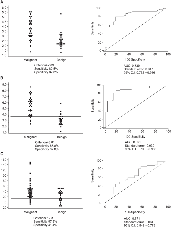
Fig. 4 (A) Plot of WB SUVs of 41 visible malignant nodes and 29 visible benign nodes (left) and ROC curve of WB SUVs (right). For a cut-off value of 2.89 WB SUV, the sensitivity was 80.5% and the specificity 82.8%. (B) Plot of Reg SUVs of the same nodes (left) and the ROC curve of Reg SUVs (right). For a cut-off value of 3.61 Reg SUV, the sensitivity was 87.8% and the specificity 82.8%. (C) Plot of percent change of SUVs (% SUV Changes) of the same nodes (left) and the ROC curve of % SUV Changes (right). For a cut-off value of 12.3 % SUV Change, the sensitivity was 87.8% and the specificity 41.4.
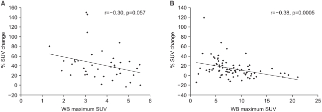
Fig. 5 (A) Correlation between WB SUVs and % SUV Changes of 41 dissected malignant nodes which were visible on FDG PET. Although there was a trend toward negative correlation, it did not reach statistical significance (r=-0.30, p=0.057). (B) Correlation between WB SUVs and % SUV Changes of 82 malignant primary lung lesions. A significant negative correlation was found (r=-0.38, p=0.0005).
Reference
-
1. Hagge RJ, Wong TZ, Coleman RE. Positron emission tomography: brain tumors and lung cancer. Radiol Clin North Am. 2001; 39:871–881. PMID: 11587059.2. Seltzer MA, Yap CS, Silverman DH, Meta J, Schiepers C, Phelps ME, et al. The impact of PET on management of lung cancer: the referring physician's perspective. J Nucl Med. 2002; 43:752–756. PMID: 12050318.3. Hoh CK, Hawkins RA, Glaspy JA, Dahlbom M, Tse NY, Hoffman EJ, et al. Cancer detection with whole-body PET using 2-[18F]Fluoro-2-deoxy-D-glucose. J Comput Assist Tomogr. 1993; 17:582–589. PMID: 8331230.4. Lowe VJ, DeLong DM, Hoffman JM, Coleman RE. Optimum scanning protocol for FDG-PET evaluation of pulmonary malignancy. J Nucl Med. 1995; 36:883–887. PMID: 7738668.5. Fischman AJ, Alpert NM. FDG-PET in oncology: there's more to it than looking at pictures. J Nucl Med. 1993; 34:6–11. PMID: 8418272.6. Hamberg LM, Hunter GJ, Alpert NM, Choi NC, Babich JW, Fischman AJ. The dose uptake ratio as an index of glucose metabolism: useful parameter or oversimplification? J Nucl Med. 1994; 35:1308–1312. PMID: 8046485.7. Lodge MA, Lucas JD, Marsden PK, Cronin BF, ODoherty MJ, Smith MA. A PET study of 18FDG uptake in soft tissue masses. Eur J Nucl Med. 1999; 26:22–30. PMID: 9933658.8. Boerner AR, Weckesser M, Herzog H, Schmitz T, Audretsch W, Nitz U, et al. Optimal scan time for fluorine-18 fluorodeoxyglucose positron emission tomography in breast cancer. Eur J Nucl Med. 1999; 26:226–230. PMID: 10079312.
Article9. Hustinx R, Smith RJ, Benard F, Rosenthal DI, Machtay M, Farber LA, et al. Dual time point fluorine-18 fluorodeoxyglucose positron emission tomography: a potential method to differentiate malignancy from inflammation and normal tissue in the head and neck. Eur J Nucl Med. 1999; 26:1345–1348. PMID: 10541835.
Article10. Zhuang H, Pourdehnad M, Lambright ES, Yamamoto AJ, Lanuti M, Li P, et al. Dual time point 18F-FDG PET imaging for differentiating malignant from inflammatory processes. J Nucl Med. 2001; 42:1412–1417. PMID: 11535734.11. Matthies A, Hickeson M, Cuchiara A, Alavi A. Dual time point 18F-FDG PET for the evaluation of pulmonary nodules. J Nucl Med. 2002; 43:871–875. PMID: 12097455.12. Kubota R, Kubota K, Yamada S, Tada M, Ido T, Tamahashi N. Microautoradiographic study for the differentiation of intratumoral macrophages, granulation tissues and cancer cells by the dynamics of fluorine-18-fluorodeoxyglucose uptake. J Nucl Med. 1994; 35:104–112. PMID: 8271030.13. Mountain CF, Dresler CM. Regional lymph node classification for lung cancer staging. Chest. 1997; 111:1718–1723. PMID: 9187199.
Article14. National Electrical manufacturers Association. NEMA Standards Publication NU2-1994. 1994. Washington DC: National Electrical Manufacturers Association.15. Reivich M, Alavi A, Wolf A, Fowler J, Russell J, Arnett C, et al. Glucose metabolic rate kinetic model parameter determination in humans: the lumped constants and rate constants for [18F]fluorodeoxyglucose and [11C]deoxyglucose. J Cereb Blood Flow Metab. 1985; 5:179–192. PMID: 3988820.16. Nolop KB, Rhodes CG, Brudin LH, Beaney RP, Krausz T, Jones T, et al. Glucose utilization in vivo by human pulmonary neoplasms. Cancer. 1987; 60:2682–2689. PMID: 3499969.17. Kubota K, Itoh M, Ozaki K, Ono S, Tashiro M, Yamaguchi K, et al. Advantage of delayed whole-body FDG-PET imaging for tumour detection. Eur J Nucl Med. 2001; 28:696–703. PMID: 11440029.
Article18. Yamada S, Kubota K, Kubota R, Ido T, Tamahashi N. High accumulation of fluorine-18-fluorodeoxyglucose in turpentine-induced inflammatory tissue. J Nucl Med. 1995; 36:1301–1306. PMID: 7790960.19. Demura Y, Tsuchida T, Ishizaki T, Mizuno S, Totani Y, Ameshima S, et al. 18F-FDG accumulation with PET for differentiation between benign and malignant lesions in the thorax. J Nucl Med. 2003; 44:540–548. PMID: 12679397.
- Full Text Links
-
- Actions
-
Cited
- CITED
-
- Close
- Share
-
- Similar articles
-
- 18F-2-Deoxy-2-Fluoro-D-Glucose Positron Emission Tomography: Computed Tomography for Preoperative Staging in Gastric Cancer Patients
- The Role of PET in Staging Non-Small Cell Lung Cancer
- Reliability of 18F-Fluorodeoxyglucose Positron Emission Tomography/Computed Tomography in the Nodal Staging of Colorectal Cancer Patients
- The Usefulness of FDG-PET/CT for the Prediction of Regional Lymph Node Metastases in Colorectal Cancer
- Staging of Lung Cancer
