Tuberc Respir Dis.
2013 Apr;74(4):187-190. 10.4046/trd.2013.74.4.187.
Nontuberculous Mycobacterial Lung Disease Caused by Mycobacterium lentiflavum in a Patient with Bronchiectasis
- Affiliations
-
- 1Division of Pulmonary and Critical Care Medicine, Department of Medicine, Samsung Medical Center, Sungkyunkwan University School of Medicine, Seoul, Korea. wjkoh@skku.edu
- 2Department of Microbiology, Chungnam National University College of Medicine, Daejeon, Korea.
- 3Department of Microbiology, Yonsei University College of Medicine, Seoul, Korea.
- KMID: 1495860
- DOI: http://doi.org/10.4046/trd.2013.74.4.187
Abstract
- We report a rare case of lung disease caused by Mycobacterium lentiflavum in a previously healthy woman. A 54-year-old woman was referred to our hospital due to chronic cough and sputum. A computed tomography scan of the chest revealed bilateral bronchiectasis with bronchiolitis in the right middle lobe and the lingular division of the left upper lobe. Nontuberculous mycobacteria were isolated twice from three expectorated sputum specimens. All isolates were identified as M. lentiflavum by multilocus sequence analysis based on rpoB, hsp65, and 16S rRNA fragments. To the best of our knowledge, this is the first documented case of M. lentiflavum lung disease in an immunocompetent adult in Korea.
MeSH Terms
Figure
-
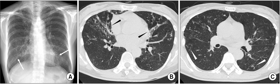
Figure 1 A 54-year-old woman with bronchiectasis and nontuberculous mycobacterial lung disease caused by Mycobacterium lentiflavum. (A) A chest radiography reveals bilateral multifocal tram-track signs (white arrows). (B) A transverse chest computed tomography (CT) scan (2.5-mm-section thickness) on the level with the right inferior pulmonary vein reveals bilateral bronchiectasis (black arrowheads) in the right middle lobe and the lingular segment of the left upper lobe. (C) A chest CT scan obtained on the level with the superior segmental bronchus of the left lower lobe reveals bilateral bronchiolitis in both lungs (white arrowheads).
Reference
-
1. Springer B, Wu WK, Bodmer T, Haase G, Pfyffer GE, Kroppenstedt RM, et al. Isolation and characterization of a unique group of slowly growing mycobacteria: description of Mycobacterium lentiflavum sp. nov. J Clin Microbiol. 1996. 34:1100–1107.2. Marshall HM, Carter R, Torbey MJ, Minion S, Tolson C, Sidjabat HE, et al. Mycobacterium lentiflavum in drinking water supplies, Australia. Emerg Infect Dis. 2011. 17:395–402.3. Haase G, Kentrup H, Skopnik H, Springer B, Bottger EC. Mycobacterium lentiflavum: an etiologic agent of cervical lymphadenitis. Clin Infect Dis. 1997. 25:1245–1246.4. Safdar A, Han XY. Mycobacterium lentiflavum, a recently identified slow-growing mycobacterial species: clinical significance in immunosuppressed cancer patients and summary of reported cases of infection. Eur J Clin Microbiol Infect Dis. 2005. 24:554–558.5. Tortoli E, Mattei R, Russo C, Scarparo C. Mycobacterium lentiflavum, an emerging pathogen? J Infect. 2006. 52:e185–e187.6. Tortoli E, Bartoloni A, Erba ML, Levre E, Lombardi N, Mantella A, et al. Human infections due to Mycobacterium lentiflavum. J Clin Microbiol. 2002. 40:728–729.7. Molteni C, Gazzola L, Cesari M, Lombardi A, Salerno F, Tortoli E, et al. Mycobacterium lentiflavum infection in immunocompetent patient. Emerg Infect Dis. 2005. 11:119–122.8. Shamaei M, Marjani M, Farnia P, Tabarsi P, Mansouri D. Human infections due to Mycobacterium lentiflavum: first report in Iran. Iran J Microbiol. 2010. 2:27–29.9. Shin S, Yoon JH, Song SH, Kim EC. Isolation of Mycobacterium lentiflavum from a patient with a lung destroyed by tuberculosis. Korean J Lab Med. 2007. 27:124–127.10. Adekambi T, Drancourt M. Dissection of phylogenetic relationships among 19 rapidly growing Mycobacterium species by 16S rRNA, hsp65, sodA, recA and rpoB gene sequencing. Int J Syst Evol Microbiol. 2004. 54(Pt 6):2095–2105.11. Telenti A, Marchesi F, Balz M, Bally F, Bottger EC, Bodmer T. Rapid identification of mycobacteria to the species level by polymerase chain reaction and restriction enzyme analysis. J Clin Microbiol. 1993. 31:175–178.12. Devulder G, Perouse de Montclos M, Flandrois JP. A multigene approach to phylogenetic analysis using the genus Mycobacterium as a model. Int J Syst Evol Microbiol. 2005. 55(Pt 1):293–302.13. Benson DA, Karsch-Mizrachi I, Lipman DJ, Ostell J, Wheeler DL. GenBank. Nucleic Acids Res. 2007. 35:D21–D25.14. Harmsen D, Rothganger J, Frosch M, Albert J. RIDOM: Ribosomal Differentiation of Medical Micro-organisms Database. Nucleic Acids Res. 2002. 30:416–417.15. Griffith DE, Aksamit T, Brown-Elliott BA, Catanzaro A, Daley C, Gordin F, et al. An official ATS/IDSA statement: diagnosis, treatment, and prevention of nontuberculous mycobacterial diseases. Am J Respir Crit Care Med. 2007. 175:367–416.
- Full Text Links
-
- Actions
-
Cited
- CITED
-
- Close
- Share
-
- Similar articles
-
- Nontuberculous Mycobacterial Lung Disease Caused by Mycobacterium terrae in a Patient with Bronchiectasis
- Nontuberculous Mycobacterial Lung Disease Caused by Mycobacterium shinjukuense: The First Reported Case in Korea
- Isolation of Mycobacterium lentiflavum from a Patient with a Lung Destroyed by Tuberculosis
- The First Korean Case of Nontuberculous Mycobacterial Lung Disease Caused by Mycobacterium abscessus Subspecies bolletii in a Patient with Bronchiectasis
- Diagnosis and treatment of nontuberculous mycobacterial lung disease
