J Korean Endocr Soc.
2008 Oct;23(5):327-331. 10.3803/jkes.2008.23.5.327.
A Case of Hyalinizing Trabecular Tumor of the Thyroid Gland Misdiagnosed as Medullary Carcinoma at Cytologic Examination
- Affiliations
-
- 1Department of Endocrinology and Metabolism, Kyung Hee University School of Medicine, Korea.
- 2Research Institute of Endocrinology, Kyung Hee University School of Medicine, Korea.
- 3Department of Pathology, Kyung Hee University School of Medicine, Korea.
- KMID: 1479225
- DOI: http://doi.org/10.3803/jkes.2008.23.5.327
Abstract
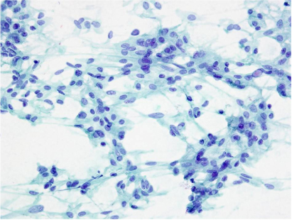
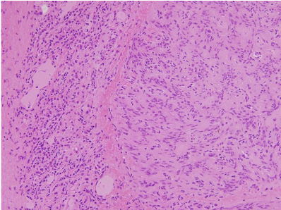
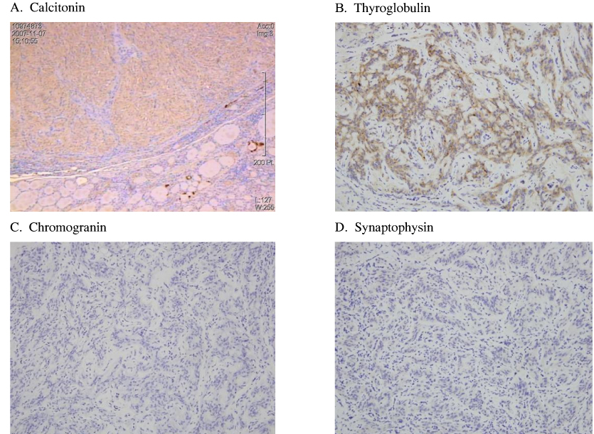
- A hyalinizing trabecular tumor (HTT) is a rare benign thyroid tumor that can present as a solitary thyroid nodule, a prominent nodule in a multinodular goiter, or as an incidental finding within a thyroidectomy specimen. The clinical importance of this entity is that it is frequently misdiagnosed as papillary carcinoma or medullary carcinoma on fine-needle aspiration cytology or histopathologic examination. The cytology of HTT is characterized by hypercellularity, nuclear grooves, nuclear pseudoinclusions, and powdery chromatin of the tumor cells, which is frequently seen in papillary carcinomas. The histologic findings of the tumor show polygonal and spindle cells arranged in a trabecular growth pattern with the presence of a variable hyalinized stroma. Calcitonin and other neuroendocrine markers can be used to differentiate HTT from medullary carcinoma. MIB-1, galectin-3, or other cytokeratin markers help to exclude papillary carcinoma. We report a patient with a thyroid tumor misdiagnosed as a medullary carcinoma on fine-needle aspiration and finally diagnosed as HTT after total thyroidectomy and immunohistochemical examination.
MeSH Terms
Figure
Reference
-
1. Carney JA, Ryan J, Goellner JR. Hyalinizing trabecular adenoma of the thyroid gland. Am J Surg Pathol. 1987. 11:583–591.2. Yim HE, Shim C, Soh EY. Hyalinizing trabecular adenoma of the thyroid-A case report. Korean J Pathol. 1998. 32:226–230.3. Lee HK, Kim HS, Hur MH, Kang SS, Lee JH, Lee SK. Hyalinizing trabecular adenoma of thyroid gland. J Korean Surg Soc. 2002. 62:87–90.4. Park KS, Kim SW, Min HS, Han WS, Noh DY, Park SH, Youn YK, Oh SK, Choe KJ. Hyalinizing trabecular adenoma of thyroid. J Korean Surg Soc. 2003. 65:572–575.5. Hirokawa M, Carney JA, Ohtsuki Y. Hyalinizing trabecular adenoma and papillary carcinoma of the thyroid gland express different cytokeratin patterns. Am J Surg Pathol. 2000. 24:877–881.6. Hirokawa M, Carney JA. Cell membrane and cytoplasmic staining for MIB-1 in hyalinizing trabecular adenoma of the thyroid gland. Am J Surg Pathol. 2000. 24:575–578.7. Gaffney RL, Carney JA, Sebo TJ, Erickson LA, Volante M, Papotti M, Lloyd RV. Galectin-3 expression in hyalinizing trabecular tumors of the thyroid gland. Am J Surg Pathol. 2003. 27:494–498.8. Salvatore G, Chiappetta G, Nikiforov YE, Decaussin-Petrucci M, Fusco A, Carney JA, Santoro M. Molecular profile of hyalinizing trabecular tumours of the thyroid: high prevalence of RET/PTC rearrangements and absence of B-raf and N-ras point mutations. Eur J Cancer. 2005. 41:816–821.9. Molberg K, Albores-Saavedra J. Hyalinizing trabecular carcinoma of the thyroid gland. Hum Pathol. 1994. 25:192–197.10. McCluggage WG, Sloan JM. Hyalinizing trabecular carcinoma of thyroid gland. Histopathology. 1996. 28:357–362.11. Papotti M, Riella P, Montemurro F, Pietribiasi F, Bussolati G. Immunophenotypic heterogeneity of hyalinizing trabecular tumours of the thyroid. Histopathology. 1997. 31:525–533.12. Gonzalez-Campora R, Fuentes-Vaamonde E, Hevia-Vazquez A, Otal-Salaverri C, Villar-Rodriguez J, Galera-Davidson H. Hyalinizing trabecular carcinoma of the thyroid gland: report of two cases of follicular cell thyroid carcinoma with hyalinizing trabecular pattern. Ultrastruct Pathol. 1998. 22:39–46.13. Evenson A, Mowschenson P, Wang H, Connolly J, Mendrinos S, Parangi S, Hasselgren PO. Hyalinizing trabecular adenoma-an uncommon thyroid tumor frequently misdiagnosed as papillary or medullary thyroid carcinoma. Am J Surg. 2007. 193:707–712.
- Full Text Links
- Actions
-
Cited
- CITED
-
- Close
- Share
- Similar articles
-
- Fine Needle Aspiration Cytology of the Hyalinizing Trabecular Adenoma of the Thyroid Gland: A Case Report
- A Case of Hyalinizing Trabecular Tumor of the Thyroid Gland
- A Case of Hyalinizing Trabecular Adenoma of the Thyroid Gland
- A Case of Hyalinizing Trabecular Adenoma of the Thyroid Gland
- The Effect of a-Tocopherol in Puromycin Aminonucleoside Induced Nephropathy in Rats


