Korean J Obstet Gynecol.
2011 Dec;54(12):836-840. 10.5468/KJOG.2011.54.12.836.
A giant endometrial polyp with tamoxifen therapy in postmenopausal woman
- Affiliations
-
- 1Department of Obstetrics and Gynecology, Pusan National University School of Medicine, Busan, Korea. ghkim@pusan.ac.kr
- 2Department of Pathology, Pusan National University School of Medicine, Busan, Korea.
- 3Department of Obstetrics and Gynecology, Busan St. Mary's Medical Center, Busan, Korea.
- KMID: 1443953
- DOI: http://doi.org/10.5468/KJOG.2011.54.12.836
Abstract
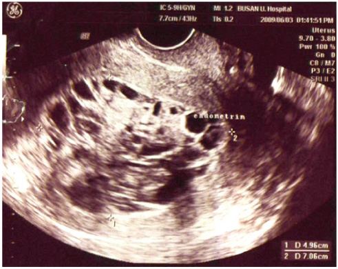
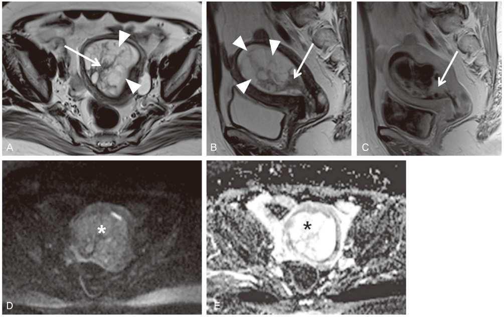
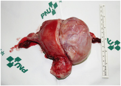
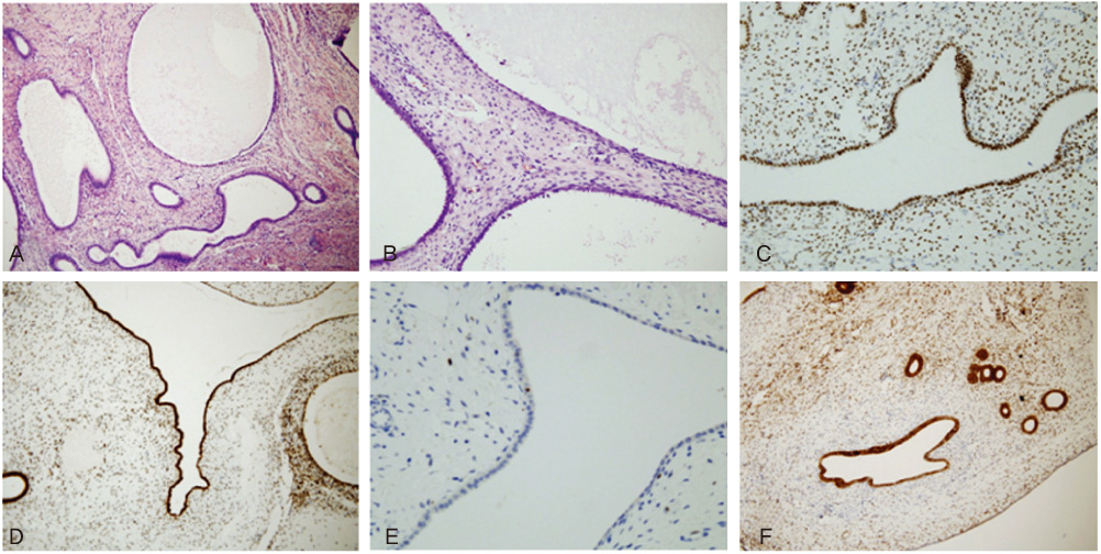
- Tamoxifen is a synthetic non-steroid anti-estrogen that has been used effectively for several years in the adjuvant treatment of breast cancer. But, the drug has been associated with development of endometrial poylp, hyperplasia and adenocarcinoma possibly mediated through its agonistic estrogen properties during the menopausal period in which estrogens are at a low level. Endometrial polyp has been described as the most common endometrial pathology in association with postmenopausal tamoxifen treatment. We present the case of woman with a giant endometrial polyp of uncommon dimension who was receiving adjuvant tamoxifen for 5 years after breast cancer surgery.
Keyword
MeSH Terms
Figure
Reference
-
1. Bergman L, Beelen ML, Gallee MP, Hollema H, Benraadt J, van Leeuwen FE. Risk and prognosis of endometrial cancer after tamoxifen for breast cancer. Comprehensive Cancer Centres' ALERT Group. Assessment of Liver and Endometrial cancer Risk following Tamoxifen. Lancet. 2000. 356:881–887.2. Erdemoglu E, Güney M, Keskin B, Mungan T. Tamoxifen and giant endometrial polyp. Eur J Gynaecol Oncol. 2008. 29:198–199.3. Killackey MA, Hakes TB, Pierce VK. Endometrial adenocarcinoma in breast cancer patients receiving antiestrogens. Cancer Treat Rep. 1985. 69:237–238.4. Lee KE, Ko YB, Noh HT, Suh KS. Endometrial pathologies in tamoxifen-treated breast cancer patients. Korean J Obstet Gynecol. 2008. 51:757–765.5. Cohen I, Azaria R, Shapira J, Yigael D, Tepper R. Significance of secondary ultrasonographic endometrial thickening in postmenopausal tamoxifen-treated women. Cancer. 2002. 94:3101–3106.6. Cohen I, Altaras MM, Shapira J, Tepper R, Rosen DJ, Cordoba M, et al. Time-dependent effect of tamoxifen therapy on endometrial pathology in asymptomatic postmenopausal breast cancer patients. Int J Gynecol Pathol. 1996. 15:152–157.7. Bonadona V, Mignotte H, Chauvin F, Lesur A, Mauriac L, Granon C, et al. Tamoxifen and endometrial cancer: the results from a multicenter case controlled study. Bull Cancer. 1996. 83:507–508.8. Fisher B, Costantino JP, Redmond CK, Fisher ER, Wickerham DL, Cronin WM. Endometrial cancer in tamoxifen-treated breast cancer patients: findings from the National Surgical Adjuvant Breast and Bowel Project (NSABP) B-14. J Natl Cancer Inst. 1994. 86:527–537.9. McGurgan P, Taylor LJ, Duffy SR, O'Donovan PJ. Does tamoxifen therapy affect the hormone receptor expression and cell proliferation indices of endometrial polyps? An immunohistochemical comparison of endometrial polyps from postmenopausal women exposed and not exposed to tamoxifen. Maturitas. 2006. 54:252–259.10. Choe BH, Choi EK, Kim YT, Kim JW, Park BW. Cases with endometrial polyp and endocervical polyp associated with tamoxifen use. Korean J Obstet Gynecol. 2000. 43:725–730.11. Franchi M, Ghezzi F, Donadello N, Zanaboni F, Beretta P, Bolis P. Endometrial thickness in tamoxifen-treated patients: an independent predictor of endometrial disease. Obstet Gynecol. 1999. 93:1004–1008.12. Neven P, De Muylder X, Van Belle Y, Vanderick G, De Muylder E. Tamoxifen and the uterus and endometrium. Lancet. 1989. 1:375.13. Mourits MJ, Van der Zee AG, Willemse PH, Ten Hoor KA, Hollema H, De Vries EG. Discrepancy between ultrasonography and hysteroscopy and histology of endometrium in postmenopausal breast cancer patients using tamoxifen. Gynecol Oncol. 1999. 73:21–26.
- Full Text Links
- Actions
-
Cited
- CITED
-
- Close
- Share
- Similar articles
-
- Cases with Endometrial Polyp and Endocervical Polyp Associated With Tamoxifen Use
- The Change of Endometrial Thickness in Tamoxifen-treated Postmenopausal Breast Cancer Patients
- Analysis of Abnormally Thickened Endometrial Patterns on Trans vaginal Sonography
- Toremifene-associated endometrial polyp: A case report and review of the literature
- Tamoxifen-associated polypoid endometriosis mimicking an ovarian neoplasm


