Image Findings in Brain Developmental Venous Anomalies
- Affiliations
-
- 1Department of Neurosurgery, Seoul Paik Hospital, Inje University College of Medicine, Seoul, Korea. hanibalkms@hanmail.net
- KMID: 1441683
- DOI: http://doi.org/10.7461/jcen.2012.14.1.37
Abstract
OBJECTIVE
Developmental venous anomalies (DVAs) are benign anatomic variations; therefore, they are usually discovered incidentally. The aim of this article was to describe radiological findings of DVAs.
METHODS
A retrospective search for DVAs of the brain was performed in 1899 patients who had undergone magnetic resonance imaging (MRI) with contrast enhancement between January 1, 2005 and April 25, 2011. We also reviewed the results of computed tomography (CT), magnetic resonance angiography (MRA), CT angiography, and transfemoral cerebral angiography (TFCA) studies performed in patients with DVAs.
RESULTS
Thirty-two DVAs were identified in 31 of the 1899 patients (1.63%). These 31 patients underwent five enhanced CTs, three MRAs, two CT angiographies, and two TFCAs. Thirty of the 32 DVAs were supratentorial (ST) and two were infratentorial (IT). All enhanced MRI studies exhibited excellent resolution of DVAs. All DVAs had only one draining vein. The venous drainage system was an IT vein in three DVAs and an ST vein in 29 DVAs. Two out of five enhanced CTs presented good visualization of the draining vein. None of the MRAs, including the source image, revealed the presence of DVAs. The two CT angiographies exhibited good resolution of DVAs. One of the two TFCAs yielded an excellent illustration of the DVA.
CONCLUSION
CT angiography and MRI with contrast enhancement yielded detailed findings of DVAs. In contrast, MRA did not identify the DVAs. Enhanced CT presented only the draining vein of DVAs.
MeSH Terms
Figure
-
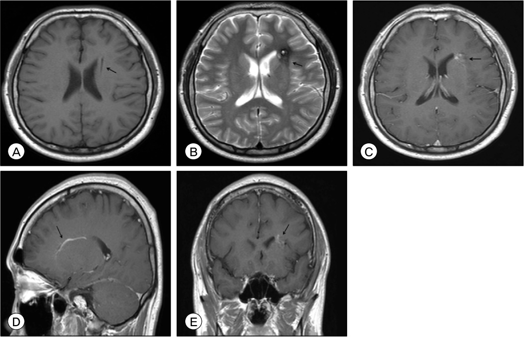
Fig. 1 A 26-year-old man presented with cerebral concussion. T1-weighted axial image (A) and T2-weighted axial image (B) show a draining vein (A, arrow) and a cavernous malformation (B, arrow) located in the left frontal lobe, respectively. T1-weighted images after contrast enhancement (C: axial, D: sagittal, E: coronal) indicate a cavernous malformation (C, arrow), a draining vein (D, arrow) into the ependymal vein and a developmental venous anomaly (E, arrow).

Fig. 2 A 29-year-old woman presented with seizure. T1-enhanced images after contrast enhancement (A: axial; B: coronal) show a draining vein (A and B, arrow) located in the right frontal lobe. T2-weighted image (C) reveals a draining vein (arrow) in the right frontal lobe. Enhanced computed tomography (D) shows a draining vein (arrow) located in the right frontal lobe. Computed tomography angiography (E) reveals a developmental venous anomaly (arrow) located in the right frontal lobe.
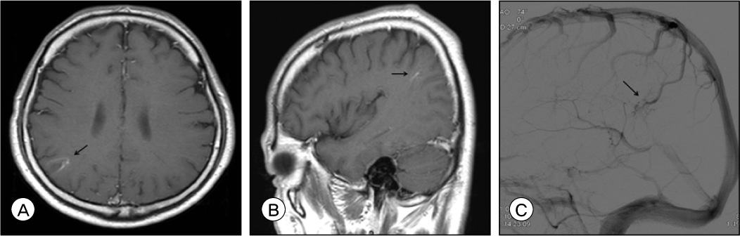
Fig. 3 A 54-year-old male underwent magnetic resonance imaging (MRI) as a routine check-up. T1-weighted images after contrast enhancement (A: axial image; B: sagittal image) show a developmental venous anomaly (DVA) (A and B, arrow) located in the right parietal lobe. Transfemoral cerebral angiography (C) reveals a DVA (arrow) with superficial draining vein into the superior sagittal sinus.
Reference
-
1. Augustyn GT, Scott JA, Olson E, Gilmor RL, Edwards MK. Cerebral venous angiomas: MR imaging. Radiology. 1985. 08. 156(2):391–395.
Article2. Hashimoto M, Yokota A, Kajiwara H, Matsuoka S, Tsukamoto Y. Venous angioma: follow-up study and therapeutic considerations. Neurol Med Chir (Tokyo). 1990. 08. 30(8):599–603.
Article3. Lasjaunias P, Burrows P, Planet C. Developmental venous anomalies (DVA): the so-called venous angioma. Neurosurg Rev. 1986. 9(3):233–242.
Article4. Lee C, Pennington MA, Kenney CM 3rd. MR evaluation of developmental venous anomalies: medullary venous anatomy of venous angiomas. AJNR Am J Neuroradiol. 1996. 01. 17(1):61–70.5. Peebles TR, Vieco PT. Intracranial developmental venous anomalies: diagnosis using CT angiography. J Comput Assist Tomogr. 1997. Jul-Aug. 21(4):582–586.6. Pereira VM, Geibprasert S, Krings T, Aurboonyawat T, Ozanne A, Toulgoat F, et al. Pathomechanisms of symptomatic developmental venous anomalies. Stroke. 2008. 12. 39(12):3201–3215.
Article7. Rammos SK, Maina R, Lanzino G. Developmental venous anomalies: current concepts and implications for management. Neurosurgery. 2009. 07. 65(1):20–29. discussion 29-30.8. Ruiz DS, Yilmaz H, Gailloud P. Cerebral developmental venous anomalies: current concepts. Ann Neurol. 2009. 09. 66(3):271–283.9. San Millán Ruíz D, Gailloud P. Cerebral developmental venous anomalies. Childs Nerv Syst. 2010. 10. 26(10):1395–1406.
Article10. Toro VE, Geyer CA, Sherman JL, Parisi JE, Brantley MJ. Cerebral venous angiomas: MR findings. J Comput Assist Tomogr. 1988. 12(6):935–940.
- Full Text Links
-
- Actions
-
Cited
- CITED
-
- Close
- Share
-
- Similar articles
-
- A Case Of Cerebellar Hemorrhage Associated with Cavernous Hemangioma and Developmental Venous Anomaly
- Associated Brain Parenchymal Abnormalities in Developmental Venous Anomalies: Evaluation with Susceptibility-weighted MR Imaging
- Developmental Venous Anomaly Presenting Intracranial Hemorrhage without Associated Vascular Anomaly
- Developmental Venous Anomalies of the Brainstem Associated with Spontaneous Vertigo: A Case Report
- Multimodal Imaging Follow-up of a Thrombosed Developmental Venous Anomaly: CT, CT Angiography and Digital Subtraction Angiography
