Multimodal Imaging Follow-up of a Thrombosed Developmental Venous Anomaly: CT, CT Angiography and Digital Subtraction Angiography
- Affiliations
-
- 1Department of Radiology, Chungbuk National University Hospital, Cheongju, Korea. shcha@chungbuk.ac.kr
- 2Department of Neurosurgery, Chungbuk National University Hospital, Cheongju, Korea.
- KMID: 1784030
- DOI: http://doi.org/10.5469/neuroint.2013.8.2.120
Abstract
- We report a rare case of thrombosed developmental venous anomaly (DVA) in a 31-year old male with hemorrhagic cerebral venous infarction at the initial clinical presentation. In this case, sequential CT, CT angiography and digital subtraction angiography demonstrated thrombotic obstruction of the venous drainage from DVA, its progressive recanalization and temporal evolution of the affected brain parenchyma. The relevant previous literatures were reviewed and summarized.
Keyword
MeSH Terms
Figure
-
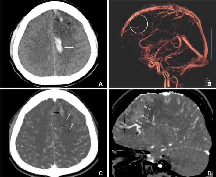
Fig. 1 CT and CT angiography (CTA) images at the day of initial clinical presentation.Multifocal hyperdense intracerebral hemorrhages (white arrow) and hypodense area (asterisk) are present in the left frontal lobe. Continuing hyperdensity (black arrow) courses from the hypodense area to the superior sagittal sinus (A). 3-dimensionally reconstructed CTA (B) shows no venous drainage to the superior sagittal sinus (white circle). Thrombosed vein (black arrow) and area of parenchymal staining, suggestive of venous congestion (white arrows) is evident on the axial source (C) and sagittal reformatted (D) images, and medullary and collecting veins in the adjacent brain (black arrowheads) are dilated and prominent (D).
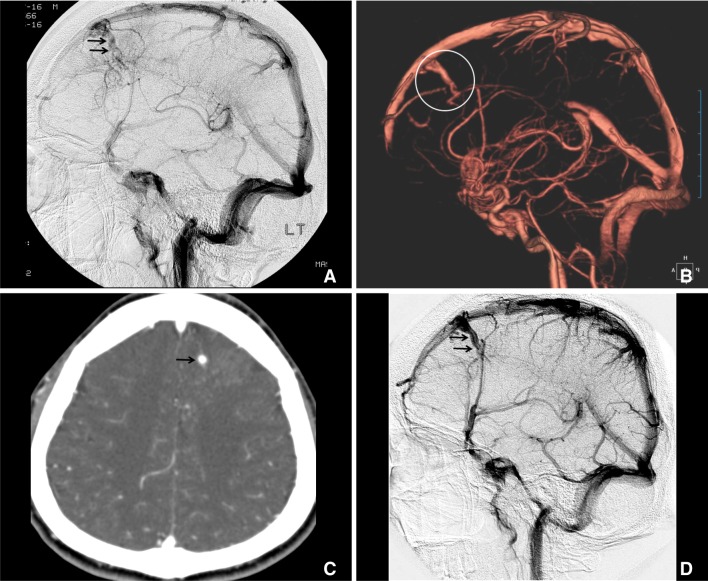
Fig. 2 Follow-up images demonstrating progressive resolution of thrombosis and recanalization of draining vein from the DVA.DSA at one month shows intravenous filling defects in the left frontal DVA (black arrows), dilated medullary veins, delayed contrast wash-out and parenchymal staining in the lateral projection image (A). CTA at two month reveals patent draining vein (white circle) on three-dimensional reconstruction (B) and axial source image (C) demonstrates corresponding contrast enhancement (black arrow). Note parenchymal staining in the left frontal lobe, evident on the initial CTA (Fig. 1C), is no longer prominent (C). 0n two-year follow-up DSA, medullary veins are not engorged and draining vein is completely recanalized (D).
Reference
-
1. Ruiz DS, Yilmaz H, Gailloud P. Cerebral developmental venous anomalies: current concepts. Ann Neurol. 2009; 66:271–283. PMID: 19798638.2. Roh JE, Cha SH, Lee SY, Lee SY, Jeon MH, Cho BS, et al. Atypical developmental venous anomaly associated with single arteriovenous fistula and intracerebral hemorrhage: a case demonstrated by superselective angiography. Korean J Radiol. 2012; 13:107–110. PMID: 22247645.
Article3. Oran I, Kiroglu Y, Yurt A, Ozer FD, Acar F, Dalbasti T, et al. Developmental venous anomaly (DVA) with arterial component: a rare cause of intracranial haemorrhage. Neuroradiology. 2009; 51:25–32. PMID: 18787814.
Article4. Fok KF, Holmin S, Alvarez H, Ozanne A, Krings T, Lasjaunias PL. Spontaneous intracerebral hemorrhage caused by an unusual association of developmental venous anomaly and arteriovenous malformation. Interv Neuroradiol. 2006; 12:113–121. PMID: 20569563.
Article5. Im SH, Han MH, Kwon BJ, Ahn JY, Jung C, Park SH, et al. Venous-predominant parenchymal arteriovenous malformation: a rare subtype with a venous drainage pattern mimicking developmental venous anomaly. J Neurosurg. 2008; 108:1142–1147. PMID: 18518718.
Article6. Sepelyak K, Gailloud P, Jordan LC. Thrombosis of a developmental venous anomaly with hemorrhagic venous infarction. Arch Neurol. 2010; 67:1028. PMID: 20697060.
Article7. Field LR, Russell EJ. Spontaneous hemorrhage from a cerebral venous malformation related to thrombosis of the central draining vein: demonstration with angiography and serial MR. AJNR Am J Neuroradiol. 1995; 16:1885–1888. PMID: 8693990.8. Yamamoto M, Inagawa T, Kamiya K, Ogasawara H, Monden S, Yano T. Intracerebral hemorrhage due to venous thrombosis in venous agioma: case report. Neurol Med Chir. 1989; 29:1044–1046.9. Seki Y, Sahara Y. Spontaneous thrombosis of a venous malformation leading to intracerebral hemorrhage: case report. Neurol Med Chir. 2007; 47:310–313.10. Masson C, Godefroy O, Leclerc X, Colombani JM, Leys D. Cerebral venous infarction following thrombosis of the draining vein of a venous angioma (developmental abnormality). Cerebrovasc Dis. 2000; 10:235–238. PMID: 10773651.
Article11. Kim P, Castellani R, Tresser N. Cerebral venous malformation complicated by spontaneous thrombosis. Childs Nerv Syst. 1996; 12:172–175. PMID: 8697463.
Article12. Merten CL, Knitelius HO, Hedde JP, Assheuer J, Bewermeyer H. Intracerebral haemorrhage from a venous angioma following thrombosis of a draining vein. Neuroradiology. 1998; 40:15–18. PMID: 9493181.
Article13. Bouchacourt E, Carpena JP, Bories J, Koussa A, Chiras J. Ischemic accident caused by thrombosis of a venous angioma. Apropos of a case. J Radiol. 1986; 67:631–635. PMID: 3795187.14. Guerrero AL, Blanco A, Arcaya J, Cach J. Venous infarct as presenting form of venous angioma of the posterior fossa. Rev Clin Esp. 1998; 198:484–485. PMID: 9737164.15. Walsh M, Parmar H, Mukherji SK, Mamourian A. Developmental venous anomaly with symptomatic thrombosis of the draining vein. J Neurosurg. 2008; 109:1119–1122. PMID: 19035729.
Article16. Gama RL, Nakayama M, Tavora DG, Bomfim RC, Carneiro TC, Pimentel LH. Thrombosed developmental venous anomaly associated with cerebral venous infarct. Arq Neuropsiquiatr. 2008; 66:560–562. PMID: 18813722.
Article17. Vieira Santos A, Saraiva P. Spontaneous isolated non-haemorrhagic thrombosis in a child with developmental venous anomaly: case report and review of the literature. Childs Nerv Syst. 2006; 22:1631–1633. PMID: 17072663.18. Lovrencic-Huzjan A, Rumboldt Z, Marotti M, Demarin V. Subarachnoid haemorrhage headache from a developmental venous anomaly. Cephalalgia. 2004; 24:763–766. PMID: 15315533.
Article19. Konan AV, Raymond J, Bourgouin P, Lesage J, Milot G, Roy D. Cerebellar infarct caused by spontaneous thrombosis of a developmental venous anomaly of the posterior fossa. AJNR Am J Neuroradiol. 1999; 20:256–258. PMID: 10094347.20. Peltier J, Toussaint P, Desenclos C, Le Gars D, Deramond H. Cerebral venous angioma of the pons complicated by nonhemorrhagic infarction. case report. J Neurosurg. 2004; 101:690–693. PMID: 15481728.
- Full Text Links
-
- Actions
-
Cited
- CITED
-
- Close
- Share
-
- Similar articles
-
- The Diagnostic Value of Digital Subtraction Angiography Considering the Pathomechanism of Symptomatic Cerebral Developmental Venous Anomaly
- A Clinical Significance of lntraarterial Digital Subtraction Angiography in Renal Diseases
- Radiologic consideration of intra-arterial digital subtraction angiography
- Cerebral Venous Thrombosis Presenting as Isolated Subarachnoid Hemorrhage
- Cerebellar Dysfunctions Associated with a Developmental Venous Anomaly
