Time Efficiency and Diagnostic Accuracy of New Automated Myocardial Perfusion Analysis Software in 320-Row CT Cardiac Imaging
- Affiliations
-
- 1Department of Radiology, Charite - Universitatsmedizin Berlin, Berlin 10117, Germany. dewey@charite.de
- KMID: 1430040
- DOI: http://doi.org/10.3348/kjr.2013.14.1.21
Abstract
OBJECTIVE
We aimed to evaluate the time efficiency and diagnostic accuracy of automated myocardial computed tomography perfusion (CTP) image analysis software.
MATERIALS AND METHODS
320-row CTP was performed in 30 patients, and analyses were conducted independently by three different blinded readers by the use of two recent software releases (version 4.6 and novel version 4.71GR001, Toshiba, Tokyo, Japan). Analysis times were compared, and automated epi- and endocardial contour detection was subjectively rated in five categories (excellent, good, fair, poor and very poor). As semi-quantitative perfusion parameters, myocardial attenuation and transmural perfusion ratio (TPR) were calculated for each myocardial segment and agreement was tested by using the intraclass correlation coefficient (ICC). Conventional coronary angiography served as reference standard.
RESULTS
The analysis time was significantly reduced with the novel automated software version as compared with the former release (Reader 1: 43:08 +/- 11:39 min vs. 09:47 +/- 04:51 min, Reader 2: 42:07 +/- 06:44 min vs. 09:42 +/- 02:50 min and Reader 3: 21:38 +/- 3:44 min vs. 07:34 +/- 02:12 min; p < 0.001 for all). Epi- and endocardial contour detection for the novel software was rated to be significantly better (p < 0.001) than with the former software. ICCs demonstrated strong agreement (> or = 0.75) for myocardial attenuation in 93% and for TPR in 82%. Diagnostic accuracy for the two software versions was not significantly different (p = 0.169) as compared with conventional coronary angiography.
CONCLUSION
The novel automated CTP analysis software offers enhanced time efficiency with an improvement by a factor of about four, while maintaining diagnostic accuracy.
Keyword
MeSH Terms
-
Aged
Analysis of Variance
Body Mass Index
Coronary Angiography
Coronary Artery Disease/*radiography
*Efficiency, Organizational
Female
Humans
Male
Middle Aged
Myocardial Perfusion Imaging/*methods
Pattern Recognition, Automated/*methods
Prospective Studies
Radiographic Image Interpretation, Computer-Assisted/*methods
*Software
Statistics, Nonparametric
Time Factors
Tomography, X-Ray Computed/*methods
Figure
-
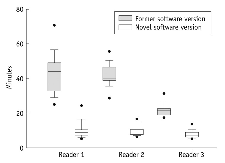
Fig. 1 Overall reading time. For novel software, overall reading time was significantly reduced (white boxplots) compared to former version (grey boxplots, p = <0.001 for all readers). Median (horizontal bar within boxplot) analysis time of Reader 1 and Reader 2 was equivalent, however Reader 3 with more experience had significantly less reading time in both novel and former software version compared to Readers 1 and 2. Black dots represent 5th/95th percentile.
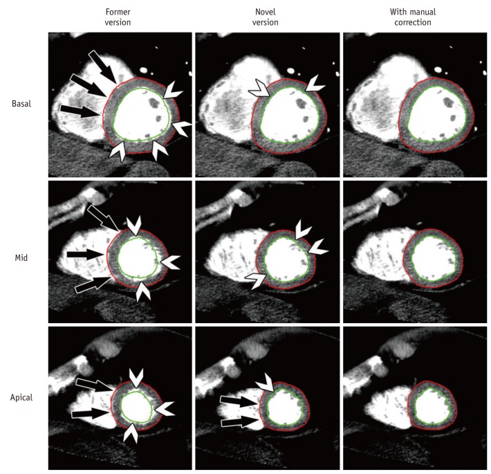
Fig. 2 Myocardial contour detection. Effects of improved automated contour detection by novel software version are shown in column 2 where precise epicardial (red) and endocardial (green) delineation is present. Only few endocardial regions need manual correction (arrowheads) and especially epicardium had to be manually corrected only in septal area of apical third of heart (bottom row, image in middle with arrows). In contrast, detection was poor (column 1) in former software version and required extensive, almost circumferential contour corrections (arrows and arrowheads) in total of 18 slices. Results after manual correction are represented in column 3 showing congruent delineation of epi- and endocardial borders with exclusion of papillary muscles and trabecular structures. All images (wl 100/ww 200) are taken from rest CTP, slice thickness is 8 mm.
Reference
-
1. Miller JM, Rochitte CE, Dewey M, Arbab-Zadeh A, Niinuma H, Gottlieb I, et al. Diagnostic performance of coronary angiography by 64-row CT. N Engl J Med. 2008; 359:2324–2336. PMID: 19038879.
Article2. Lee HJ, Kim JS, Kim YJ, Hur J, Yoo KJ, Choe KO, et al. Diagnostic accuracy of 64-slice multidetector computed tomography for selecting coronary artery bypass graft surgery candidates. J Thorac Cardiovasc Surg. 2011; 141:571–577. PMID: 20416891.
Article3. Moon JH, Park EA, Lee W, Yin YH, Chung JW, Park JH, et al. practice: a single institution’s experience. Korean J Radiol. 2011; 12:308–318. PMID: 21603290.4. Dewey M, Zimmermann E, Deissenrieder F, Laule M, Dübel HP, Schlattmann P, et al. Noninvasive coronary angiography by 320-row computed tomography with lower radiation exposure and maintained diagnostic accuracy: comparison of results with cardiac catheterization in a head-to-head pilot investigation. Circulation. 2009; 120:867–875. PMID: 19704093.5. So A, Wisenberg G, Islam A, Amann J, Romano W, Brown J, et al. Non-invasive assessment of functionally relevant coronary artery stenoses with quantitative CT perfusion: preliminary clinical experiences. Eur Radiol. 2012; 22:39–50. PMID: 21938441.
Article6. Bastarrika G, Ramos-Duran L, Schoepf UJ, Rosenblum MA, Abro JA, Brothers RL, et al. Adenosine-stress dynamic myocardial volume perfusion imaging with second generation dual-source computed tomography: Concepts and first experiences. J Cardiovasc Comput Tomogr. 2010; 4:127–135. PMID: 20430344.
Article7. George RT, Arbab-Zadeh A, Cerci RJ, Vavere AL, Kitagawa K, Dewey M, et al. Diagnostic performance of combined noninvasive coronary angiography and myocardial perfusion imaging using 320-MDCT: the CT angiography and perfusion methods of the CORE320 multicenter multinational diagnostic study. AJR Am J Roentgenol. 2011; 197:829–837. PMID: 21940569.
Article8. Ko SM, Choi JW, Song MG, Shin JK, Chee HK, Chung HW, et al. Myocardial perfusion imaging using adenosine-induced stress dual-energy computed tomography of the heart: comparison with cardiac magnetic resonance imaging and conventional coronary angiography. Eur Radiol. 2011; 21:26–35. PMID: 20658242.
Article9. Cerqueira MD, Verani MS, Schwaiger M, Heo J, Iskandrian AS. Safety profile of adenosine stress perfusion imaging: results from the Adenoscan Multicenter Trial Registry. J Am Coll Cardiol. 1994; 23:384–389. PMID: 8294691.
Article10. Voigtländer T, Schmermund A, Bramlage P, Elsässer A, Magedanz A, Kauczor HU, et al. The adverse events and hemodynamic effects of adenosine-based cardiac MRI. Korean J Radiol. 2011; 12:424–430. PMID: 21852902.
Article11. Ko BS, Cameron JD, Meredith IT, Leung M, Antonis PR, Nasis A, et al. Computed tomography stress myocardial perfusion imaging in patients considered for revascularization: a comparison with fractional flow reserve. Eur Heart J. 2012; 33:67–77. PMID: 21810860.
Article12. Choi JH, Min JK, Labounty TM, Lin FY, Mendoza DD, Shin DH, et al. Intracoronary transluminal attenuation gradient in coronary CT angiography for determining coronary artery stenosis. JACC Cardiovasc Imaging. 2011; 4:1149–1157. PMID: 22093264.
Article13. Reimann AJ, Tsiflikas I, Brodoefel H, Scheuering M, Rinck D, Kopp AF, et al. Efficacy of computer aided analysis in detection of significant coronary artery stenosis in cardiac using dual source computed tomography. Int J Cardiovasc Imaging. 2009; 25:195–203. PMID: 18821077.
Article14. Dewey M, Schnapauff D, Laule M, Lembcke A, Borges AC, Rutsch W, et al. Multislice CT coronary angiography: evaluation of an automatic vessel detection tool. Rofo. 2004; 176:478–483. PMID: 15088170.15. Okada DR, Ghoshhajra BB, Blankstein R, Rocha-Filho JA, Shturman LD, Rogers IS, et al. Direct comparison of rest and adenosine stress myocardial perfusion CT with rest and stress SPECT. J Nucl Cardiol. 2010; 17:27–37. PMID: 19936863.
Article16. Tamarappoo BK, Dey D, Nakazato R, Shmilovich H, Smith T, Cheng VY, et al. Comparison of the extent and severity of myocardial perfusion defects measured by CT coronary angiography and SPECT myocardial perfusion imaging. JACC Cardiovasc Imaging. 2010; 3:1010–1019. PMID: 20947046.
Article17. Halliburton SS, Abbara S, Chen MY, Gentry R, Mahesh M, Raff GL, et al. SCCT guidelines on radiation dose and dose-optimization strategies in cardiovascular CT. J Cardiovasc Comput Tomogr. 2011; 5:198–224. PMID: 21723512.
Article18. Hoffmann MH, Lessick J, Manzke R, Schmid FT, Gershin E, Boll DT, et al. Automatic determination of minimal cardiac motion phases for computed tomography imaging: initial experience. Eur Radiol. 2006; 16:365–373. PMID: 16021450.
Article19. Cootes TF, Taylor CJ, Cooper DH, Graham J. Active shape modelstheir training and application. Comput Vis Image Underst. 1995; 61:38–59.
Article20. Rogers IS, Cury RC, Blankstein R, Shapiro MD, Nieman K, Hoffmann U, et al. Comparison of postprocessing techniques for the detection of perfusion defects by cardiac computed tomography in patients presenting with acute ST-segment elevation myocardial infarction. J Cardiovasc Comput Tomogr. 2010; 4:258–266. PMID: 20579617.
Article21. Valdiviezo C, Ambrose M, Mehra V, Lardo AC, Lima JA, George RT. Quantitative and qualitative analysis and interpretation of CT perfusion imaging. J Nucl Cardiol. 2010; 17:1091–1100. PMID: 20924735.
Article22. Cerqueira MD, Weissman NJ, Dilsizian V, Jacobs AK, Kaul S, Laskey WK, et al. Standardized myocardial segmentation and nomenclature for tomographic imaging of the heart. A statement for healthcare professionals from the Cardiac Imaging Committee of the Council on Clinical Cardiology of the American Heart Association. Int J Cardiovasc Imaging. 2002; 18:539–542. PMID: 12135124.
Article23. Shrout PE, Fleiss JL. Intraclass correlations: uses in assessing rater reliability. Psychol Bull. 1979; 86:420–428. PMID: 18839484.
Article24. Dey D, Schepis T, Marwan M, Slomka PJ, Berman DS, Achenbach S. Automated three-dimensional quantification of noncalcified coronary plaque from coronary CT angiography: comparison with intravascular US. Radiology. 2010; 257:516–522. PMID: 20829536.
Article25. Dey D, Cheng VY, Slomka PJ, Nakazato R, Ramesh A, Gurudevan S, et al. Automated 3-dimensional quantification of noncalcified and calcified coronary plaque from coronary CT angiography. J Cardiovasc Comput Tomogr. 2009; 3:372–382. PMID: 20083056.
Article26. Sugiura T, Takeguchi T, Sakata Y, Nitta S, Okazaki T, Matsumoto N, et al. Automatic model-based contour detection of left ventricle myocardium from cardiac CT images. Int J Comput Assist Radiol Surg. 2012; [Epub ahead of print].
Article27. Müller M, Teige F, Schnapauff D, Hamm B, Dewey M. Evaluation of right ventricular function with multidetector computed tomography: comparison with magnetic resonance imaging and analysis of inter- and intraobserver variability. Eur Radiol. 2009; 19:278–289. PMID: 18704431.
Article28. Dewey M, Müller M, Eddicks S, Schnapauff D, Teige F, Rutsch W, et al. Evaluation of global and regional left ventricular function with 16-slice computed tomography, biplane cineventriculography, and two-dimensional transthoracic echocardiography: comparison with magnetic resonance imaging. J Am Coll Cardiol. 2006; 48:2034–2044. PMID: 17112993.29. Mo YH, Jaw FS, Wang YC, Jeng CM, Peng SF. Effects of propranolol on the left ventricular volume of normal subjects during CT coronary angiography. Korean J Radiol. 2011; 12:319–326. PMID: 21603291.
Article30. Boehm T, Alkadhi H, Roffi M, Willmann JK, Desbiolles LM, Marincek B, et al. Time-effectiveness, observer-dependence, and accuracy of measurements of left ventricular ejection fraction using 4-channel MDCT. Rofo. 2004; 176:529–537. PMID: 15088177.31. Schlosser T, Pagonidis K, Herborn CU, Hunold P, Waltering KU, Lauenstein TC, et al. Assessment of left ventricular parameters using 16-MDCT and new software for endocardial and epicardial border delineation. AJR Am J Roentgenol. 2005; 184:765–773. PMID: 15728595.
Article32. Plumhans C, Keil S, Ocklenburg C, Mühlenbruch G, Behrendt FF, Günther RW, et al. Comparison of manual, semi- and fully automated heart segmentation for assessing global left ventricular function in multidetector computed tomography. Invest Radiol. 2009; 44:476–482. PMID: 19561515.
Article33. Dewey M, Müller M, Teige F, Hamm B. Evaluation of a semiautomatic software tool for left ventricular function analysis with 16-slice computed tomography. Eur Radiol. 2006; 16:25–31. PMID: 15965660.
Article34. Ko SM, Kim YW, Han SW, Seo JB. Early and delayed myocardial enhancement in myocardial infarction using two-phase contrast-enhanced multidetector-row CT. Korean J Radiol. 2007; 8:94–102. PMID: 17420626.
Article35. Kachenoura N, Veronesi F, Lodato JA, Corsi C, Mehta R, Newby B, et al. Volumetric quantification of myocardial perfusion using analysis of multi-detector computed tomography 3D datasets: comparison with nuclear perfusion imaging. Eur Radiol. 2010; 20:337–347. PMID: 19711083.
Article36. Patel AR, Lodato JA, Chandra S, Kachenoura N, Ahmad H, Freed BH, et al. Detection of myocardial perfusion abnormalities using ultra-low radiation dose regadenoson stress multidetector computed tomography. J Cardiovasc Comput Tomogr. 2011; 5:247–254. PMID: 21723516.
Article37. George RT, Arbab-Zadeh A, Miller JM, Kitagawa K, Chang HJ, Bluemke DA, et al. Adenosine stress 64- and 256-row detector computed tomography angiography and perfusion imaging: a pilot study evaluating the transmural extent of perfusion abnormalities to predict atherosclerosis causing myocardial ischemia. Circ Cardiovasc Imaging. 2009; 2:174–182. PMID: 19808590.38. Lubbers DD, Kuijpers D, Bodewes R, Kappert P, Kerkhof M, van Ooijen PM, et al. Inter-observer variability of visual analysis of "stress"-only adenosine first-pass myocardial perfusion imaging in relation to clinical experience and reading criteria. Int J Cardiovasc Imaging. 2011; 27:557–562. PMID: 20882414.
Article
- Full Text Links
-
- Actions
-
Cited
- CITED
-
- Close
- Share
-
- Similar articles
-
- State of the Art of Imaging Equipment and Tools for Nuclear Cardiology
- Stress Testing and Imaging Protocols for Myocardial Perfusion Studies
- Clinical Applications of CT Myocardial Perfusion Imaging
- Correlation Between Functional Myocardial Perfusion Imaging and Anatomical Cardiac CT in a Case of Myocardial Bridging
- Myocardial Contractility, Perfusion, and Viability Analysis Using Multidetector CT in Patients with Ischemic Heart Disease
