Endoscope-Assisted Trans-Sphenoidal Approach for Treatment of Sternberg's Canal
- Affiliations
-
- 1Health Sciences Department and Operative Unit of Neurosurgery, University of L'Aquila, San Salvatore Hospital, L'Aquila, Italy. giuliano.maselli@tin.it
- 2Operative Unit of Neurosurgery, San Salvatore Hospital, L'Aquila, Italy.
- KMID: 1426267
- DOI: http://doi.org/10.3340/jkns.2012.52.6.555
Abstract
- We report an uncommon case of a 45-year-old woman who presented with spontaneous rhinorrhea. A computed tomography (CT) scan of the head revealed an abnormally large sphenoid sinus associated with a parasellar bony defect (Sternberg's canal) through which magnetic resonance imaging could detect an encephalocele of the right temporal lobe. An endoscope-assisted trans-sphenoidal approach was performed and, with the aid of image guided surgery, reduction of the encephalocele was obtained and followed by surgical repair of the dural and bony defects. The postoperative course was uneventful and the cerebrospinal fluid fistula was closed as confirmed by the postoperative CT scan and by the absence of rhinorrhea. After three years of monitoring the patient remained asymptomatic.
MeSH Terms
Figure
-
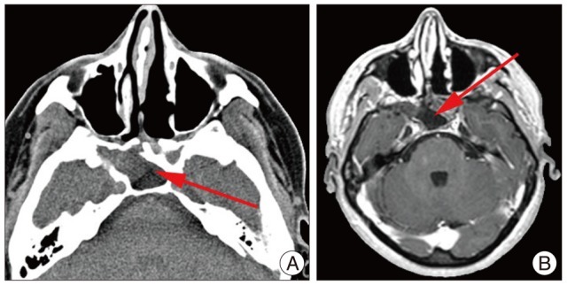
Fig. 1 A : High resolution computed tomography of facial massix revealing an accessory cell posterior to the right maxillary sinus. B : Magnetic resonance imaging with contrast medium demonstrating peripheral enhancement of the right sphenoidal encephalocele.
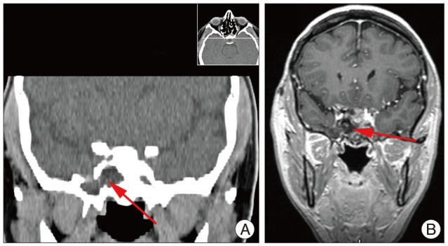
Fig. 2 A : High resolution computed tomography of facial massix; coronal reconstruction scan revealing the persistent Sternberg's canal. B : Magnetic resonance imaging with contrast medium coronal scan demonstrating peripheral enhancement of the right sphenoidal encephalocele.
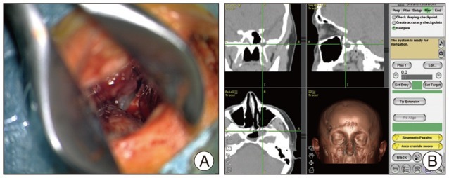
Fig. 3 A : Endoscope-assisted trans-sphenoidal approach. B : Neuronavigation image confirms the bony defect.
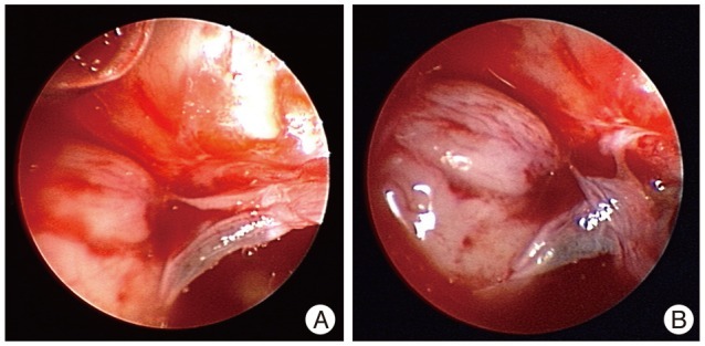
Fig. 4 A : Endoscopic image of the right sphenoidal encephalocele with cerebrospinal fluid fistula in the endoscopic-assisted trans-sphenoidal approach. B : Endoscopic-assisted trans-sphenoidal approach : a particular of the cerebrospinal liquor outflow under brain pulsation.
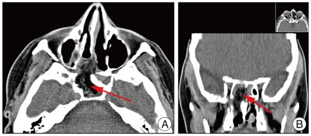
Fig. 5 A : Postoperative high resolution computed tomography of facial massix demonstrating the reduction of the encephalocele and the complete closure of the defect by duroplasty. B : Postoperative high resolution computed tomography of facial massix; coronal reconstruction scan demonstrating the complete reconstruction by a duroplasty.
Reference
-
1. Albernaz MS, Horton WD, Adkins WY, Garen PD. Intrasphenoidal encephalocele. Otolaryngol Head Neck Surg. 1991; 104:279–281. PMID: 1901161.
Article2. Barañano CF, Curé J, Palmer JN, Woodworth BA. Sternberg's canal : fact or fiction? Am J Rhinol Allergy. 2009; 23:167–171. PMID: 19401043.3. Buchfelder M, Fahlbusch R, Huk WJ, Thierauf P. Intrasphenoidal encephaloceles--a clinical entity. Acta Neurochir (Wien). 1987; 89:10–15. PMID: 3324649.
Article4. Castelnuovo P, Dallan I, Pistochini A, Battaglia P, Locatelli D, Bignami M. Endonasal endoscopic repair of Sternberg's canal cerebrospinal fluid leaks. Laryngoscope. 2007; 117:345–349. PMID: 17277632.
Article5. Catala M. [Embryology of the sphenoid bone]. J Neuroradiol. 2003; 30:196–200. PMID: 14566186.6. Cheung DK, Attia EL, Kirkpatrick DA, Marcarian B, Wright B. An anatomic and CT scan study of the lateral wall of the sphenoid sinus as related to the transnasal transethmoid endoscopic approach. J Otolaryngol. 1993; 22:63–68. PMID: 8515518.7. DeBartolo HM, Vrabec D. Sphenoid encephalocele : report of a case. Arch Otolaryngol. 1977; 103:172–174. PMID: 836247.8. Landreneau FE, Mickey B, Coimbra C. Surgical treatment of cerebrospinal fluid fistulae involving lateral extension of the sphenoid sinus. Neurosurgery. 1998; 42:1101–1104. discussion 1104-1105. PMID: 9588555.
Article9. Lopatin AS, Kapitanov DN, Potapov AA. Endonasal endoscopic repair of spontaneous cerebrospinal fluid leaks. Arch Otolaryngol Head Neck Surg. 2003; 129:859–863. PMID: 12925345.
Article10. Myssiorek D, Cohen NL. Intrasphenoidal meningoencephalocele : a case report. Am J Otolaryngol. 1987; 8:391–394. PMID: 3434678.11. Pasquini E, Sciarretta V, Farneti G, Mazzatenta D, Modugno GC, Frank G. Endoscopic treatment of encephaloceles of the lateral wall of the sphenoid sinus. Minim Invasive Neurosurg. 2004; 47:209–213. PMID: 15346316.
Article12. Schick B, Brors D, Prescher A. Sternberg's canal--cause of congenital sphenoidal meningocele. Eur Arch Otorhinolaryngol. 2000; 257:430–432. PMID: 11073192.13. Shetty PG, Shroff MM, Fatterpekar GM, Sahani DV, Kirtane MV. A retrospective analysis of spontaneous sphenoid sinus fistula : MR and CT findings. AJNR Am J Neuroradiol. 2000; 21:337–342. PMID: 10696020.
- Full Text Links
-
- Actions
-
Cited
- CITED
-
- Close
- Share
-
- Similar articles
-
- Trans-sphenoidal Approach to the Supraclinoid Internal Carotid Artery for Endovascular Access in a Cadaver
- Endoscope-Assisted Hairline Approach for Head and Neck Masses: A Review
- Trigemino-cardiac reflex: occurrence of asystole during trans-sphenoidal adenomectomy: a case report
- Sphenoid Sinus Mucocele(Case Report)
- Prevalence and Clinical Implications of Lateral Wall Dehiscence in the Sphenoid Sinus: Sternberg’s Canal
