Effects of Intraluminal Irradiation with Holmium-166 for TIPS Stenosis: Experimental Study in a Swine Model
- Affiliations
-
- 1Department of Diagnostic Radiology, Kyung Hee University Medical Center, Seoul, Korea. ohjh6108@hanmail.net
- 2Department of Nuclear Medicine, Kyung Hee University Medical Center, Seoul, Korea.
- 3Department of Pathology, Kyung Hee University Medical Center, Seoul, Korea.
- 4Department of Diagnostic Radiology, Kang Dong Sacred Heart Hospital, Hallym University, Seoul, Korea.
- 5Department of Cardiology, Kyung Hee University Medical Center, Seoul, Korea.
- KMID: 1126836
- DOI: http://doi.org/10.3348/kjr.2007.8.2.127
Abstract
OBJECTIVE
We wanted to evaluate the effectiveness of intraluminal irradiation with Holmium-166 (166Ho) for reducing the pseudointimal hyperplasia (PIH) in the transjugular intrahepatic portosystemic shunt (TIPS) tract in a swine model. MATERIALS AND METHODS: TIPS was performed in 12 domestic pigs, after the creation of portal hypertension by intraportal injection of a mixture of N-butyl-2-cyanoacrylate (NBCA) and lipiodol. Five pigs first underwent intraluminal irradiation (30 Gy) in the parenchymal tract with using a 166Ho solution-filled balloon catheter, and this was followed by the placement of a nitinol stent in the TIPS tract. For the seven control pigs, the balloon was filled with saline and contrast media mixture. Two weeks later, follow-up portography and histological analysis were performed. RESULTS: TIPS was successfully performed in all twelve pigs with achieving artificially induced portal hypertension. Portography performed two weeks after TIPS showed the patent tracts in the TIPS tracts that were irradiated with 166Ho (5/5, 100%), whereas either completely (5/6, 83.3%) or partially (1/6, 16.7%) occluded TIPS were seen in the seven pigs of the nonirradiated control group, except in one pig that experienced periprocedural death due to bleeding. Histological analysis showed a statistically significant difference for the maximal PIH (irradiated: 32.8%, nonirradiated: 76.0%, p < 0.001) between the two groups. CONCLUSION: Intraluminal irradiation with 30 Gy of 166Ho for TIPS significantly improved the TIPS patency in a swine model of portal hypertension during a 2-week period of follow-up.
Keyword
MeSH Terms
Figure
-
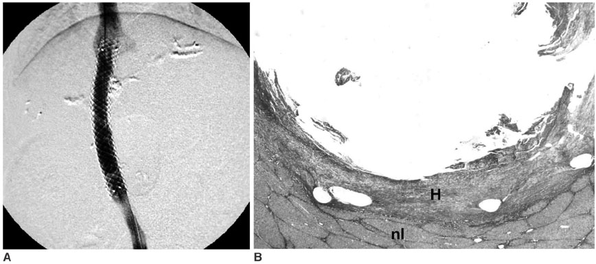
Fig. 1 Follow-up studies done two weeks after TIPS with 166Ho irradiation. A. Portography demonstrates good patency of the shunt without narrowing or occlusion. B. Microscopic examination (Masson-trichrome stain, ×10) of the specimen obtained from the parenchymal tract of the patent stent reveals that the luminal surface is lined by a relatively thin, uniform layer of pseudointimal hyperplasia. Pseudointimal hyperplasia (H) is well delineated from the normal liver parenchymal layer (nl).
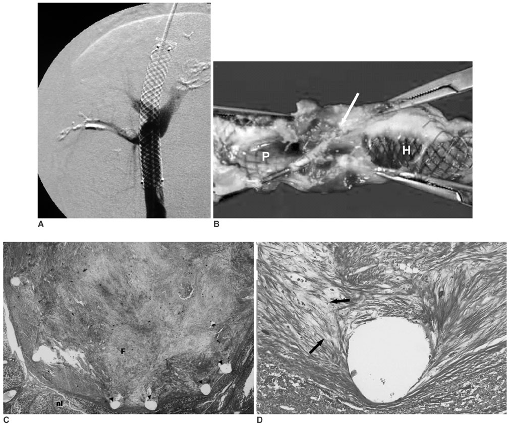
Fig. 2 Follow-up studies done two weeks after TIPS without 166Ho irradiation in the control group. A. Portography via the transjugular approach demonstrates the complete occlusion of the shunt. B. The corresponding gross specimen before removal of the stent wire shows marked ingrowth of fibrotic tissue (arrow) within the parenchymal tract, which is in contrast to the lack of significant narrowing at both venous ends (H, hepatic venous end; P, portal venous end). C. Transverse section (Masson-trichrome stain, ×10) through the parenchymal tract of TIPS reveals the complete luminal occlusion with the most superficial layer of fibrin (F), thick granulation tissue and the inflammatory reaction. The removed portions of the wires (arrowheads) are seen as holes. These stent holes are seen adjacent to the normal parenchymal tissue (nl), which is clearly demarcated from the PIH. D. Close-up (Masson-trichrome stain, ×100) of the PIH shows numerous spindle-shaped myofibroblasts (arrows) and the abundant collagen matrix.
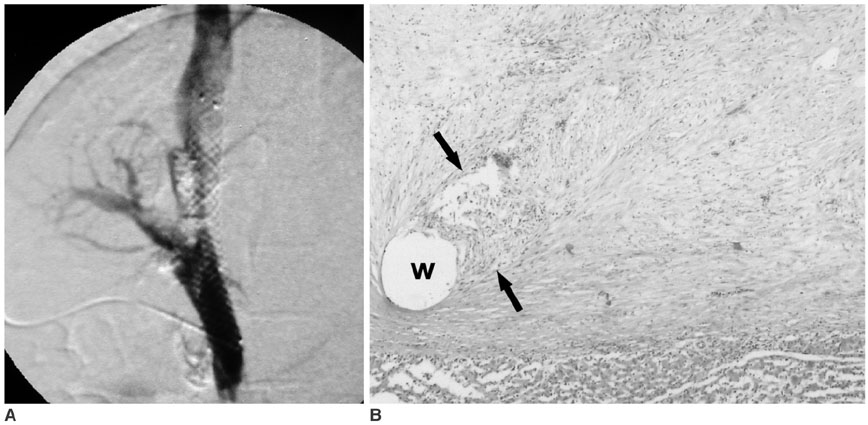
Fig. 3 Follow-up studies done two weeks after TIPS without 166Ho irradiation in the control group. A. Portography via the superior mesenteric vein, which was exposed by a skin incision, shows partial occlusion of the shunt at the level of parenchymal tract of TIPS. B. Microscopic cross section revealed a homogeneous layer of granulation tissue that's composed of an abundant acellular matrix, myofibroblasts and numerous inflammatory cells (between arrows) adjacent the stent wire (w).
Reference
-
1. Saxon RS, Ross PL, Mendel-Hartvig J, Barton RE, Benner K, Flora K, et al. Transjugular intrahepatic portosystemic shunt patency and the importance of stenosis location in the development of recurrent symptoms. Radiology. 1998. 207:683–693.2. Haskal ZJ, Pentecost MJ, Soulen MC, Shlansky-Goldberg RD, Baum RA, Cope C. Transjugular intrahepatic portosystemic shunt stenosis and revision: early and midterm results. AJR Am J Roentgenol. 1994. 163:439–444.3. Terayama N, Matsui O, Kadoya M, Yoshikawa J, Gabata T, Miyayama S, et al. Transjugular intrahepatic portosystemic shunt: histologic and immunohistochemical study of autopsy cases. Cardiovasc Intervent Radiol. 1997. 20:457–461.4. Ducoin H, El-Khoury J, Rousseau H, Barange K, Peron JM, Pierragi MT, et al. Histopathologic analysis of transjugular intrahepatic portosystemic shunts. Hepatology. 1997. 25:1064–1069.5. Teng GJ, Bettmann MA, Hoopes PJ, Wagner RJ, Park BH, Yang L, et al. Transjugular intrahepatic portosystemic shunt: effect of bile leak on smooth muscle cell proliferation. Radiology. 1998. 209:799–805.6. Condado JA, Waksman R, Calderas C, Saucedo J, Lansky A. Two-year follow-up after intracoronary gamma radiation therapy. Cardiovasc Radiat Med. 1999. 1:30–35.7. Verin V, Urban P, Popowski Y, Schwager M, Nouet P, Dorsaz PA, et al. Feasibility of intracoronary beta-irradiation to reduce restenosis after balloon angioplasty. A clinical pilot study. Circulation. 1997. 95:1138–1144.8. Wardeh AJ, Kay IP, Sabate M, Coen VL, Gijzel AL, Ligthart JM, et al. Beta-particle-emitting radioactive stent implantation. A safety and feasibility study. Circulation. 1999. 100:1684–1689.9. Kim W, Jeong MH, Park OY, Rhew JY, Bom HS, Choi SJ, et al. Effects of beta-radiation using a holmium-166 coated balloon on neointimal hyperplasia in a porcine coronary stent restenosis model. Circ J. 2003. 67:625–629.10. Carter AJ, Laird JR, Bailey LR, Hoopes TG, Farb A, Fischell DR, et al. Effects of endovascular radiation from a beta-particle-emitting stent in a porcine coronary restenosis model. A dose-response study. Circulation. 1996. 94:2364–2368.11. Lessie T, Yoon HC, Nelson HA, Fillmore DJ, Baldwin GN, Miller FJ. Intraluminal irradiation for TIPS stenosis: preliminary results in a swine model. J Vasc Interv Radiol. 1999. 10:899–906.12. Rajendran JG, Eary JF, Bensinger W, Durack LD, Vernon C, Fritzberg A. High-dose 166Ho-DOTMP in myeloablative treatment of multiple myeloma: pharmacokinetics, biodistribution, and absorbed dose estimation. J Nucl Med. 2002. 43:1383–1390.13. Firestone RB. Table of isotopes. 1996. New York: Wiley-Interscience.14. Klugherz BD, Meneveau NF, Kolansky DM, Herrmann HC, Schiele F, Matthai WH Jr, et al. Predictors of clinical outcome following percutaneous intervention for in-stent restenosis. Am J Cardiol. 2000. 85:1427–1431.15. Mazur W, Ali MN, Khan MM, Dabaghi SF, DeFelice CA, Paradis P Jr, et al. High dose rate intracoronary radiation for inhibition of neointimal formation in the stented and balloon-injured porcine models of restenosis: angiographic, morphometric, and histopathologic analyses. Int J Radiat Oncol Biol Phys. 1996. 36:777–788.16. Joh CW, Park CH, Kang HJ, Oh YT, Chun , Kim HS, et al. Measurement of radiation absorbed dose in endovascular Ho-166 brachytherapy using a balloon angio-catheter. Nucl Med Commun. 2000. 21:959–964.17. Hong YD, Park KB, Jang BS, Choi SJ, Choi SM, Kim YM. Holmium-166-DTPA as a liquid source for endovascular brachytherapy. Nucl Med Biol. 2002. 29:833–839.18. Kichikawa K, Saxon RR, Nishimine K, Nishida N, Uchida BT. Experimental TIPS with spiral Z-stents in swine with and without induced portal hypertension. Cardiovasc Intervent Radiol. 1997. 20:197–203.19. Nishimine K, Saxon RR, Kichikawa K, Mendel-Hartvig J, Timmermans HA, Shim HJ, et al. Improved transjugular intrahepatic portosystemic shunt patency with PTFE-covered stent-grafts: experimental results in swine. Radiology. 1995. 196:341–347.20. Pavcnik D, Saxon RR, Kubota Y, Tanihata H, Uchida BT, Corless C, et al. Attempted induction of chronic portal venous hypertension with polyvinyl alcohol particles in swine. J Vasc Interv Radiol. 1997. 8:123–128.21. Palmaz JC, Garcia F, Sibbitt RR, Tio FO, Kopp DT, Schwesinger W, et al. Expandable intrahepatic portacaval shunt stents in dogs with chronic portal hypertension. AJR Am J Roentgenol. 1986. 147:1251–1254.22. Song JK, Gobin YP, Duckwiler GR, Murayama Y, Frazee JG, Martin NA, et al. N-butyl 2-cyanoacrylate embolization of spinal dural arteriovenous fistulae. AJNR Am J Neuroradiol. 2001. 22:40–47.23. Denys A, Lacombe C, Schneider F, Madoff DC, Doenz F, Qanadli SD, et al. Portal vein embolization with N-butyl cyanoacrylate before partial hepatectomy in patients with hepatocellular carcinoma and underlying cirrhosis or advanced fibrosis. J Vasc Interv Radiol. 2005. 16:1667–1674.
- Full Text Links
-
- Actions
-
Cited
- CITED
-
- Close
- Share
-
- Similar articles
-
- Feasibility of Endovascular Radiation Therapy Using Holmium-166 Filled Balloon Catheter in a Swine Hemodialysis Fistula Model: Preliminary Results
- Efficacy of a Dexamethasone-Eluting Nitinol Stent on the Inhibition of Pseudointimal Hyperplasia in a Transjugular Intrahepatic Portosystemic Shunt: An Experimental Study in a Swine Model
- beta-irradiation (166Ho patch)-induced skin injury in mini-pigs: effects on NF-kappaB and COX-2 expression in the skin
- Therapeutic Effects of Holmium-166 Chitosan Complex in Rat Brain Tumor Model
- Hepatic Parenchymal Changes After Percutaneous Injection of Holmium-166 in Rabbit
