CT, MR, and Angiography Findings of a Solitary Fibrous Tumor of the Larynx: a Case Report
- Affiliations
-
- 1Department of Radiology, Kangdong Seong-Sim Hospital, Hallym University College of Medicine, Seoul, Korea. chkcsk@empal.com
- 2Department of Pathology, Kangdong Seong-Sim Hospital, Hallym University College of Medicine, Seoul, Korea.
- KMID: 1118881
- DOI: http://doi.org/10.3348/kjr.2008.9.6.568
Abstract
- This report details the CT, MR, and angiography findings of a solitary fibrous tumor involving the larynx of a 34-year-old man. A precontrast CT scan revealed a well-defined isodense mass in the submucosal region of the supraglottic larynx. The tumor appeared as a mixed intensity lesion on the T1- and T2-weighted MR images. A T2-weighted MR image showed a central, round, and low signal intensity area within the mass. For both the CT and MR images, the mass demonstrated heterogeneous enhancement following the administration of contrast material. The angiography showed a hypervascular tumor with heterogeneous persistent staining.
Keyword
MeSH Terms
Figure
-
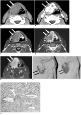
Fig. 1 Images of 34-year-old man presenting foreign body sensation in throat. A. Precontrast CT scan shows well-marginated mass (arrows) which is hypodense relative to muscle on right side of supraglottic larynx. B. Postcontrast CT scan demonstrates heterogeneous enhancement of mass (arrows). C. Axial T1-weighted MR image (800/15 [TR/TE]) shows well-defined mass (arrows) which is hyperintense compared to adjacent muscle in supraglottic larynx. Note preserved surrounding fat planes. D. Axial T2-weighted MR image (4000/80 [TR/TE]) at same level as C demonstrates mixture of low and high signal intensities in mass (arrows). Also, round hypointense area (small arrows) is noted in posterior region of mass. E. Axial T1-weighted, postcontrast, fat-saturated image (1100/10 [TR/TE]) shows strong enhancement of mass (arrows). Note moderate enhancement (small arrows) in posterior region of mass, corresponding to round hypointense area on T2-weighted images. F. Early arterial (left) and late phase (right) images of right common carotid angiography demonstrate hypervascular tumor with persistent staining (arrows) in anterior neck region. G. Photomicrograph of surgical specimen reveal that tumor is composed of uniform spindle cells in solid pattern. In addition, prominent vessels with a branching pattern are also seen (Hematoxylin & Eosin staining; original magnification ×100). Immunohistochemical study (inset) reveals neoplastic cells which are strongly positive (brown color) for CD34 monoclonal antibody (original magnification ×100).
Reference
-
1. Kim TA, Brunberg JA, Pearson JP, Ross DA. Solitary fibrous tumor of the paranasal sinuses: CT and MR appearance. AJNR Am J Neuroradiol. 1996. 17:1767–1772.2. Jeong AK, Lee HK, Kim SY, Cho KJ. Solitary fibrous tumor of the parapharyngeal space: MR imaging findings. AJNR Am J Neuroradiol. 2002. 23:473–475.3. Kim HJ, Lee HK, Seo JJ, Kim HJ, Shin JH, Jeong AK, et al. MR imaging of solitary fibrous tumors in the head and neck. Korean J Radiol. 2005. 6:136–142.4. Dunfee BL, Sakai O, Spiegel JH, Pistey R. Solitary fibrous tumor of the buccal space. AJNR Am J Neuroradiol. 2005. 26:2114–2116.5. Benlyazid A, Lescanne E, Lefrancq T, Fetissoff F, Beutter P. Solitary fibrous tumor of the larynx: report of a case. J Laryngol Otol. 1998. 112:286–289.6. Fan CY, Van Hemert RL, Thomas JR, Breau RL. Atypical solitary fibrous tumor of the larynx. Otolaryngol Head Neck Surg. 2006. 134:880–882.7. Dotto JE, Ahrens W, Lesnik DJ, Kowalski D, Sasaki C, Flynn S. Solitary fibrous tumor of the larynx: a case report and review of the literature. Arch Pathol Lab Med. 2006. 130:213–216.8. Alobid I, Alós L, Maldonado M, Menéndez LM, Bernal-Sprekelsen M. Laryngeal solitary fibrous tumor treated with CO2 laser excision: case report. Eur Arch Otorhinolaryngol. 2005. 262:286–288.9. Stomeo F, Padovani D, Bozzo C, Pastore A. Laryngeal solitary fibrous tumor. Auris Nasus Larynx. 2007. 34:405–408.10. England DM, Hochholzer L, McCarthy MJ. Localized benign and malignant fibrous tumours of the pleura. A clinicopathologic review of 233 cases. Am J Surg Pathol. 1989. 13:640–658.11. Alawi F, Stratton D, Freedman PD. Solitary fibrous tumor of the oral soft tissues: a clinicopathologic and immunohistochemical study of 16 cases. Am J Surg Pathol. 2001. 25:900–910.12. Liang GS, Loevner LA, Kumar P. Laryngeal rhabdomyoma involving the paraglottic space. AJR Am J Roentgenol. 2000. 174:1285–1287.
- Full Text Links
-
- Actions
-
Cited
- CITED
-
- Close
- Share
-
- Similar articles
-
- Solitary Fibrous Tumor of the Adrenal Gland: A Case Report
- An Ancillary CT Finding of Intrapulmonary Solitary Fibrous Tumor: A Case Report
- Solitary Fibrous Tumor of the Pancreas: Imaging Findings
- Imaging Findings of a Solitary Fibrous Tumor in Pancreas: A Case Report
- Myxoid Solitary Fibrous Tumor of the Retroperitoneum: MRI Findings with the Pathologic Correlation
