J Korean Soc Radiol.
2017 Feb;76(2):121-125. 10.3348/jksr.2017.76.2.121.
An Ancillary CT Finding of Intrapulmonary Solitary Fibrous Tumor: A Case Report
- Affiliations
-
- 1Department of Radiology, CHA Bundang Medical Center, CHA University, Seongnam, Korea. rhochest@cha.ac.kr
- 2Department of Pathology, CHA Bundang Medical Center, CHA University, Seongnam, Korea.
- KMID: 2367859
- DOI: http://doi.org/10.3348/jksr.2017.76.2.121
Abstract
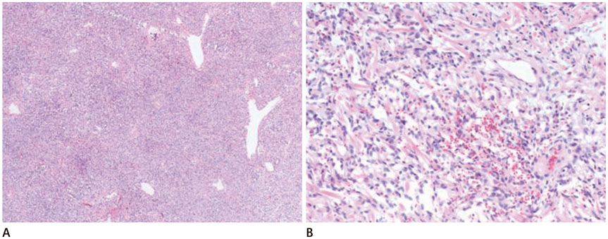
- Intrapulmonary solitary fibrous tumor is extremely rare. A few reports have presented typical CT findings such as well-defined, variable"”sized, heterogeneously or homogenously well-enhanced intrapulmonary nodules. We report herein a rare case of intrapulmonary solitary fibrous tumor that showed typical clinical and CT features, and we also provide an ancillary CT finding that shows a distinguishable tubular vascular structure within the nodule. The tubular vascular structure was conjoined to the proximal pulmonary vein. In this study, we highlight an ancillary CT finding reported for the first time for the diagnosis of a patient with intrapulmonary solitary fibrous tumor.
Figure
Reference
-
1. Chang YL, Lee YC, Wu CT. Thoracic solitary fibrous tumor: clinical and pathological diversity. Lung Cancer. 1999; 23:53–60.2. Chick JF, Chauhan NR, Madan R. Solitary fibrous tumors of the thorax: nomenclature, epidemiology, radiologic and pathologic findings, differential diagnoses, and management. AJR Am J Roentgenol. 2013; 200:W238–W248.3. Patsios D, Hwang DM, Chung TB. Intraparenchymal solitary fibrous tumor of the lung: an uncommon cause of a pulmonary nodule. J Thorac Imaging. 2006; 21:50–53.4. Meroni S, Funicelli L, Rampinelli C, Galetta D, Bonello L, Spaggiari L, et al. Solitary fibrous tumours: unusual aspects of a rare disease. Hippokratia. 2012; 16:269–274.5. Rao N, Colby TV, Falconieri G, Cohen H, Moran CA, Suster S. Intrapulmonary solitary fibrous tumors: clinicopathologic and immunohistochemical study of 24 cases. Am J Surg Pathol. 2013; 37:155–166.6. Kanamori Y, Hashizume K, Sugiyama M, Motoi T, Fukayama M, Ida K, et al. Intrapulmonary solitary fibrous tumor in an eight-year-old male. Pediatr Pulmonol. 2005; 40:261–264.7. Lee D, Haam SJ, Choi SE, Park CH, Kim TH. Calcifying fibrous tumor of the pleura: a rare case with an unusual presentation on CT and MRI. J Korean Soc Radiol. 2015; 72:123–127.8. Kawaguchi K, Taniguchi T, Usami N, Yokoi K. Intrapulmonary solitary fibrous tumor. Gen Thorac Cardiovasc Surg. 2011; 59:61–64.9. Wignall OJ, Moskovic EC, Thway K, Thomas JM. Solitary fibrous tumors of the soft tissues: review of the imaging and clinical features with histopathologic correlation. AJR Am J Roentgenol. 2010; 195:W55–W62.10. Gengler C, Guillou L. Solitary fibrous tumour and haemangiopericytoma: evolution of a concept. Histopathology. 2006; 48:63–74.
- Full Text Links
- Actions
-
Cited
- CITED
-
- Close
- Share
- Similar articles
-
- Intrapulmonary Solitary Fibrous Tumor Masquerade Sigmoid Adenocarcinoma Metastasis
- A Case of Intrapulmonary Solitary Fibrous Tumor: A case report
- Solitary Fibrous Tumor of the Adrenal Gland: A Case Report
- Imaging Findings of a Solitary Fibrous Tumor in Pancreas: A Case Report
- A Case of Solitary Fibrous Tumor in the Cheek


