Myxoid Solitary Fibrous Tumor of the Retroperitoneum: MRI Findings with the Pathologic Correlation
- Affiliations
-
- 1Department of Radiology, Chonnam National University Hospital, Gwangju, Korea. kjradsss@dreamwiz.com
- 2Department of Radiology, Chonnam National University Hwasun Hospital, Chonnam, Korea.
- KMID: 1758463
- DOI: http://doi.org/10.3348/kjr.2008.9.3.279
Abstract
- We report here on a case of solitary fibrous tumor of the retroperitoneum, and the tumor displayed a predominantly myxoid histology. A 56-year-old man presented with an incidentally detected retroperitoneal mass. On the MR images, the mass was observed as having iso-signal intensity on the T1-weighted images and high signal intensity on the fat-saturated T2-weighted images. The mass showed intense enhancement on the Gd-DTPA enhanced T1-weighted images. At surgery, a well-defined solid mass was found in the left retroperitoneum. The histological diagnosis was made as solitary fibrous tumor with a predominantly myxoid histology.
MeSH Terms
Figure
-
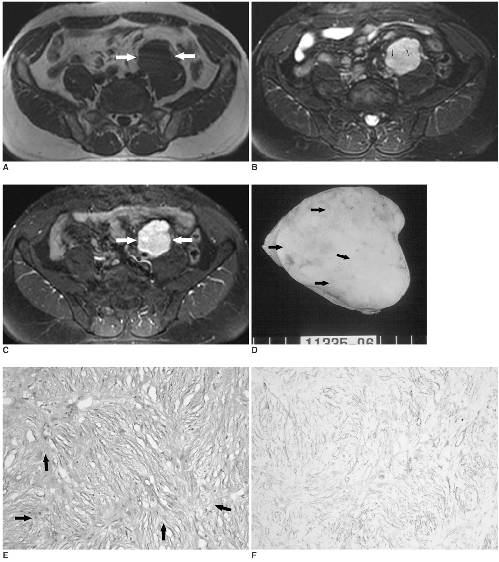
Fig. 1 56-year-old man presenting with incidentally detected retroperitoneal mass. A. Axial T1-weighted MR image shows well-defined, lobulated mass that is isointense to adjacent muscle (arrows). B. Axial T2-weighted MR image with fat saturation shows that mass is heterogeneously hyperintense to muscle. There are several hypointense streaks (arrows) within mass. C. Axial Gd-DTPA enhanced T1-weighted MR image demonstrates strong enhancement of mass that abuts on external iliac vessel and psoas muscle (arrows). D. Sectioned gross pathologic specimen demonstrates yellowish white cut surface with both soft myxoid areas (arrows) and rubbery fibrous areas. E. Photomicrograph shows elongated spindle cells dispersed in heterogeneous stromal matrix with myxoid areas (arrows) and collagenous areas (original magnification; Hematoxylin-Eosin staining, × 100). F. Immunohistochemical stain for CD 34. Tumor cells show diffuse strong reactivity for CD 34 (original magnification, × 100).
Reference
-
1. Gengler C, Guillou L. Solitary fibrous tumor and haemangiopericytoma: evolution of a concept. Histopathology. 2006. 48:63–74.2. Vossough A, Torigian DA, Zhang PJ, Siegelman ES, Banner MP. Extrathoracic solitary fibrous tumor of the pelvic peritoneum with central malignant degeneration on CT and MRI. J Magn Reson Imaging. 2005. 22:684–686.3. Johnson TR, Pedrosa I, Goldsmith J, Dewolf WC, Rofsky NM. Magnetic resonance imaging findings in solitary fibrous tumor of the kidney. J Comput Assist Tomogr. 2005. 29:481–483.4. de Saint Aubain Somerhausen N, Rubin BP, Fletcher CD. Myxoid solitary fibrous tumor: a study of seven cases with emphasis on differential diagnosis. Mod Pathol. 1999. 12:463–471.5. Wei YC, Li CF, Sung MT, Chen YT, Ko SF, Eng HL, et al. Primary myxoid solitary fibrous tumor involving the seminal vesicle. Pathol Int. 2006. 56:642–644.6. Goodlad JR, Fletcher CD. Solitary fibrous tumor arising at unusual sites: analysis of a series. Histopathology. 1991. 19:515–522.7. Kim HJ, Lee HK, Seo JJ, Kim HJ, Shin JH, Jeong AK, et al. MR imaging of solitary fibrous tumors in the head and neck. Korean J Radiol. 2005. 6:136–142.8. Chun HJ, Byun JY, Jung SE, Kim KH, Shinn KS. Benign solitary fibrous tumor of the pre-sacral space: MRI findings. Br J Radiol. 1998. 71:677–679.9. Nagase T, Adachi I, Yamada T, Murakami N, Morita K, Yoshino Y, et al. Solitary fibrous tumor in the pelvic cavity with hypoglycemia: report of a case. Surg Today. 2005. 35:181–184.10. Gold JS, Antonescu CR, Hajdu C, Ferrone CR, Hussain M, Lewis JJ, et al. Clinicopathologic correlates of solitary fibrous tumors. Cancer. 2002. 94:1057–1068.
- Full Text Links
-
- Actions
-
Cited
- CITED
-
- Close
- Share
-
- Similar articles
-
- Solitary Fibrous Tumor in Retroperitoneum
- Myxoid Solitary Fibrous Tumor on the Scalp
- Malignant Solitary Fibrous Tumor of Retroperitoneum Mimicking Gastric Submucosal Tumor
- Intramedullary Solitary Fibrous Tumor of Cervicothoracic Spinal Cord
- Dedifferentiated Liposarcoma of the Retroperitoneum: A case report
