Evaluation of partial cranial cruciate ligament rupture with positive contrast computed tomographic arthrography in dogs
- Affiliations
-
- 1Veterinary Medical Center, College of Veterinary Medicine, Chungbuk National University, Cheongju 361-763, Korea. dwchang@cbnu.ac.kr
- 2Department of Veterinary Medicine, College of Veterinary Medicine, Chungnam National University, Daejeon 305-764, Korea.
- 3Department of Veterinary Medicine, College of Veterinary Medicine, Gyeongsang National University, Jinju 660-701, Korea.
- KMID: 1104915
- DOI: http://doi.org/10.4142/jvs.2008.9.4.395
Abstract
- Computed tomographic arthrography (CTA) of four cadaveric canine stifles was performed before and after partial cranial cruciate ligament rupture in order to verify the usefulness of CTA examination for the diagnosis of partial cranial cruciate ligament rupture. To obtain the sequential true transverse image of a cranial cruciate ligament, the computed tomography gantry was angled such that the scanning plane was parallel to the fibula. True transverse images of cranial cruciate ligaments were identified on every sequential image, beginning just proximal to the origin of the cranial cruciate ligament distal to the tibial attachment, after the administration of iodinated contrast medium. A significant decrease in the area of the cranial cruciate ligament was identified on CTA imaging after partial surgical rupture of the cranial cruciate ligament. This finding implies that CTA can be used for assessing partial cranial cruciate ligament ruptures in dogs.
Keyword
MeSH Terms
Figure
-
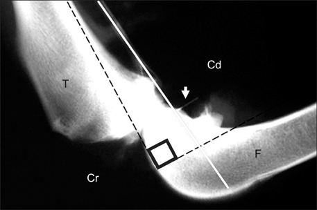
Fig. 1 Lateral radiograph of the stifle showing the relationship between stifle angle and the cranial cruciate ligament. Metal landmarks implanted in the cranial cruciate ligament (arrow) are shown. The cranial cruciate ligament and the fibula cross at a right angle when the stifle is flexed at 90 degrees. Cr: cranial, Cd: caudal, F: femur, T: tibia.
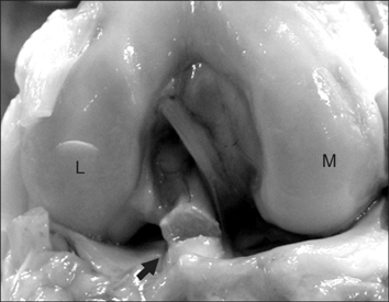
Fig. 2 Photograph of the canine cadaveric stifle joint illustrating the cranial cruciate ligament. Experimental cranial cruciate ligament rupture is identified (arrow). L: lateral, M: medial.

Fig. 3 Two-millimeter sequential transverse computed tomographic arthrography images were scanned parallel to the fibula, femoral attachment (A), and tibial attachment (I). The cranial cruciate ligament (black arrow) transverse images and caudal cruciate ligament sagittal images (white arrow) were clearly identified. L: lateral, M: medial.
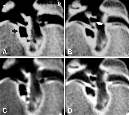
Fig. 4 Comparison of the pre-operative conventional view (A) and tracing view (C) with the post-operative conventional view (B) and tracing view (D). The transverse area of the cranial cruciate ligament image (black arrow) was decreased by 25% on the post-operative image. The small gas artifact (white arrow) was considered a normal finding. L: lateral, M: medial.
Reference
-
1. Arnoczky SP, Marshall JL. The cruciate ligaments of the canine stifle: an anatomical and functional analysis. Am J Vet Res. 1977. 38:1807–1814.2. Atilola MA, Pennock PW, Sumner-Smith G. Evaluation of analytical grade of metrizamide for canine stifle arthrography. J Am Vet Med Assoc. 1984. 185:436–439.3. Baird DK, Hathcock JT, Rumph PF, Kincaid SA, Visco DM. Low-field magnetic resonance imaging of the canine stifle joint: normal anatomy. Vet Radiol Ultrasound. 1998. 2:87–97.
Article4. Banfield CM, Morrison WB. Magnetic resonance arthrography of the canine stifle joint: technique and applications in eleven military dogs. Vet Radiol Ultrasound. 2000. 41:200–213.
Article5. Elliott DA. Nelson RW, editor. Disorders of metabolism. Small Animal Internal Medicine. 2003. St. Louis: Mosby;818.6. Gnudi G, Bertoni G. Echographic examination of the stifle joint affected by cranial cruciate ligament rupture in the dog. Vet Radiol Ultrasound. 2001. 42:266–270.
Article7. Hayashi K, Manley PA, Muir P. Cranial cruciate ligament pathophysiology in dogs with cruciate disease: a review. J Am Anim Hosp Assoc. 2004. 40:385–390.
Article8. Samii VF, Dyce J. Computed tomographic arthrography of the normal canine stifle. Vet Radiol Ultrasound. 2004. 45:402–406.
Article9. Scavelli TD, Schrader SC, Matthiesen DT, Skorup DE. Partial rupture of the cranial cruciate ligament of the stifle in dogs: 25 cases (1982-1988). J Am Vet Med Assoc. 1990. 196:1135–1138.10. Seong Y, Eom K, Lee H, Lee J, Park J, Lee K, Jang K, Oh T, Yoon J. Ultrasonographic evaluation of cranial cruciate ligament rupture via dynamic intra-articular saline injection. Vet Radiol Ultrasound. 2005. 46:80–82.
Article11. Vande Berg BC, Lecouvet FE, Poilvache P, Dubuc JE, Maldague B, Malghem J. Anterior cruciate ligament tears and associated meniscal lesions: assessment at dual-detector spiral CT arthrography. Radiology. 2002. 223:403–409.
Article12. Vande Berg BC, Lecouvet FE, Poilvache P, Maldague B, Malghem J. Spiral CT arthrography of the knee: technique and value in the assessment of internal derangement of the knee. Eur Radiol. 2002. 12:1800–1810.
Article13. Vasseur PB. Slatter D, editor. Stifle joint. Textbook of Small Animal Surgery. 2003. 3rd ed. Philadelphia: Saunders;2090–2133.14. Whitehair JG, Vasseur PB, Willits NH. Epidemiology of cranial cruciate ligament rupture in dogs. J Am Vet Med Assoc. 1993. 203:1016–1019.
- Full Text Links
-
- Actions
-
Cited
- CITED
-
- Close
- Share
-
- Similar articles
-
- Feasibility of utilizing the patellar ligament angle for assessing cranial cruciate ligament rupture in dogs
- Extension Block by the Posterior Cruciate Ligament Partial Rupture in the Knee: A Case Report
- Biceps femoris muscle transposition for treatment of cranial cruciate ligament rupture in small breed dogs
- Clinical Study on the Arthrography of the Knee in Ten Cases
- Cranial cruciate ligament structure in relation to the tibial plateau slope and intercondylar notch width in dogs
