J Vet Sci.
2010 Sep;11(3):185-189. 10.4142/jvs.2010.11.3.185.
64-Channel multi-detector row CT angiographic evaluation of the micropigs for potential living donor lung transplantation
- Affiliations
-
- 1College of Veterinary Medicine, Biotherapy Human Resources Center (BK21), Chonnam National University, Gwangju 500-757, Korea. hjhan@chonnam.ac.kr
- 2Department of Radiology, Chonnam National University Hospital, Chonnam National University Medical School, Gwangju 501-746, Korea.
- 3College of Veterinary Medicine, Seoul National University, Seoul 151-742, Korea.
- KMID: 1093491
- DOI: http://doi.org/10.4142/jvs.2010.11.3.185
Abstract
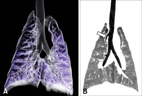
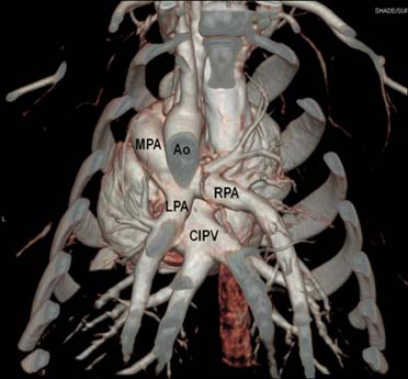
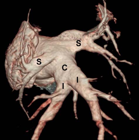
- Micropigs are the most likely source animals for xenotransplantation. However, an appropriate method for evaluating the lung of micropigs had not been established. Therefore, this study was performed to evaluate the feasibility of 64-channel multi-detector row computed tomography (MDCT) to measure the diameter of the pulmonary arteries and the lung volume in micropigs. The mean diameters of the trachea, and left and right bronchi were 1.6 +/- 0.17, 1.18 +/- 0.14, and 1.1 +/- 0.11 cm, respectively. The mean diameters of the main, right, and left pulmonary arteries were 1.38 +/- 0.09, 1.07 +/- 0.26, and 0.98 +/- 0.13 cm and the diameters of right, left, and common inferior pulmonary veins were 0.97 +/- 0.20, 0.76 +/- 0.20, and 1.99 +/- 0.26 cm, respectively. The mean lung volume was 820.3 +/- 77.11 mL. The data presented in this study suggest that the MDCT may be a noninvasive, rapid, and accurate investigational method for pulmonary anatomy in living lung donors.
MeSH Terms
Figure
Reference
-
1. Abecassis M, Adams M, Adams P, Arnold RM, Atkins CR, Barr ML, Bennett WM, Bia M, Briscoe DM, Burdick J, Corry RJ, Davis J, Delmonico FL, Gaston RS, Harmon W, Jacobs CL, Kahn J, Leichtman A, Miller C, Moss D, Newmann JM, Rosen LS, Siminoff L, Spital A, Starnes VA, Thomas C, Tyler LS, Williams L, Wright FH, Youngner S. Consensus statement on the live organ donor. JAMA. 2000. 284:2919–2926.2. Bowdish ME, Barr ML, Starnes VA. Living lobar transplantation. Chest Surg Clin N Am. 2003. 13:505–524.
Article3. Butler J. The Emerging role of multi-detector computed tomography in heart failure. J Card Fail. 2007. 13:215–226.
Article4. Cantu E, Parker W, Platt JL, Duane Davis R. Pulmonary xenotransplantation: rapidly progressing into the unknown. Am J Transplant. 2004. 4:Suppl 6. 25–35.
Article5. Cohen RG, Barr ML, Schenkel FA, DeMeester TR, Wells WJ, Starnes VA. Living-related donor lobectomy for bilateral lobar transplantation in patients with cystic fibrosis. Ann Thorac Surg. 1994. 57:1423–1427.6. Daggett CW, Yeatman M, Lodge AJ, Chen EP, Linn SS, Gullotto C, Frank MM, Platt JL, Davis RD. Total respiratory support from swine lungs in primate recipients. J Thorac Cardiovasc Surg. 1998. 115:19–27.
Article7. Dooldeniya MD, Warrens AN. Xenotransplantation: where are we today? J R Soc Med. 2003. 96:111–117.
Article8. Evans RW, Orians CE, Ascher NL. The potential supply of organ donors. An assessment of the efficacy of organ procurement efforts in the United States. JAMA. 1992. 267:239–246.
Article9. Foley WD. Special focus session: multidetector CT: abdominal visceral imaging. Radiographics. 2002. 22:701–719.10. Grude M, Juergens KU, Wichter T, Paul M, Fallenberg EM, Muller JG, Heindel W, Breithardt G, Fischbach R. Evaluation of global left ventricular myocardial function with electrocardiogram-gated multidetector computed tomography: comparison with magnetic resonance imaging. Invest Radiol. 2003. 38:653–661.
Article11. Hoffman EA, Simon BA, McLennan G. State of the Art. A structural and functional assessment of the lung via multidetector-row computed tomography: phenotyping chronic obstructive pulmonary disease. Proc Am Thorac Soc. 2006. 3:519–532.
Article12. Hampton T, Armstrong S, Russell WJ. Estimating the diameter of the left main bronchus. Anaesth Intensive Care. 2000. 28:540–542.
Article13. Juergens KU, Maintz D, Grude M, Boese JM, Heimes B, Fallenberg EM, Heindel W, Fischbach R. Multi-detector row computed tomography of the heart: does a multisegment reconstruction algorithm improve left ventricular volume measurements? Eur Radiol. 2005. 15:111–117.
Article14. Kalender WA, Polacin A. Physical performance characteristics of spiral CT scanning. Med Phys. 1991. 18:910–915.
Article15. Kaplon RJ, Platt JL, Kwiatkowski PA, Edwards NM, Xu H, Shah AS, Masroor S, Michler RE. Absence of hyperacute rejection in pig-to-primate orthotopic pulmonary xenografts. Transplantation. 1995. 59:410–416.
Article16. Karazincir S, Balci A, Seyfeli E, Akoğlu S, Babayiğit C, Akgül F, Yalçin F, Eğilmez E. CT assessment of main pulmonary artery diameter. Diagn Interv Radiol. 2008. 14:72–74.17. Lambrigts D, Sachs DH, Cooper DK. Discordant organ xenotransplantation in primates: world experience and current status. Transplantation. 1998. 66:547–561.18. Logan JS. Prospects for xenotransplantation. Curr Opin Immunol. 2000. 12:563–568.
Article19. Pierson RN 3rd, Kasper-Konig W, Tew DN, Young VK, Dunning JJ, Horsley J, Carey NR, Wallwork J, White DJ. Hyperacute lung rejection in a pig-to-human transplant model: the role of anti-pig antibody and complement. Transplantation. 1997. 63:594–603.
Article20. Prokop M. General principles of MDCT. Eur J Radiol. 2003. 45:Suppl 1. S4–S10.
Article21. Starnes VA, Barr ML, Cohen RG. Lobar transplantation: indications, technique, and outcome. J Thorac Cardiovasc Surg. 1994. 108:403–411.22. Stern EJ, Graham CM, Webb WR, Gamsu G. Normal trachea during forced expiration: dynamic CT measurements. Radiology. 1993. 187:27–31.
Article23. Takase B, Nagata M, Matsui T, Kihara T, Kameyama A, Hamabe A, Noya K, Satomura K, Ishihara M, Kurita A, Ohsuzu F. Pulmonary vein dimensions and variation of branching pattern in patients with paroxysmal atrial fibrillation using magnetic resonance angiography. Jpn Heart J. 2004. 45:81–92.
Article24. Wintersperger BJ, Herzog P, Jakobs T, Reiser MF, Becker CR. Initial experience with the clinical use of a 16 detector row CT system. Crit Rev Comput Tomogr. 2002. 43:283–316.25. Yeatman M, Daggett CW, Lau CL, Byrne GW, Logan JS, Platt JL, Davis RD. Human complement regulatory proteins protect swine lungs from xenogeneic injury. Ann Thorac Surg. 1999. 67:769–775.
Article
- Full Text Links
- Actions
-
Cited
- CITED
-
- Close
- Share
- Similar articles
-
- Imaging evaluation of the liver using multi-detector row computed tomography in micropigs as potential living liver donors
- Multi-Slice Spiral CT of Living-Related Liver Transplantation in Children: Pictorial Essay
- Multi-Detector Row CT of the Central Airway Disease
- Multidetector computed tomographic angiography evaluation of micropig major systemic vessels for xenotransplantation
- Mediastinal and Hilar Lymphadenopathy: Cross-Referenced Anatomy on Axial and Coronal Images Displayed by Using Multi-detector row CT


