Rapidly Growing Bilateral Pseudoangiomatous Stromal Hyperplasia of the Breast
- Affiliations
-
- 1Department of Radiology, Uijeongbu St. Mary's Hospital, College of Medicine, The Catholic University of Korea, Gyeonggi-do 480-717, Korea. tiger@catholic.ac.kr
- 2Department of Clinical Pathology, Uijeongbu St. Mary's Hospital, College of Medicine, The Catholic University of Korea, Gyeonggi-do 480-717, Korea.
- KMID: 946276
- DOI: http://doi.org/10.3348/kjr.2010.11.3.355
Abstract
- A tumoral pseudoangiomatous stromal hyperplasia (PASH) that causes huge breast enlargement is very rare. Only two cases of huge tumoral PASHs have been reported in the English medical literature. We report here on a surgically confirmed case of bilateral huge tumoral PASH in a 47-year-old woman, and we present the imaging and histopathology findings. We also review the relevant medical literature.
MeSH Terms
-
Angiomatosis/*pathology/surgery/ultrasonography
Biopsy, Needle
Breast/cytology/pathology/surgery
Breast Diseases/*pathology/surgery/ultrasonography
Contrast Media/diagnostic use
Diagnosis, Differential
Female
Gadolinium DTPA/diagnostic use
Humans
Hyperplasia
Image Enhancement/methods
Magnetic Resonance Imaging/methods
Mammography/methods
Middle Aged
Stromal Cells/pathology
Figure
-
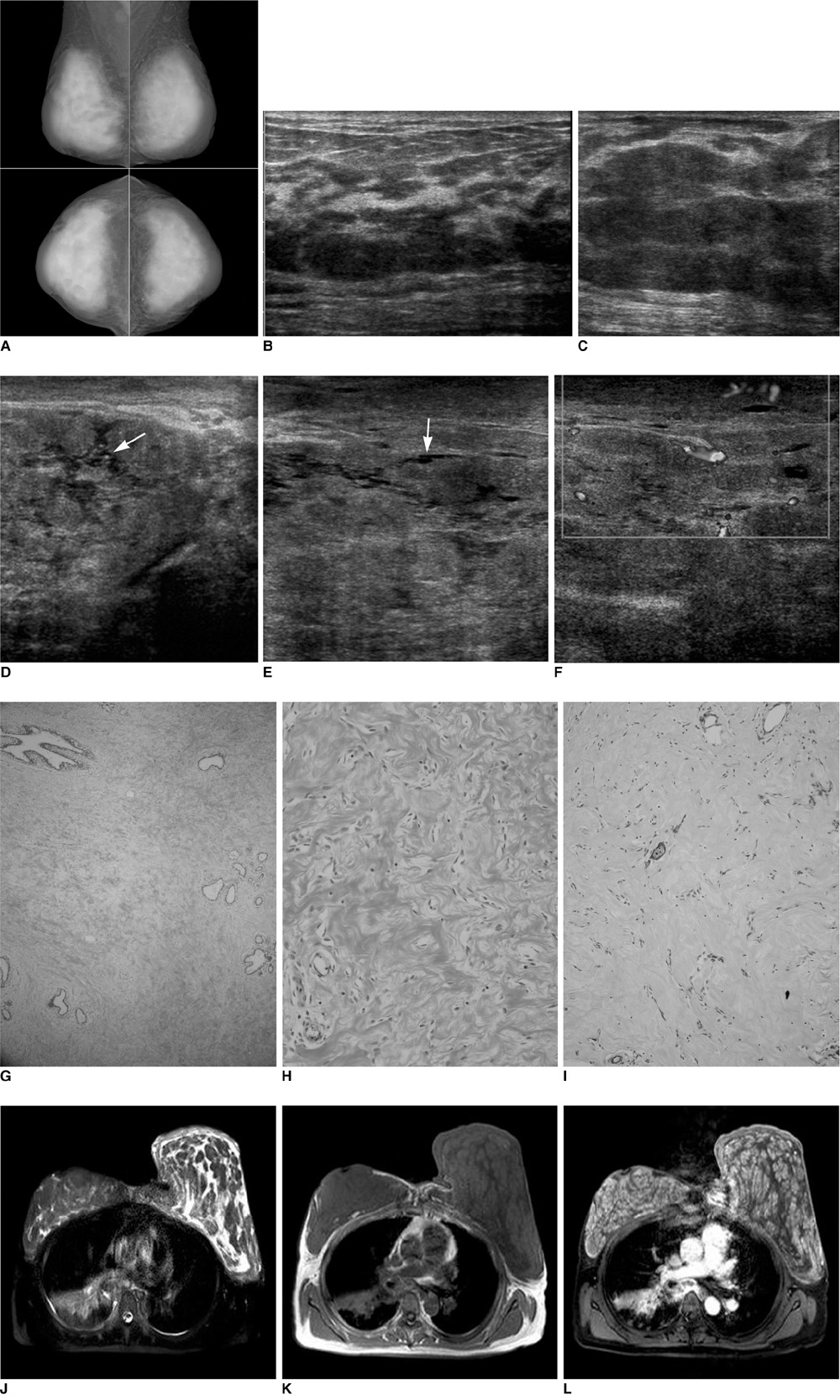
Fig. 1 Bilateral pseudoangiomatous stromal hyperplasia of breasts in 47-year-old woman. A. Initial mammography shows huge enlargement of both breasts. Parenchymal pattern is extremely dense. There is reticular density around parenchyma as well as skin thickening. B, C. Sonography of right (B) and left (C) breasts shows skin thickening and low echoes similar to those caused by lobular thickening of glandular layer. Left breast is more prominent than right breast. D-F. Sonography obtained six months after shows substantial progression of glandular layer thickening in patient's left breast. Note presence of conglomerations of tiny cystic spaces (arrows) and abundant color flow indicating hyperemic change. G. Classic features of pseudoangiomatous stromal hyperplasia, i.e., dense, fibrous tissue separates lobular architecture with loose, supporting stroma (Hematoxylin & Eosin staining, ×40). H. Higher magnification view shows cleft lined by endothelial-like, spindle cells in context of stromal hyperplasia (Hematoxylin & Eosin staining, ×200). I. Immunohistochemistry preparation reveals CD 34 reactivity of pseudovascular space lined by myoepithelial cells (Hematoxylin & Eosin staining, ×100). J-L. MRI examination of breast shows high-signal spaces between diffuse and nodular low signals on fat-saturated half-fourier acquisition single-shot turbo spin-echo T2 (J). Entire breast reveals low signal on 2D flash T1. Fibrous hyperplasia with cystic spaces is suggested (K). Low signal on T2 or dense, fibrous tissue shows homogeneous enhancement on fat-saturated T1 after contrast infusion (L). Dynamic study using dedicated coil could not be performed.
Reference
-
1. Hargaden GC, Yeh ED, Georgian-Smith D, Moore RH, Rafferty EA, Halpern EF, et al. Analysis of the mammographic and sonographic features of pseudoangiomatous stromal hyperplasia. AJR Am J Roentgenol. 2008. 191:359–363.2. Ibrahim RE, Sciotto CG, Weidner N. Pseudoangiomatous hyperplasia of mammary stroma. Some observations regarding its clinicopathologic spectrum. Cancer. 1989. 63:1154–1160.3. Polger MR, Denison CM, Lester S, Meyer JE. Pseudoangiomatous stromal hyperplasia: mammographic and sonographic appearances. AJR Am J Roentgenol. 1996. 166:349–352.4. Cohen MA, Morris EA, Rosen PP, Dershaw DD, Liberman L, Abramson AF. Pseudoangiomatous stromal hyperplasia: mammographic, sonographic, and clinical patterns. Radiology. 1996. 198:117–120.5. Piccoli CW, Feig SA, Palazzo JP. Developing asymmetric breast tissue. Radiology. 1999. 211:111–117.6. Mercado CL, Naidrich SA, Hamele-Bena D, Fineberg SA, Buchbinder SS. Pseudoangiomatous stromal hyperplasia of the breast: sonographic features with histopathologic correlation. Breast J. 2004. 10:427–432.7. Teh HS, Chiang SH, Leung JW, Tan SM, Mancer JF. Rapidly enlarging tumoral pseudoangiomatous stromal hyperplasia in a 15-year-old patient: distinguishing sonographic and magnetic resonance imaging findings and correlation with histologic findings. J Ultrasound Med. 2007. 26:1101–1106.8. Baskin H, Layfield L, Morrell G. MRI appearance of pseudoangiomatous stromal hyperplasia causing asymmetric breast enlargement. Breast J. 2007. 13:209–210.
- Full Text Links
-
- Actions
-
Cited
- CITED
-
- Close
- Share
-
- Similar articles
-
- Radiologic Imaging Findings of Bilateral Infiltrating Pseudoangiomatous Stromal Hyperplasia of the Breasts: A Case Report
- Pseudoangiomatous Stromal Hyperplasia of the Breast Appearing as a Giant Mass: A Case Report
- Bilateral gigantomastia due to benign breast tumors: a case series and brief review focusing on bilateral diffuse pseudoangiomatous stromal hyperplasia
- Pseudoangiomatous Stromal Hyperplasia of the Breast in a Female Adolescent Presenting as Bilateral Gigantomastia
- Huge Bilateral Breast Hamartoma Accompanied with Pseudoangiomatous Stromal Hyperplasia
