J Korean Soc Radiol.
2013 Apr;68(4):329-332. 10.3348/jksr.2013.68.4.329.
Radiologic Imaging Findings of Bilateral Infiltrating Pseudoangiomatous Stromal Hyperplasia of the Breasts: A Case Report
- Affiliations
-
- 1Department of Radiology, Kangnam Sacred Heart Hospital, College of Medicine, Hallym University, Seoul, Korea. jskyoung@hallym.or.kr
- KMID: 1425149
- DOI: http://doi.org/10.3348/jksr.2013.68.4.329
Abstract
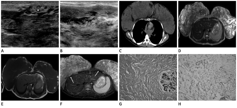
- Pseudoangiomatous stromal hyperplasia (PASH), first described by Vuitch et al., is a rare benign lesion, shows the proliferation of the breast stromal tissue. We experienced a case of a 19-year-old woman with bilateral PASH manifesting as marked enlargement of the breasts for a duration of several months. Morphologic evaluation of imaging modalities was done, including ultrasound, computed tomography and magnetic resonance images. Here, we describe bilateral PASH in a young girl presenting as bilateral breast gigantism from a main point of radiologic image finding.
MeSH Terms
Figure
Reference
-
1. Goel NB, Knight TE, Pandey S, Riddick-Young M, de Paredes ES, Trivedi A. Fibrous lesions of the breast: imaging-pathologic correlation. Radiographics. 2005. 25:1547–1559.2. Celliers L, Wong DD, Bourke A. Pseudoangiomatous stromal hyperplasia: a study of the mammographic and sonographic features. Clin Radiol. 2010. 65:145–149.3. Choi YJ, Ko EY, Kook S. Diagnosis of pseudoangiomatous stromal hyperplasia of the breast: ultrasonography findings and different biopsy methods. Yonsei Med J. 2008. 49:757–764.4. Ryu EM, Whang IY, Chang ED. Rapidly growing bilateral pseudoangiomatous stromal hyperplasia of the breast. Korean J Radiol. 2010. 11:355–358.5. Hargaden GC, Yeh ED, Georgian-Smith D, Moore RH, Rafferty EA, Halpern EF, et al. Analysis of the mammographic and sonographic features of pseudoangiomatous stromal hyperplasia. AJR Am J Roentgenol. 2008. 191:359–363.6. Singh KA, Lewis MM, Runge RL, Carlson GW. Pseudoangiomatous stromal hyperplasia. A case for bilateral mastectomy in a 12-year-old girl. Breast J. 2007. 13:603–660.7. Sng KK, Tan SM, Mancer JF, Tay KH. The contrasting presentation and management of pseudoangiomatous stromal hyperplasia of the breast. Singapore Med J. 2008. 49:e82–e85.8. Teh HS, Chiang SH, Leung JW, Tan SM, Mancer JF. Rapidly enlarging tumoral pseudoangiomatous stromal hyperplasia in a 15-year-old patient: distinguishing sonographic and magnetic resonance imaging findings and correlation with histologic findings. J Ultrasound Med. 2007. 26:1101–1106.9. Baskin H, Layfield L, Morrell G. MRI appearance of pseudoangiomatous stromal hyperplasia causing asymmetric breast enlargement. Breast J. 2007. 13:209–210.
- Full Text Links
- Actions
-
Cited
- CITED
-
- Close
- Share
- Similar articles
-
- Pseudoangiomatous Stromal Hyperplasia of the Breast Appearing as a Giant Mass: A Case Report
- Huge Bilateral Breast Hamartoma Accompanied with Pseudoangiomatous Stromal Hyperplasia
- Rapidly Growing Bilateral Pseudoangiomatous Stromal Hyperplasia of the Breast
- Bilateral gigantomastia due to benign breast tumors: a case series and brief review focusing on bilateral diffuse pseudoangiomatous stromal hyperplasia
- Treatment of Pseudoangiomatous Stromal Hyperplasia of the Breast: Implant-Based Reconstruction with a Vascularized Dermal Sling


