Korean J Ophthalmol.
2010 Jun;24(3):139-142. 10.3341/kjo.2010.24.3.139.
Evaluation of Corneal Biomechanical Properties Following Penetrating Keratoplasty Using the Ocular Response Analyzer
- Affiliations
-
- 1Department of Ophthalmology, Seoul National University Hospital, Seoul, Korea. wrwee@snu.ac.kr
- 2Seoul Artificial Eye Center, Seoul National University Hospital Clinical Research Institute, Seoul, Korea.
- KMID: 945982
- DOI: http://doi.org/10.3341/kjo.2010.24.3.139
Abstract
- PURPOSE
To evaluate corneal biomechanical properties in eyes that had previously undergone penetrating keratoplasty (PK) using the ocular response analyzer (ORA). METHODS: We recruited 26 patients who had received unilateral PK. Corneal hysteresis (CH), corneal resistance factor (CRF), Goldmann-correlated intraocular pressure (IOPg), and cornea-compensated intraocular pressure (IOPcc) were measured with the ORA and were compared to the measurements from the contralateral eyes that did not undergo PK. RESULTS: The CH was 8.95+/-2.59 mmHg in eyes that underwent PK and 9.78+/-1.45 mmHg in the contralateral eyes that did not undergo PK (p=0.077). The CRF was 10.26+/-2.64 mmHg in post-PK eyes and 9.75+/-1.45 mmHg in the contralateral eyes (p=0.509), and the CH-CRF was significantly smaller in post-PK eyes (-1.31+/-2.32 mmHg in post-PK eyes vs. 0.03+/-0.88 mmHg in fellow eyes, p=0.016). The IOPg and IOPcc were significantly higher in the PK group than they were in the control group. The IOPcc's were 20.81+/-7.81 mmHg and 16.27+/-2.49 mmHg in post-PK and control eyes, respectively (p=0.011); and the IOPg's were 19.22+/-7.34 mmHg and 15.07+/-3.03 mmHg in post-PK and control eyes, respectively (p=0.019). The IOPcc-g's were 1.59+/-2.81 mmHg and 1.21+/-1.30 mmHg in post-PK and control eyes, respectively (p=0.412), and the central corneal thickness (CCT)'s were 489.11+/-90.60 microm and 556.24+/-42.84 microm in post-PK and control eyes, respectively (p=0.068). CONCLUSIONS: Following PK, CH tended to decrease while CRF tended to increase, significantly decreasing CH-CRF. A significantly higher intraocular pressure and a thinner CCT following PK may have contributed to the observed changes in these corneal biomechanical parameters.
MeSH Terms
Figure
-

Fig. 1 Correlations of corneal hysteresis (CH) and corneal resistance factor (CRF) with cornea-compensated intraocular pressure (IOPcc) and Goldmann-correlated intraocular pressure (IOPg).
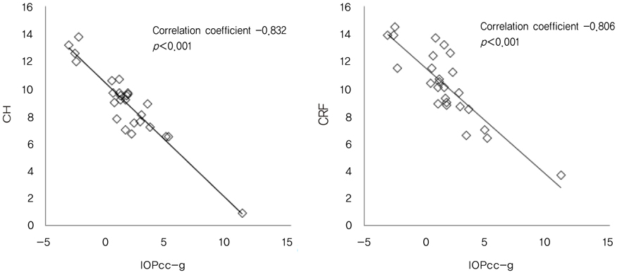
Fig. 2 Correlations of cornea-compensated intraocular pressure and Goldmann-correlated intraocular pressure (IOPcc-g) with corneal hysteresis (CH) and corneal resistance factor (CRF).
Reference
-
1. Luce DA. Determining in vivo biomechanical properties of the cornea with an ocular response analyzer. J Cataract Refract Surg. 2005. 31:156–162.2. Ortiz D, Pinero D, Shabayek MH, et al. Corneal biomechanical properties in normal, post-laser in situ keratomileusis, and keratoconic eyes. J Cataract Refract Surg. 2007. 33:1371–1375.3. Pepose JS, Feigenbaum SK, Qazi MA, et al. Changes in corneal biomechanics and intraocular pressure following LASIK using static, dynamic, and noncontact tonometry. Am J Ophthalmol. 2007. 143:39–47.4. Medeiros FA, Weinreb RN. Evaluation of the influence of corneal biomechanical properties on intraocular pressure measurements using the ocular response analyzer. J Glaucoma. 2006. 15:364–370.5. Lam A, Chen D, Chiu R, Chui WS. Comparison of IOP measurements between ORA and GAT in normal Chinese. Optom Vis Sci. 2007. 84:909–914.6. Sullivan-Mee M, Billingsley SC, Patel AD, et al. Ocular Response Analyzer in subjects with and without glaucoma. Optom Vis Sci. 2008. 85:463–470.7. Moreno-Montanes J, Maldonado MJ, Garcia N, et al. Reproducibility and clinical relevance of the ocular response analyzer in nonoperated eyes: corneal biomechanical and tonometric implications. Invest Ophthalmol Vis Sci. 2008. 49:968–974.8. Kirwan C, O'Malley D, O'Keefe M. Corneal hysteresis and corneal resistance factor in keratoectasia: findings using the Reichert ocular response analyzer. Ophthalmologica. 2008. 222:334–337.9. Shah S, Laiquzzaman M, Bhojwani R, et al. Assessment of the biomechanical properties of the cornea with the ocular response analyzer in normal and keratoconic eyes. Invest Ophthalmol Vis Sci. 2007. 48:3026–3031.10. del Buey MA, Cristobal JA, Ascaso FJ, et al. Biomechanical properties of the cornea in Fuchs' corneal dystrophy. Invest Ophthalmol Vis Sci. 2009. 50:3199–3202.11. Shen M, Fan F, Xue A, et al. Biomechanical properties of the cornea in high myopia. Vision Res. 2008. 48:2167–2171.12. Gatinel D, Chaabouni S, Adam PA, et al. Corneal hysteresis, resistance factor, topography, and pachymetry after corneal lamellar flap. J Refract Surg. 2007. 23:76–84.13. Kucumen RB, Yenerel NM, Gorgun E, et al. Corneal biomechanical properties and intraocular pressure changes after phacoemulsification and intraocular lens implantation. J Cataract Refract Surg. 2008. 34:2096–2098.14. Touboul D, Roberts C, Kerautret J, et al. Correlations between corneal hysteresis, intraocular pressure, and corneal central pachymetry. J Cataract Refract Surg. 2008. 34:616–622.
- Full Text Links
-
- Actions
-
Cited
- CITED
-
- Close
- Share
-
- Similar articles
-
- The Use of a Dynamic Scheimpflug Analyzer to Measure Changes of Post-keratoplasty Corneal Biomechanical Properties
- Cataract Extraction after Penetrating Keratoplasty
- Corneal Biomechanical Properties of Normal Tension Glaucoma in Young Patients Evaluated with the Ocular Response Analyzer
- Change in Corneal Biomechanical Parameters in Diabetes Mellitus
- Early Corneal-Thickness Changes after Penetrating Keratoplasty
