Impression Cytology of Herpetic Simplex Keratitis in Rabbits
- Affiliations
-
- 1Department of Ophthalmology, Hanyang University College of Medicine, Seoul, Korea. fovea@hanyang.ac.kr
- KMID: 754404
- DOI: http://doi.org/10.3341/kjo.2005.19.2.96
Abstract
- PURPOSE
To use impression cytology to examine the structural changes in corneal epithelial cells infected with the herpes simplex virus in rabbit eyes. METHODS: Corneal surfaces of 7 rabbits were scratched using a 25-gauge needle. Herpes simplex virus (type 1, Kos strain) was inoculated to the injured cornea. As the corneal diseases were observed using slit lamp biomicroscopy, impression cytology was performed for 18 days after inoculation. Specimens were stained with hematoxylin-eosin and examined using optical microscopy. RESULTS: Corneal lesions consisted mainly of round epithelial cells, inflammatory cells, ballooning cells, multinucleated giant cells, and various inclusion bodies. Over time, the corneal epithelial cells peeled away as a result of corneal edema in the corneal lesions. Dendritic lesions were also observed. In the recovery phase, the number of detached cells and infiltrated inflammatory cells decreased. CONCLUSIONS: It was presumed that dendritic lesions might have been formed at the scratched cornea region, thereby aggravating the epithelial cells falling off as a result of the infiltration of inflammatory cells. These cytopathologic effects occur in experimental herpes simplex keratitis.
MeSH Terms
Figure
-
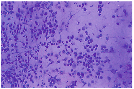
Fig. 1 Many inflammatory cells and degenerated corneal epithelial cells have infiltrated the corneal lesion, 3 days following inoculation of the herpes simplex virus (Impression cytology, H-E stain ×100).

Fig. 2 Balloon cells (arrow) can be seen in the area of the detached and degenerated corneal epithelium, which had swollen cytoplasm and a pale nucleus, 3 days following inoculation of the herpes simplex virus (Impression cytology, H-E stain ×100).
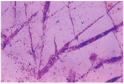
Fig. 3 Epithelial cells transferred from the lesion exhibited widening of the scratching wound lesion, 5 days following inoculation of the herpes simplex virus (Impression cytology, H-E stain ×100) (H-E stain ×40).

Fig. 4 Higher magnification of the dendritic keratitis lesion, 7 days following inoculation of the herpes simplex virus. There were an increased numbers of balloon cells (arrow), degenerated superficial cells with bizarre nucleus and vacuolated cells (arrowhead), 7 days following inoculation of the herpes simplex virus(Impression cytology, H-E stain ×400).

Fig. 5 The specimen obtained from the center of an actively infected lesion, showing syncytia (arrow) and epithelial cells including a dense basophilic nucleus. Snake cells (arrowhead) were observed, 11 days following inoculation of the herpes simplex virus (Impression cytology, H-E stain ×400).
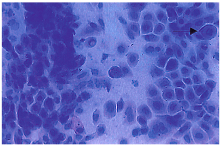
Fig. 6 Intranuclear inclusions (arrow) in a specimen of dendritic keratitis, 11 days following inoculation of the herpes simplex virus(Impression cytology, H-E stain ×400).

Fig. 7 Detached superficial and wing epithelial cells are evident, 15 days following inoculation of the herpes simplex virus (Impression cytology, H-E stain ×400).
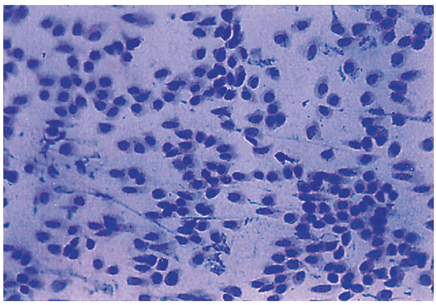
Fig. 8 The detached epithelial cells are still evident and the number of inflammatory cells was decreased, 18 days following inoculation of the herpes simplex virus (Impression cytology, H-E stain ×100).
Reference
-
1. Hyndiuk RA, Glasser D. Tabbara KF, editor. Herpes simplex keratitis. Infections of the Eye. 1986. 1st ed. Boston: Little Brown;1204–1223.2. Pepose JS, Leib DA. Stuart M, editor. Herpes simplex virus diseases. Ocular infection and immunity. 1996. v.1:1st ed. London: Mosby;chap. 71.3. Dawson CR, Togni B. Herpes simplex eye infections: clinical manifestations, pathogenesis and management. Surv Ophthalmol. 1976. 21:121–135.4. Lee KY, Chung MR, Ko MK. Scanning Electron Microscopic Observations of Sequential Alterations of Type 1 HSV Keratitis in Rabbits. J Korean Ophthalmol Soc. 2004. 45:1174–1180.5. Egbert PR, Lauber S, Maurice DM. A simple conjunctival biopsy. Am J Ophthalmol. 1977. 84:798–801.6. Arora I, Singhvi S. Impression debridement of corneal lesions. Ophthalmology. 1994. 101:1935–1941.7. Maskin SL, Heitman KF, Lawton AW, Yee RW. Diagnostic impression cytology for external eye disease. Cornea. 1989. 8:270–279.8. Nelson DJ. Impression cytology. Cornea. 1988. 7:71–85.9. Simon MW, Miller DMT, Pflugefelder SC. Comparison of immunocytology to tissue culture for diagnosis of presumed herpesvirus dendritic epithelial keratitis. Ophthalmology. 1992. 99:1408–1420.10. Tabery HM. Epithelial changes in early primary herpes simplex virus keratitis. Photomicrographic observations in a case of human infection. Acta Ophthalmol Scand. 2000. 78:706–709.11. Kim DC, Ko MK. Slit - Lamp Examination of the Experimentally Induced HSV - 1 Keratitis. J Korean Ophthalmol Soc. 1998. 29:251–255.12. Kwon JW, Han JH, Ko MK, Kim JW. Ultrastructure of Corneal Epithelial Cells Following Inoculation of Herpes Simplex Virus in Rabbit. J Korean Ophthalmol Soc. 2001. 42:349–354.13. Wander AH, Centifanto YM, Kaufman HE. Strain specificity of clinical isolates of herpes simplex virus. Arch Ophthalmol. 1980. 98:1458–1461.14. Metcalf JF, Michaelis BA. Herpetic keratitis in inbred mice. Invest Ophthalmol Vis Sci. 1984. 25:1222–1225.15. Lee YJ, Ko MK, Kim JG. The Ultrastructure of Rabbit Keratocyte Infected by Herpes Simplex Virus. J Korean Ophthalmol Soc. 2000. 41:28–33.16. Knop E, Reale E. Fine structure and significance of snakelike chromatin in conjunctival epithelial cells. Invest Ophthalmol Vis Sci. 1994. 35:711–719.17. Maudgal PC, Missotten L. Histopathology of human superficial herpes simplex keratitis. Br J Ophthalmol. 1978. 62:46–52.18. Vrabec F, Darrell RW. The corneal epithelium in experimental herpetic keratitis in rabbits. Doc Ophthalmol. 1970. 29:213–224.19. McKee AP. The biology of herpes simplex virus. Invest Ophthalmol. 1963. 2:490–497.
- Full Text Links
-
- Actions
-
Cited
- CITED
-
- Close
- Share
-
- Similar articles
-
- The Result of Isnlation of Herps Simplex Virus with Viral Culture Method in Herpes Simplex Keratitis
- 3 Cases of Latanoprost Associated Herpes Simplex Keratitis
- A Case of Herpetic Simplex Keratitis after Application of 0.015% Tafluprost Eye Drops
- Clinical Evaluations of Recurrence after Keratoplasty in Herpes Simplex Keratitis
- Three Cases of Secondary Fungal Infection in Herpes Simplex Keratitis
