Obstet Gynecol Sci.
2025 Jan;68(1):79-89. 10.5468/ogs.24236.
Significance and limitations of routine p16/Ki-67 immunohistochemistry as a diagnostic tool for high-grade squamous intraepithelial lesions of the uterine cervix
- Affiliations
-
- 1Department of Diagnostic Pathology, Kumamoto University Hospital, Kumamoto, Japan
- 2Department of Surgical Pathology, Japan Community Health Care Organization Kumamoto General Hospital, Kumamoto, Japan
- 3Department of Pathology and Cell Biology, University of the Ryukyus, Okinawa, Japan
- KMID: 2563940
- DOI: http://doi.org/10.5468/ogs.24236
Abstract
Objective
To evaluate the diagnostic utility and limitations of routine p16 and Ki-67 immunohistochemistry (IHC) in detecting high-grade squamous intraepithelial lesions (HSILs) in the uterine cervix.
Methods
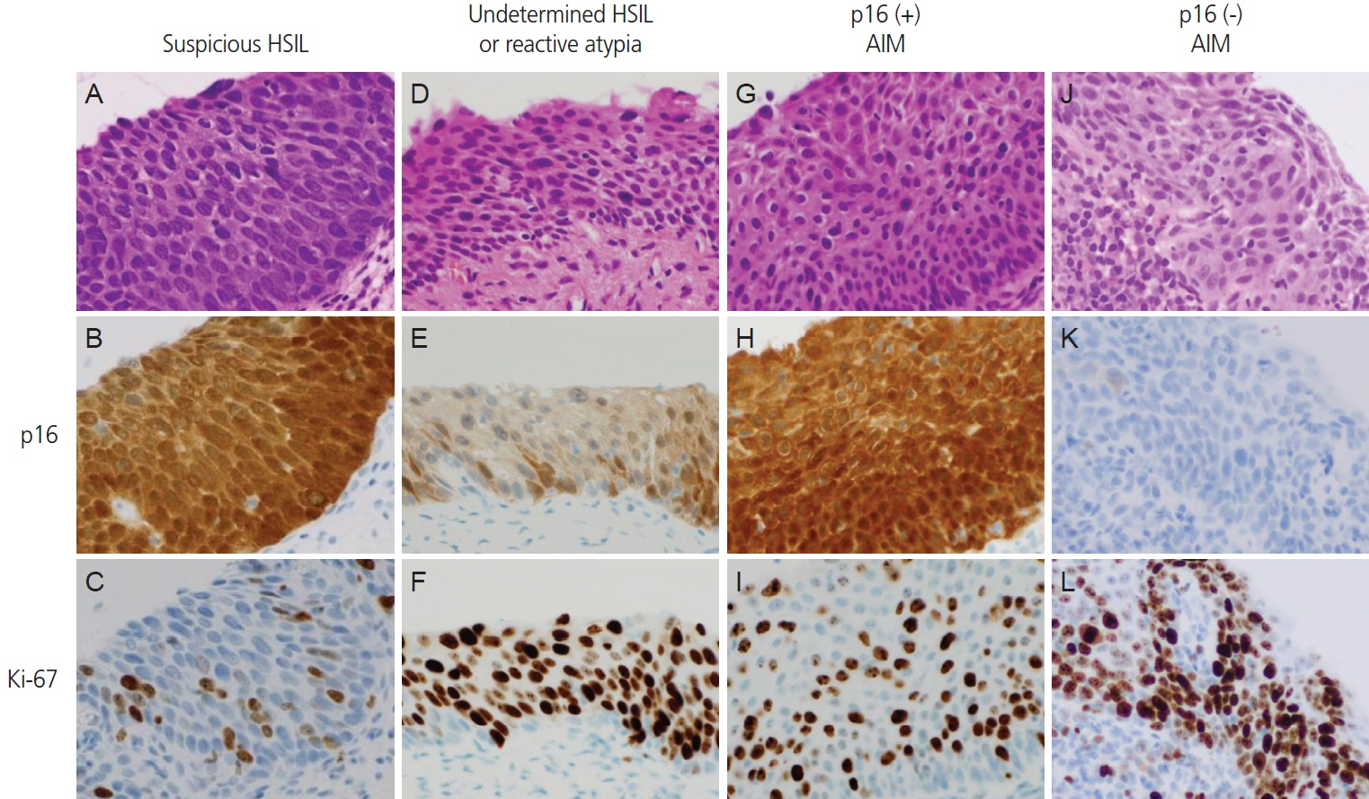
We reviewed 2,061 cervical biopsy records, including 271 morphologically indeterminate squamous lesions, evaluated using p16/Ki-67 IHC for HSIL detection or exclusion. HSIL was diagnosed based on p16 positivity and a high Ki-67 labeling index (Ki-LI). In cases that remained inconclusive after IHC, follow-up histological and/or cytological outcomes were assessed.
Results
p16/Ki-67 IHC established a definitive diagnosis of either HSIL or non-HSIL in 74.2% (201/271) of morphologically indeterminate cases, whereas 25.8% (70/271) remained inconclusive. p16/Ki-67 IHC contributed to diagnosing 120 HSIL cases, representing 11.9% (120/1,011) of all HSILs cases and 44.3% (120/271) of morphologically indeterminate cases. Among the 70 inconclusive cases, 58 had available follow-up data, of which 22 were subsequently diagnosed with HSIL, including 12 within 1 month of the initial biopsy. HSIL outcomes were more frequent in cases with suspicious HSIL on the initial biopsy (66.7% [12/18]). Based on the p16/Ki-LI status observed in the initial biopsy, patients with HSIL outcomes were categorized into three groups: p16-positive/low Ki-LI (54.2% [13/24]), p16-negative/high Ki-LI (50.0% [5/10]), and p16-negative/low Ki-LI (16.7% [4/24]). Multiple comparisons revealed a significant difference between the p16-positive/low Ki-LI and p16-negative/low Ki-LI groups (Benjamini-Yekutieli adjusted P=0.0435), while other comparisons were not significant.
Conclusion
p16/Ki-67 IHC significantly improved the diagnostic performance for HSIL. In cases that remain inconclusive after IHC, IHC-based risk stratification offers a valuable approach for surveillance, thus mitigating delays in HSIL diagnosis.
Keyword
Figure
Reference
-
References
1. Sung H, Ferlay J, Siegel RL, Laversanne M, Soerjomataram I, Jemal A, et al. Global cancer statistics 2020: GLOBOCAN estimates of incidence and mortality worldwide for 36 cancers in 185 countries. CA Cancer J Clin. 2021; 71:209–49.
Article2. Ferlay J, Ervik M, Lam F, Colombet M, Mery L, Piñeros M, et al. Global cancer observatory: cancer today; 2020 [Internet]. Lyon: International Agency for Research on Cancer;c2023. [cited 2023 Nov 3]. Available from: https://gco.iarc.who.int/today.3. Lin S, Gao K, Gu S, You L, Qian S, Tang M, et al. Worldwide trends in cervical cancer incidence and mortality, with predictions for the next 15 years. Cancer. 2021; 127:4030–9.
Article4. Schiffman M, Doorbar J, Wentzensen N, de Sanjosé S, Fakhry C, Monk BJ, et al. Carcinogenic human papillomavirus infection. Nat Rev Dis Primers. 2016; 2:16086.
Article5. Klaes R, Friedrich T, Spitkovsky D, Ridder R, Rudy W, Petry U, et al. Overexpression of p16(INK4A) as a specific marker for dysplastic and neoplastic epithelial cells of the cervix uteri. Int J Cancer. 2001; 92:276–84.
Article6. Darragh TM, Colgan TJ, Thomas Cox J, Heller DS, Henry MR, Luff RD, et al. The lower anogenital squamous terminology standardization project for HPV-associated lesions: background and consensus recommendations from the College of American Pathologists and the American Society for Colposcopy and Cervical Pathology. Int J Gynecol Pathol. 2013; 32:76–115.
Article7. Iaconis L, Hyjek E, Ellenson LH, Pirog EC. p16 and Ki-67 immunostaining in atypical immature squamous metaplasia of the uterine cervix: correlation with human papillomavirus detection. Arch Pathol Lab Med. 2007; 131:1343–9.8. Galgano MT, Castle PE, Atkins KA, Brix WK, Nassau SR, Stoler MH. Using biomarkers as objective standards in the diagnosis of cervical biopsies. Am J Surg Pathol. 2010; 34:1077–87.
Article9. Kim TH, Han JH, Shin E, Noh JH, Kim HS, Song YS. Clinical implication of p16, Ki-67, and proliferating cell nuclear antigen expression in cervical neoplasia: improvement of diagnostic accuracy for high-grade squamous intraepithelial lesion and prediction of resection margin involvement on conization specimen. J Cancer Prev. 2015; 20:70–7.
Article10. Lim S, Lee MJ, Cho I, Hong R, Lim SC. Efficacy of p16 and Ki-67 immunostaining in the detection of squamous intraepithelial lesions in a high-risk HPV group. Oncol Lett. 2016; 11:1447–52.
Article11. Miralpeix E, Solé-Sedeño JM, Mancebo G, Lloveras B, Bellosillo B, Carreras R, et al. Value of p16(INK4a) and Ki-67 immunohistochemical staining in cervical intraepithelial neoplasia grade 2 biopsies as biomarkers for cervical intraepithelial neoplasia grade 3 in cone results. Anal Quant Cytopathol Histpathol. 2016; 38:1–8.12. Silva DC, Gonçalves AK, Cobucci RN, Mendonça RC, Lima PH, Cavalcanti G Júnior. Immunohistochemical expression of p16, Ki-67 and p53 in cervical lesions - a systematic review. Pathol Res Pract. 2017; 213:723–9.
Article13. Shi Q, Xu L, Yang R, Meng Y, Qiu L. Ki-67 and p16 proteins in cervical cancer and precancerous lesions of young women and the diagnostic value for cervical cancer and precancerous lesions. Oncol Lett. 2019; 18:1351–5.
Article14. Liu J, Su S, Liu Y. The value of Ki67 for the diagnosis of LSIL and the problems of p16 in the diagnosis of HSIL. Sci Rep. 2022; 12:7613.
Article15. Singh P, Kaushik S, Thakur B, Acharya S, Bhardwaj A, Bahal N. Evaluation of variable p16 immunostaining patterns, Ki-67 indices and HPV status in cervical SILs and squamous cell carcinomas: an institutional experience. Indian J Pathol Microbiol. 2023; 66:63–9.16. Banet N, Mao Q, Chu S, Ruhul Quddus M. Comparison of human papillomavirus RNA in situ hybridization and p16 immunostaining in diagnostically challenging high-grade squamous intraepithelial lesions in the background of atrophy. Arch Pathol Lab Med. 2023; 147:323–30.
Article17. WHO Classification of Tumours Editorial Board. Female genital tumours. 5th ed. Lyon: International Agency for Research on Cancer;2020.18. Kurman RJ, Carcangiu ML, Herrington S, Young RH. WHO classification of tumours of female reproductive organs. 4th ed. Lyon: IARC;2014.19. Kanda Y. Investigation of the freely available easy-to-use software ‘EZR’ for medical statistics. Bone Marrow Transplant. 2013; 48:452–8.
Article20. Massad LS, Einstein MH, Huh WK, Katki HA, Kinney WK, Schiffman M, et al. 2012 updated consensus guidelines for the management of abnormal cervical cancer screening tests and cancer precursors. J Low Genit Tract Dis. 2013; 17:S1–27.
Article21. Ebina Y, Mikami M, Nagase S, Tabata T, Kaneuchi M, Tashiro H, et al. Japan Society of Gynecologic Oncology guidelines 2017 for the treatment of uterine cervical cancer. Int J Clin Oncol. 2019; 24:1–19.
Article22. Singh C, Manivel JC, Truskinovsky AM, Savik K, Amirouche S, Holler J, et al. Variability of pathologists’ utilization of p16 and Ki-67 immunostaining in the diagnosis of cervical biopsies in routine pathology practice and its impact on the frequencies of cervical intraepithelial neoplasia diagnoses and cytohistologic correlations. Arch Pathol Lab Med. 2014; 138:76–87.
Article23. Clinton LK, Miyazaki K, Ayabe A, Davis J, Tauchi-Nishi P, Shimizu D. The LAST guidelines in clinical practice: implementing recommendations for p16 use. Am J Clin Pathol. 2015; 144:844–9.24. Thrall MJ. Effect of lower anogenital squamous terminology recommendations on the use of p16 immunohistochemistry and the proportion of high-grade diagnoses in cervical biopsy specimens. Am J Clin Pathol. 2016; 145:524–30.
Article25. Nishio S, Fujii T, Nishio H, Kameyama K, Saito M, Iwata T, et al. p16(INK4a) immunohistochemistry is a promising biomarker to predict the outcome of low grade cervical intraepithelial neoplasia: comparison study with HPV genotyping. J Gynecol Oncol. 2013; 24:215–21.
Article26. Huang EC, Tomic MM, Hanamornroongruang S, Meserve EE, Herfs M, Crum CP. p16ink4 and cytokeratin 7 immunostaining in predicting HSIL outcome for low-grade squamous intraepithelial lesions: a case series, literature review and commentary. Mod Pathol. 2016; 29:1501–10.
Article27. da Costa LBE, Triglia RM, Andrade LALA. p16INK4a, cytokeratin 7, and Ki-67 as potential markers for low-grade cervical intraepithelial neoplasia progression. J Low Genit Tract Dis. 2017; 21:171–6.
Article28. Ding L, Song L, Zhao W, Li X, Gao W, Qi Z, et al. Predictive value of p16INK4a, Ki-67 and ProExC immunoqualitative features in LSIL progression into HSIL. Exp Ther Med. 2020; 19:2457–66.
Article29. Sagasta A, Castillo P, Saco A, Torné A, Esteve R, Marimon L, et al. p16 staining has limited value in predicting the outcome of histological low-grade squamous intraepithelial lesions of the cervix. Mod Pathol. 2016; 29:51–9.
Article30. Ouh YT, Park JJ, Kang M, Kim M, Song JY, Shin SJ, et al. Discrepancy between cytology and histology in cervical cancer screening: a multicenter retrospective study (KGOG 1040). J Korean Med Sci. 2021; 36:e164.
Article31. Mazurec K, Trzeszcz M, Mazurec M, Streb J, Halon A, Jach R. Should we use risk selection tests for HPV 16 and/or 18 positive cases: comparison of p16/Ki67 and cytology. J Med Virol. 2024; 96:e29500.
Article32. Shalaby NA, Tudorescu-Morjan C, Manole CG, Iacata AA, Popovici ML, Grecu LI, et al. Correlation between high-risk HPV infection and p16/Ki-67 abnormalities in Pap samples in a South Eastern Europe cohort. J Med Virol. 2024; 96:e29524.
Article33. Sepodes B, Rebelo T, Santos F, Oliveira D, Catalão C, Águas F, et al. Optimization of HPV-positive women triage with p16/Ki67 dual staining cytology in an organized cervical cancer screening program in the center region of Portugal. Eur J Obstet Gynecol Reprod Biol. 2024; 302:111–5.
Article34. Jareemit N, Horthongkham N, Therasakvichya S, Viriyapak B, Inthasorn P, Benjapibal M, et al. Human papillomavirus genotype distribution in low-grade squamous intraepithelial lesion cytology, and its immediate risk for high-grade cervical lesion or cancer: a single-center, cross-sectional study. Obstet Gynecol Sci. 2022; 65:335–45.
Article
- Full Text Links
- Actions
-
Cited
- CITED
-
- Close
- Share
- Similar articles
-
- Studies on the Expression of the p16 (INK4A), p53, and Ki-67 Labeling Index in Inflammatory and Neoplastic Diseases of the Uterine Cervix
- Immunocytochemical Staining for p16 of Atypical Squamous Cells in Cervicovaginal Smear
- Endocervical Glandular Lesions in Invasive and Intraepithelial Squamous Neoplasms of the Uterine Cervix
- Expression of p63 and p53 Proteins in Premalignant Lesions and Invasive Squamous Cell Carcinomas of the Uterine Cervix
- Cervical preneoplasia biomarkers: a conundrum for the community based gynecologic surgical pathologist


