Ewha Med J.
2024 Oct;47(4):e73. 10.12771/emj.2024.e73.
Clinical practice guidelines for the diagnosis and treatment of scabies in Korea: Part 1. Epidemiology, clinical manifestations, and diagnosis — a secondary publication
- Affiliations
-
- 1Department of Dermatology, Jeonbuk National University Medical School, Jeonju, Korea
- 2Department of Dermatology, Kyung Hee University Hospital at Gangdong, Kyung Hee University School of Medicine, Seoul, Korea
- 3Department of Dermatology, Uijeongbu St. Mary’s Hospital, The Catholic University of Korea, Seoul, Korea
- 4Department of Dermatology, Incheon St. Mary’s Hospital, The Catholic University of Korea, Seoul, Korea
- 5Department of Dermatology, Korea University Guro Hospital, Seoul, Korea
- 6Department of Dermatology, Inha University Hospital, Incheon, Korea
- KMID: 2561424
- DOI: http://doi.org/10.12771/emj.2024.e73
Abstract
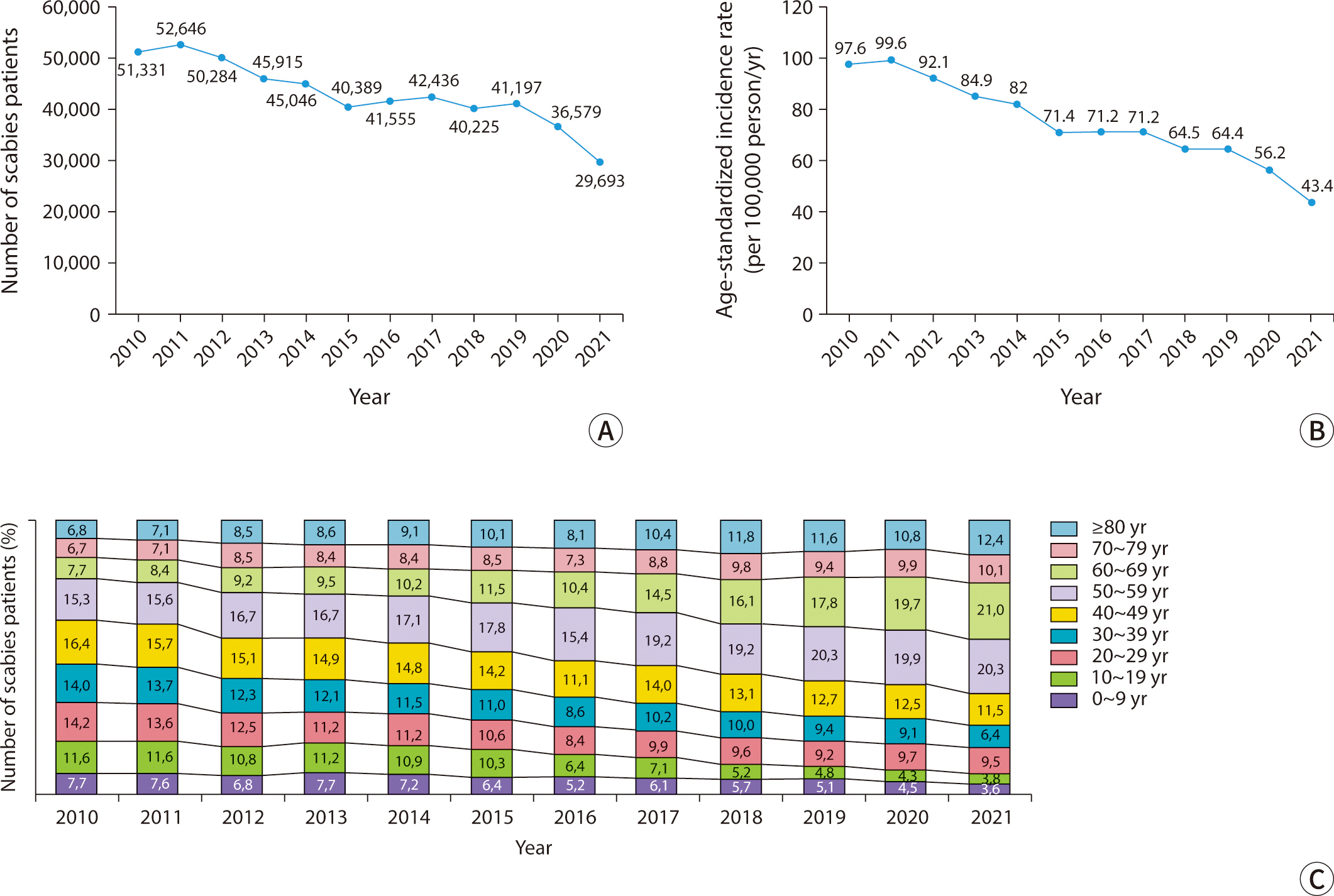
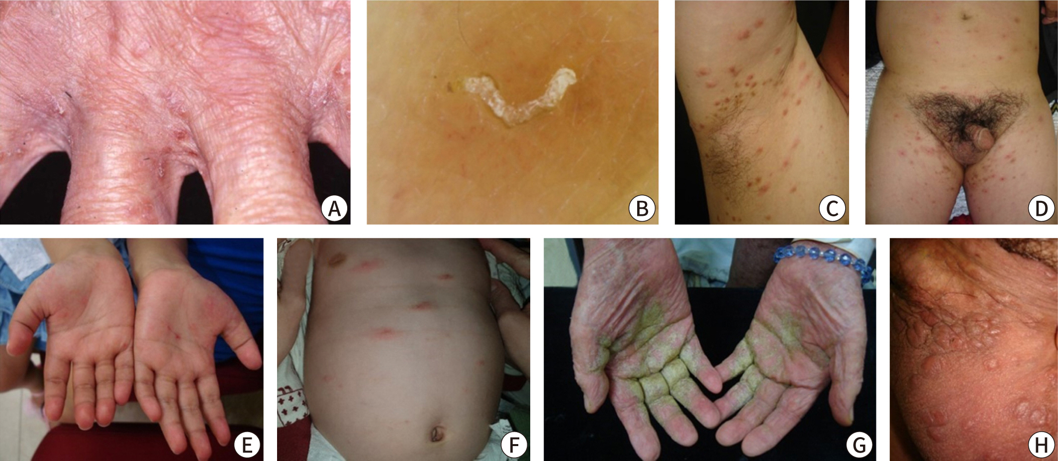
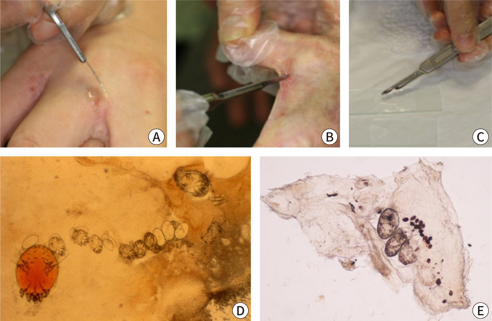
- Scabies is a skin disease caused by the parasite Sarcoptes scabiei var. hominis, which is primarily transmitted via direct skin or sexual contact or, less commonly, via contact with infested fomites. In Korea, the incidence of scabies has decreased from approximately 50,000 cases per year in 2010 to about 30,000 cases per year in 2021. However, outbreaks are consistently observed in residential facilities, such as nursing homes, especially among older adults. The clinical manifestations of scabies vary based on the patient’s age, health status, the number of mites, and the route of transmission. Typical symptoms of classic scabies include intense nocturnal itching and characteristic skin rashes (burrows and erythematous papules), with a predilection for the interdigital web spaces, inner wrists, periumbilical areas, axillae, and genital areas. In contrast, older adults with immunodeficiency or neurological disorders may exhibit hyperkeratotic scaly lesions or an atypical distribution with mild to no itching (crusted scabies). The diagnosis of scabies is based on clinical symptoms and the results of diagnostic tests aimed at identifying the presence of the parasite. While a history of close contact and characteristic clinical findings suggest scabies, confirmation of the diagnosis requires detecting scabies mites, eggs, or scybala. This can be achieved through light microscopy of skin samples, noninvasive dermoscopy, and other high-resolution in vivo imaging techniques.
Keyword
Figure
Cited by 1 articles
Reference
-
References
1. Zhang W, Zhang Y, Luo L, Huang W, Shen X, Dong X, et al. Trends in prevalence and incidence of scabies from 1990 to 2017: findings from the global burden of disease study 2017. Emerg Microbes Infect. 2020; 9(1):813–816. DOI: 10.1080/22221751.2020.1754136. PMID: 32284022. PMCID: PMC7241492.
Article2. Engelman D, Yoshizumi J, Hay RJ, Osti M, Micali G, Norton S, et al. The 2020 International Alliance for the Control of Scabies Consensus Criteria for the diagnosis of scabies. Br J Dermatol. 2020; 183(5):808–820. DOI: 10.1111/bjd.18943. PMID: 32034956. PMCID: PMC7687112.
Article3. Executive Committee of Guideline for the Diagnosis and Treatment of Scabies. Guideline for the diagnosis and treatment of scabies in Japan (third edition). J Dermatol. 2017; 44(9):991–1014. DOI: 10.1111/1346-8138.13896. PMID: 28561292.4. Salavastru CM, Chosidow O, Boffa MJ, Janier M, Tiplica GS. European guideline for the management of scabies. J Eur Acad Dermatol Venereol. 2017; 31(8):1248–1253. DOI: 10.1111/jdv.14351. PMID: 28639722.
Article5. Korea Disease Control and Prevention Agency. Guide for prevention and management of scabies and head lice. Cheongju: Korea Disease Control and Prevention Agency;2018.6. Korea Disease Control and Prevention Agency. Guide for prevention and management of scabies in convalescent hospital. Cheongju: Korea Disease Control and Prevention Agency;2019.7. Cho BK. Reemerging skin disease caused by arthropods I: scabies. J Korean Med Assoc. 2011; 54(5):511–520. DOI: 10.5124/jkma.2011.54.5.511.
Article8. Lee WK, Cho BK. Taxonomical approach to scabies mites of human and animals and their prevalence in Korea. Korean J Parasitol. 1995; 33(2):85–94. DOI: 10.3347/kjp.1995.33.2.85. PMID: 7551808.
Article9. Kim JH, Cheong HK. Epidemiologic trends and seasonality of scabies in South Korea, 2010-2017. Korean J Parasitol. 2019; 57:399–404. DOI: 10.3347/kjp.2019.57.4.399. PMID: 31533406. PMCID: PMC6753298.
Article10. Lee SK, Kim JH, Kim MS, Lee UH. Risk factors for scabies treatment resistance: a retrospective cohort study. J Eur Acad Dermatol Venereol. 2022; 36(1):126–132. DOI: 10.1111/jdv.17713. PMID: 34592030.
Article11. Jannic A, Bernigaud C, Brenaut E, Chosidow O. Scabies itch. Dermatol Clin. 2018; 36(3):301–308. DOI: 10.1016/j.det.2018.02.009. PMID: 29929601.
Article12. Romani L, Steer AC, Whitfeld MJ, Kaldor JM. Prevalence of scabies and impetigo worldwide: a systematic review. Lancet Infect Dis. 2015; 15(8):960–967. DOI: 10.1016/S1473-3099(15)00132-2. PMID: 26088526.13. Chandler DJ, Fuller LC. A review of scabies: an infestation more than skin deep. Dermatology. 2019; 235(2):79–90. DOI: 10.1159/000495290. PMID: 30544123.
Article14. Kim HS, Hashimoto T, Fischer K, Bernigaud C, Chosidow O, Yosipovitch G. Scabies itch: an update on neuroimmune interactions and novel targets. J Eur Acad Dermatol Venereol. 2021; 35(9):1765–1776. DOI: 10.1111/jdv.17334. PMID: 33960033.15. Walton SF, Oprescu FI. Immunology of scabies and translational outcomes: identifying the missing links. Curr Opin Infect Dis. 2013; 26(2):116–122. DOI: 10.1097/QCO.0b013e32835eb8a6. PMID: 23385638.16. Chosidow O. Clinical practice: scabies. N Engl J Med. 2006; 354(16):1718–1727. DOI: 10.1056/NEJMcp052784. PMID: 16625010.17. Chosidow O. Scabies and pediculosis. Lancet. 2000; 355(9206):819–826. DOI: 10.1016/S0140-6736(99)09458-1. PMID: 10711939.
Article18. Arlian LG, Morgan MS. A review of Sarcoptes scabiei: past, present and future. Parasit Vectors. 2017; 10(1):297. DOI: 10.1186/s13071-017-2234-1. PMID: 28633664. PMCID: PMC5477759.19. Paller AS. Scabies in infants and small children. Semin Dermatol. 1993; 12(1):3–8.20. Wong SSY, Woo PCY, Yuen K. Unusual laboratory findings in a case of Norwegian scabies provided a clue to diagnosis. J Clin Microbiol. 2005; 43(5):2542–2544. DOI: 10.1128/JCM.43.5.2542-2544.2005. PMID: 15872307. PMCID: PMC1153733.
Article21. Roberts LJ, Huffam SE, Walton SF, Currie BJ. Crusted scabies: clinical and immunological findings in seventy-eight patients and a review of the literature. J Infect. 2005; 50(5):375–381. DOI: 10.1016/j.jinf.2004.08.033. PMID: 15907543.
Article22. Das A, Bar C, Patra A. Norwegian scabies: rare cause of erythroderma. Indian Dermatol Online J. 2015; 6(1):52–54. DOI: 10.4103/2229-5178.148951. PMID: 25657922. PMCID: PMC4314893.
Article23. Devi GC, Hazarika N. Erythroderma secondary to crusted scabies. BMJ Case Rep. 2021; 14(12):248000. DOI: 10.1136/bcr-2021-248000. PMID: 34906960. PMCID: PMC8671912.
Article24. DePaoli RT, Marks VJ. Crusted (Norwegian) scabies: treatment of nail involvement. J Am Acad Dermatol. 1987; 17(1):136–139. DOI: 10.1016/S0190-9622(87)80543-1. PMID: 2956293.25. Lin S, Farber J, Lado L. A case report of crusted scabies with methicillin-resistant Staphylococcus aureus bacteremia. J Am Geriatr Soc. 2009; 57(9):1713–1714. DOI: 10.1111/j.1532-5415.2009.02412.x. PMID: 19895437.
Article26. Sil A, Mondal S, Bhanja DB, Datta M, Mukherjee GS, Chakraborty S. Pruritic genital nodules. Pediatr Neonatol. 2020; 61(2):243–244. DOI: 10.1016/j.pedneo.2019.12.004. PMID: 31901313.
Article27. Heukelbach J, Feldmeier H. Scabies. Lancet. 2006; 367(9524):1767–1774. DOI: 10.1016/S0140-6736(06)68772-2. PMID: 16731272.
Article28. Aydıngöz İE, Mansur AT. Canine scabies in humans: a case report and review of the literature. Dermatology. 2011; 223(2):104–106. DOI: 10.1159/000327378. PMID: 21540570.
Article29. Currie BJ, McCarthy JS. Permethrin and ivermectin for scabies. N Engl J Med. 2010; 362(8):717–725. DOI: 10.1056/NEJMct0910329. PMID: 20181973.
Article30. Diab HM. Scabies incognito: diagnostic value of dermoscopy-guided microscopic examination. J Egypt Women Dermatol Soc. 2017; 14(1):56–60. DOI: 10.1097/01.EWX.0000490006.68953.19.31. Sunderkötter C, Feldmeier H, Fölster-Holst R, Geisel B, Klinke-Rehbein S, Nast A, et al. S1 guidelines on the diagnosis and treatment of scabies: short version. J Dtsch Dermatol Ges. 2016; 14(11):1155–1167. DOI: 10.1111/ddg.13130. PMID: 27879074.32. Park SY, Roh JY, Lee JY, Kim DW, Yoon TJ, Sim WY, et al. A clinical and epidemiological study of scabies in Korea: a multicenter prospective study. Korean J Dermatol. 2014; 52(7):457–464.33. Tsoi SK, Lake SJ, Thean LJ, Matthews A, Sokana O, Kama M, et al. Estimation of scabies prevalence using simplified criteria and mapping procedures in three Pacific and southeast Asian countries. BMC Public Health. 2021; 21(1):2060. DOI: 10.1186/s12889-021-12039-2. PMID: 34758806. PMCID: PMC8579609.34. Walker SL, Collinson S, Timothy J, Zayzay SK, Kollie KK, Candy N, et al. A community-based validation of the International Alliance for the Control of Scabies Consensus Criteria by expert and non-expert examiners in Liberia. PLoS Negl Trop Dis. 2020; 14(10):0008717. DOI: 10.1371/journal.pntd.0008717. PMID: 33017426. PMCID: PMC7732067.35. Leung V, Miller M. Detection of scabies: a systematic review of diagnostic methods. Can J Infect Dis Med Microbiol. 2011; 22(4):143–146. DOI: 10.1155/2011/698494. PMID: 23205026. PMCID: PMC3222761.36. Woodley D, Saurat JH. The Burrow Ink test and the scabies mite. J Am Acad Dermatol. 1981; 4(6):715–722. DOI: 10.1016/S0190-9622(81)80204-6. PMID: 6787101.
Article37. Muller G, Jacobs PH, Moore NE. Scraping for human scabies: a better method for positive preparations. Arch Dermatol. 1973; 107(1):70. DOI: 10.1001/archderm.1973.01620160042011. PMID: 4630016.38. Walter B, Heukelbach J, Fengler G, Worth C, Hengge U, Feldmeier H. Comparison of dermoscopy, skin scraping, and the adhesive tape test for the diagnosis of scabies in a resource-poor setting. Arch Dermatol. 2011; 147(4):468–473. DOI: 10.1001/archdermatol.2011.51. PMID: 21482897.
Article39. Cho BK, Lee WK. Mite and tick related dermatoses. Seoul: Seoheung;2004.40. Lacarrubba F, Musumeci ML, Caltabiano R, Impallomeni R, West DP, Micali G. High-magnification videodermatoscopy: a new noninvasive diagnostic tool for scabies in children. Pediatr Dermatol. 2001; 18(5):439–441. DOI: 10.1046/j.1525-1470.2001.01973.x. PMID: 11737693.
Article41. Argenziano G, Fabbrocini G, Delfino M. Epiluminescence microscopy: a new approach to in vivo detection of Sarcoptes scabiei. Arch Dermatol. 1997; 133(6):751–753. DOI: 10.1001/archderm.1997.03890420091011. PMID: 9197830.
Article42. Dupuy A, Dehen L, Bourrat E, Lacroix C, Benderdouche M, Dubertret L, et al. Accuracy of standard dermoscopy for diagnosing scabies. J Am Acad Dermatol. 2007; 56(1):53–62. DOI: 10.1016/j.jaad.2006.07.025. PMID: 17190621.43. Park JH, Kim CW, Kim SS. The diagnostic accuracy of dermoscopy for scabies. Ann Dermatol. 2012; 24(2):194–199. DOI: 10.5021/ad.2012.24.2.194. PMID: 22577271. PMCID: PMC3346911.
Article44. Li FZ, Chen S. Diagnostic accuracy of dermoscopy for scabies. Korean J Parasitol. 2020; 58(6):669–674. DOI: 10.3347/kjp.2020.58.6.669. PMID: 33412771. PMCID: PMC7806431.
Article45. Shoukat Q, Rizvi A, Wahood W, Coetzee S, Wrench A. Sight the mite: a meta-analysis on the diagnosis of scabies. Cureus. 2023; 15(1):34390. DOI: 10.7759/cureus.34390.46. Ueda T, Katsura Y, Sasaki A, Minagawa D, Amoh Y, Shirai K. Gray-edged line sign of scabies burrow. J Dermatol. 2021; 48(2):190–198. DOI: 10.1111/1346-8138.15650. PMID: 33063894. PMCID: PMC7894142.
Article47. Arora P, Rudnicka L, Sar-Pomian M, Wollina U, Jafferany M, Lotti T, et al. Scabies: a comprehensive review and current perspectives. Dermatol Ther. 2020; 33(4):13746. DOI: 10.1111/dth.13746.48. Lacarrubba F, Verzì AE, Micali G. Detailed analysis of in vivo reflectance confocal microscopy for Sarcoptes scabiei hominis. Am J Med Sci. 2015; 350(5):414. DOI: 10.1097/MAJ.0000000000000336. PMID: 25211585.
Article49. Cinotti E, Labeille B, Cambazard F, Biron AC, Chol C, Leclerq A, et al. Videodermoscopy compared to reflectance confocal microscopy for the diagnosis of scabies. J Eur Acad Dermatol Venereol. 2016; 30(9):1573–1577. DOI: 10.1111/jdv.13676. PMID: 27168425.
Article50. Banzhaf CA, Themstrup L, Ring HC, Welzel J, Mogensen M, Jemec GBE. In vivo imaging of Sarcoptes scabiei infestation using optical coherence tomography. Case Rep Dermatol. 2013; 5(2):156–162. DOI: 10.1159/000352066. PMID: 23874291. PMCID: PMC3712805.
Article51. Head ES, Macdonald EM, Ewert A, Apisarnthanarax P. Sarcoptes scabiei in histopathologic sections of skin in human scabies. Arch Dermatol. 1990; 126(11):1475–1477. DOI: 10.1001/archderm.1990.01670350089015. PMID: 2122813.
Article52. Arlian LG, Feldmeier H, Morgan MS. The potential for a blood test for scabies. PLoS Negl Trop Dis. 2015; 9(10):0004188. DOI: 10.1371/journal.pntd.0004188. PMID: 26492406. PMCID: PMC4619658.
Article53. David N, Rajamanoharan S, Tang A. Are sexually transmitted infections associated with scabies? Int J STD AIDS. 2002; 13(3):168–170. DOI: 10.1258/0956462021924848. PMID: 11860692.
Article54. Bae M, Kim JY, Jung J, Cha HH, Jeon NY, Lee HJ, et al. Diagnostic value of the molecular detection of Sarcoptes scabiei from a skin scraping in patients with suspected scabies. PLoS Negl Trop Dis. 2020; 14(4):0008229. DOI: 10.1371/journal.pntd.0008229. PMID: 32255795. PMCID: PMC7164670.55. Wong SSY, Poon RWS, Chau S, Wong SCY, To KKW, Cheng VCC, et al. Development of conventional and real-time quantitative PCR assays for diagnosis and monitoring of scabies. J Clin Microbiol. 2015; 53(7):2095–2102. DOI: 10.1128/JCM.00073-15. PMID: 25903566. PMCID: PMC4473232.
Article56. Hahm JE, Kim CW, Kim SS. The efficacy of a nested polymerase chain reaction in detecting the cytochrome c oxidase subunit 1 gene of Sarcoptes scabiei var. hominis for diagnosing scabies. Br J Dermatol. 2018; 179(4):889–895. DOI: 10.1111/bjd.16657. PMID: 29624634.
Article
- Full Text Links
- Actions
-
Cited
- CITED
-
- Close
- Share
- Similar articles
-
- Clinical Practice Guidelines for the Diagnosis and Treatment of Scabies in Korea: Part 1. Epidemiology, Clinical Manifestations, and Diagnosis
- Clinical practice guidelines for the diagnosis and treatment of scabies in Korea: Part 2. Treatment and prevention — a secondary publication
- Clinical Practice Guidelines for the Diagnosis and Treatment of Scabies in Korea: Part 2. Treatment and Prevention
- Guideline for the diagnosis and treatment of scabies
- Epidemiology and Characteristics of Scabies Infections in the Ulsan City: A 10-Year Retrospective Study


