J Yeungnam Med Sci.
2024 Oct;41(4):312-317. 10.12701/jyms.2024.00556.
A 32-year-old man with plexiform schwannoma of the thyroid gland: a case report
- Affiliations
-
- 1Division of Endocrinology and Metabolism, Department of Internal Medicine, Yeungnam University College of Medicine, Daegu, Korea
- 2Department of Pathology, Yeungnam University College of Medicine, Daegu, Korea
- 3Department of Otorhinolaryngology-Head and Neck Surgery, Yeungnam University College of Medicine, Daegu, Korea
- KMID: 2560493
- DOI: http://doi.org/10.12701/jyms.2024.00556
Abstract
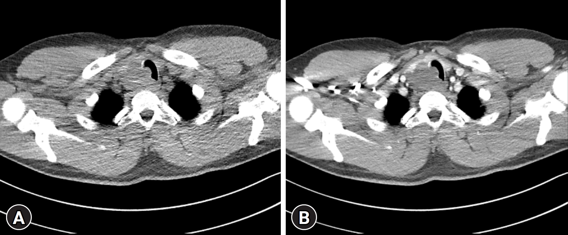
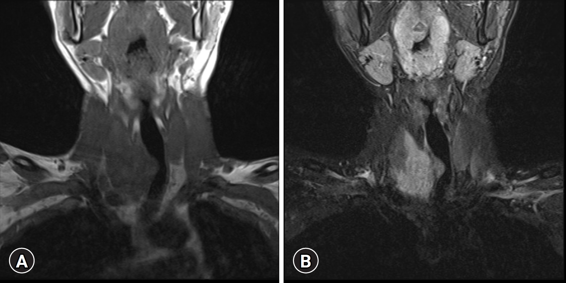
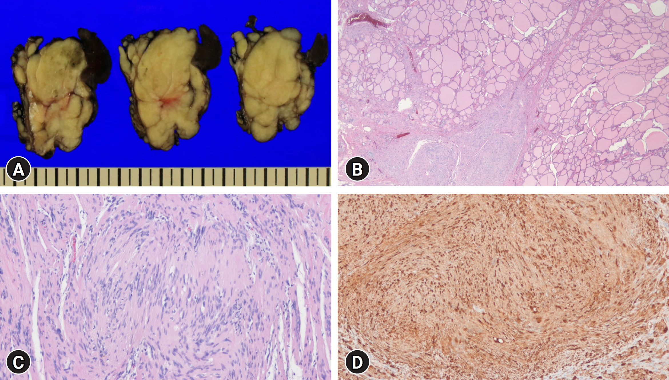
- Plexiform schwannomas representing a rare subset, comprise 5% of all schwannomas. However, their occurrence in the thyroid gland is exceptionally rare. A 32-year-old male presented with an incidentally discovered, asymptomatic thyroid mass. Imaging revealed an approximately 5 cm heterogeneous solid mass on the right thyroid lobe extending to the upper mediastinum and directly invading the upper trachea. Under the suspicion of thyroid malignancy, the patient underwent right thyroidectomy. Histological examination confirmed a plexiform schwannoma with S100-positive spindle cells. Currently, the patient is undergoing outpatient follow-up, with no reported complications. To our knowledge, this is the first documented case of plexiform schwannoma of the thyroid gland within the English literature. This case highlights the diverse and unpredictable clinical manifestations of thyroid masses, emphasizing the importance of a multidisciplinary approach for diagnosing and managing rare entities, such as thyroid gland schwannomas.
Figure
Reference
-
References
1. Colreavy MP, Lacy PD, Hughes J, Bouchier-Hayes D, Brennan P, O'Dwyer AJ, et al. Head and neck schwannomas: a 10 year review. J Laryngol Otol. 2000; 114:119–24.2. De Simone B, Del Rio P, Sianesi M. Schwannoma mimicking a neoplastic thyroid nodule. Updates Surg. 2014; 66:85–7.3. WHO Classification of Tumours Editorial Board. Soft tissue and bone tumours. 5th ed. Lyon (France): International Agency for Research on Cancer;2020.4. Dhar H, Dabholkar JP, Kandalkar BM, Ghodke R. Primary thyroid schwannoma masquerading as a thyroid nodule. J Surg Case Rep. 2014; 2014:rju094.5. Nagavalli S, Yehuda M, McPhaul LW, Gianoukakis AG. A cervical schwannoma masquerading as a thyroid nodule. Eur Thyroid J. 2017; 6:216–20.6. Abbarah S, Abbarh S, AlHarthi B. Unusual thyroid nodule: a case of symptomatic thyroid schwannoma. Cureus. 2020; 12:e11425.7. Aoki T, Kumeda S, Iwasa T, Inokawa K, Hori T, Makiuchi M. Primary neurilemoma of the thyroid gland: report of a case. Surg Today. 1993; 23:265–8.8. Vázquez-Benítez G, Pérez-Campos A, Masgrau NA, Pérez-Barrios A. Unexpected tumor: primary asymptomatic schwannoma in thyroid gland. Endocr Pathol. 2016; 27:46–9.9. Berg JC, Scheithauer BW, Spinner RJ, Allen CM, Koutlas IG. Plexiform schwannoma: a clinicopathologic overview with emphasis on the head and neck region. Hum Pathol. 2008; 39:633–40.10. Williams EA, Ravindranathan A, Gupta R, Stevers NO, Suwala AK, Hong C, et al. Novel SOX10 indel mutations drive schwannomas through impaired transactivation of myelination gene programs. Neuro Oncol. 2023; 25:2221–36.11. Wali AA, Yang R, Merbs SL, Rodriguez FJ, Eberhart CG, Lucas CG. Orbital SOX10-mutant schwannoma with plexiform growth: expanding the histopathological spectrum of a new molecular group. J Neuropathol Exp Neurol. 2023; 82:963–5.12. Zhao HN, Ma BY, Yan F, Peng YL. Multimodal ultrasound imaging of primary thyroid schwannoma: a case report. Medicine (Baltimore). 2021; 100:e25517.13. Liu HL, Yu SY, Li GK, Wei WI. Extracranial head and neck Schwannomas: a study of the nerve of origin. Eur Arch Otorhinolaryngol. 2011; 268:1343–7.14. Simo D, Selvaggi F, Cieri M, Angelucci D, Claudi R, Giuliani C, et al. Surgical approach for nodular neck lesions mimicking primitive thyroid neoplasms Report of three cases. Ann Ital Chir. 2014; 85:474–8.15. Woodruff JM, Scheithauer BW, Kurtkaya-Yapicier O, Raffel C, Amr SS, LaQuaglia MP, et al. Congenital and childhood plexiform (multinodular) cellular schwannoma: a troublesome mimic of malignant peripheral nerve sheath tumor. Am J Surg Pathol. 2003; 27:1321–9.16. Prieto-Granada CN, Wiesner T, Messina JL, Jungbluth AA, Chi P, Antonescu CR. Loss of H3K27me3 expression is a highly sensitive marker for sporadic and radiation-induced MPNST. Am J Surg Pathol. 2016; 40:479–89.17. Moroni AL, Righini C, Faure C, Serra-Tosio G, Lefournier V. CT Features of an unusual calcified schwannoma of the superior laryngeal nerve. AJNR Am J Neuroradiol. 2007; 28:981–2.18. Crist J, Hodge JR, Frick M, Leung FP, Hsu E, Gi MT, et al. Magnetic resonance imaging appearance of schwannomas from head to toe: a pictorial review. J Clin Imaging Sci. 2017; 7:38.19. Das A, Bhalla AS, Sharma R, Kumar A, Thakar A, Goyal A. Diffusion-weighted imaging in extracranial head and neck schwannomas: a distinctive appearance. Indian J Radiol Imaging. 2016; 26:231–6.20. Yang F, Chen XX, Wu HL, Zhu JF, Chen Y, Yu LF, et al. Sonographic features and diagnosis of peripheral schwannomas. J Clin Ultrasound. 2017; 45:127–33.


