Procedural errors detected by cone beam tomography in cases with indication for retreatment: in vivo cross-sectional study
- Affiliations
-
- 1Postgraduate Program in Dentistry, University of Grande Rio (UNIGRANRIO), Rio de Janeiro, RJ, Brazil
- 2Postgraduate Program in Dentistry, Cayetano Heredia University, Lima, Peru
- 3Department of Dental Research, Faculty of Dentistry, Iguaçu University (UNIG), Nova Iguaçu, RJ, Brazil
- KMID: 2559739
- DOI: http://doi.org/10.5395/rde.2024.49.e26
Abstract
Objectives
This study aimed to investigate the frequency and type of endodontic procedural errors in cases indicated for retreatment through cone-beam computed tomography (CBCT) analysis.
Materials and Methods
The sample consisted of 96 CBCT scans, encompassing 122 permanent teeth with fully formed roots. Errors included perforation, instrument fracture, canal transportation, missed canals, and inadequate apical limit of filling. Additionally, potential risk factors were analyzed and subjected to statistical modeling.
Results
The most frequent procedural error observed was the inadequate apical limit of filling, followed by canal transportation, perforation, missed canal, and instrument fracture. Statistically significant associations were identified between various procedural errors and specific factors. These include canal transportation and root canal wall, with the buccal wall being the most commonly affected; missed canal and tooth type, particularly the palatine and second mesiobuccal canal canals; inadequate apical limit of filling and root curvature, showing a higher deviation to the mesial direction in severely curved canals; inadequate apical limit of filling and the presence of calcifications, with underfilling being the most frequent; canal transportation and periapical lesion, notably with deviation to the buccal direction; and the direction of perforation and periapical lesion, most frequently occurring to buccal direction.
Conclusions
CBCT emerges as a valuable tool in identifying procedural errors and associated factors, crucial for their prevention and management.
Figure
-
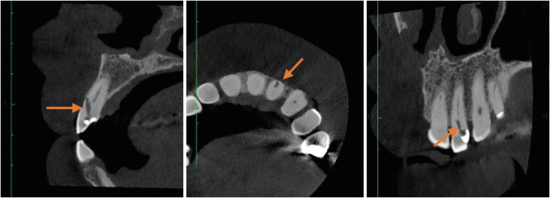
Figure 1 Dynamic cone-beam computed tomography analysis for identification of perforation in the right lateral incisor.
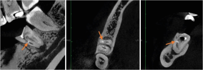
Figure 2 Dynamic cone-beam computed tomography analysis for identification of fractured instrument in lower left first molar.
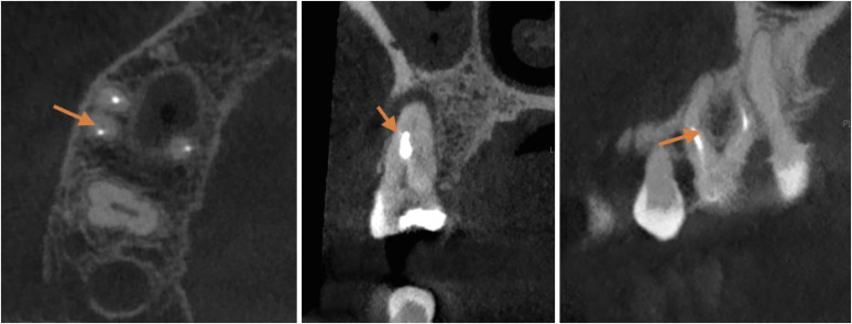
Figure 3 Dynamic cone-beam computed tomography analysis for identification of canal transportation in upper right first molar.
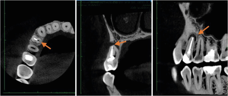
Figure 4 Dynamic analysis of cone-beam computed tomography for identification of missed canal.
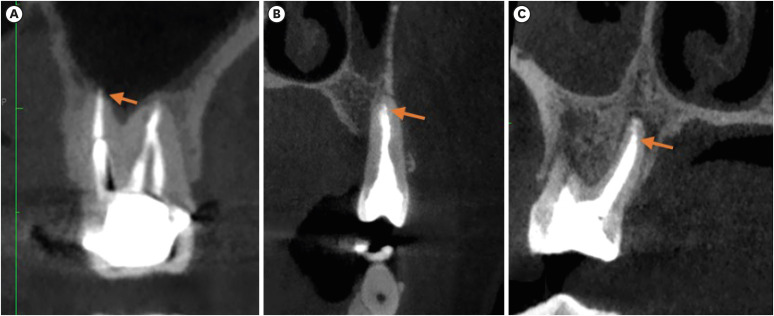
Figure 5 Identification of (A) molar with an overfilled canal, (B) premolar at the appropriate level and (C) palatal canal of an underfilled upper molar.
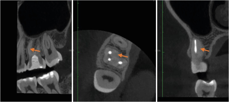
Figure 6 Identification of second mesiobuccal canal calcification in upper left first molar.
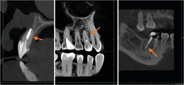
Figure 7 Identification of anterior, premolar and molar tooth type.
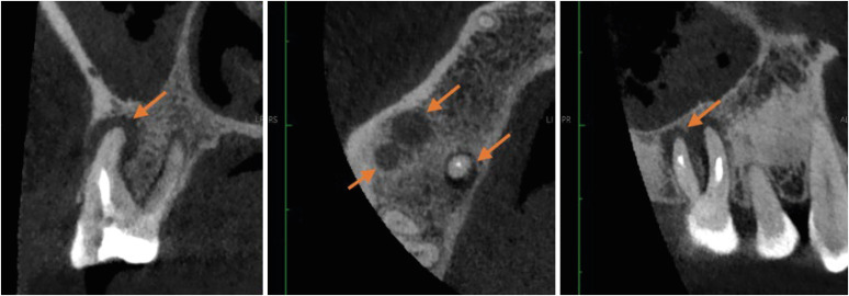
Figure 8 Identification of periapical lesion in upper right first molar.
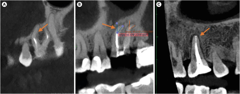
Figure 9 Identification of type of curvature, (A) moderate, (B) severe and (C) straight.
Reference
-
1. Toia CC, Khoury RD, Corazza BJM, Orozco EIF, Valera MC. Effectiveness of 1-visit and 2-visit endodontic retreatment of teeth with persistent/secondary endodontic infection: a randomized clinical trial with 18 months of follow-up. J Endod. 2022; 48:4–14. PMID: 34555421.2. Gaêta-Araujo H, Fontenele RC, Nascimento EHL, Nascimento MDCC, Freitas DQ, de Oliveira-Santos C. Association between the root canal configuration, endodontic treatment technical errors, and periapical hypodensities in molar teeth: a cone-beam computed tomographic study. J Endod. 2019; 45:1465–1471. PMID: 31575389.3. Alamoudi RA, Alharbi AH, Farie GA, Fahim O. The value of assessing case difficulty and its effect on endodontic iatrogenic errors: a retrospective cross-sectional study. Libyan J Med. 2020; 15:1688916. PMID: 31694490.4. Yagmoor M, Bakhsh A, Mandourah O, Alsofi L. Management of a radiopaque foreign body associated with a lower first premolar: a case report. Clin Case Rep. 2022; 10:e05465. PMID: 35223019.5. da Cunha LZV, Solda C, Padoin K, Rigo L. Endodontic procedural errors: analysis of images from cone beam computed tomography. Forensic Imaging. 2022; 28:200493.6. Siqueira JF Jr, Rôças IN. Clinical implications and microbiology of bacterial persistence after treatment procedures. J Endod. 2008; 34:1291–1301.e3. PMID: 18928835.7. Peña-Bengoa F, Cáceres C, Niklander SE, Meléndez P. Association between second mesiobuccal missed canals and apical periodontitis in maxillary molars of a Chilean subpopulation. J Clin Exp Dent. 2023; 15:e173–e176. PMID: 37008247.8. Ballikaya E, Koc N, Avcu N, Cehreli ZC. The quality of root canal treatment and periapical status of permanent teeth in Turkish children and teens: a retrospective CBCT study. Oral Radiol. 2022; 38:405–415. PMID: 34714509.9. Nouroloyouni A, Salem Milani A, Etminan A, Noorolouny S, Tavakkol E, Mikaieli Xiavi H, et al. Cone-beam computed tomography assessment of quality of endodontic treatment and prevalence of procedural errors in mandibular molars. Int J Clin Pract. 2023; 2023:3558974. PMID: 37251955.10. Alghamdi NS, Algarni YA, Ain TS, Alfaifi HM, AlQarni AA, Mashyakhi JQ, et al. Endodontic mishaps during root canal treatment performed by undergraduate dental students: an observational study. Medicine (Baltimore). 2021; 100:e27757. PMID: 34964733.11. Fayad MI, Nair M, Levin MD, Benavides E, Rubinstein RA, Barghan S, et al. AAE and AAOMR joint position statement: use of cone beam computed tomography in endodontics 2015 update. Oral Surg Oral Med Oral Pathol Oral Radiol. 2015; 120:508–512. PMID: 26346911.12. Costa FF, Pacheco-Yanes J, Siqueira JF Jr, Oliveira ACS, Gazzaneo I, Amorim CA, et al. Association between missed canals and apical periodontitis. Int Endod J. 2019; 52:400–406. PMID: 30284719.13. Karabucak B, Bunes A, Chehoud C, Kohli MR, Setzer F. Prevalence of apical periodontitis in endodontically treated premolars and molars with untreated canal: a cone-beam computed tomography study. J Endod. 2016; 42:538–541. PMID: 26873567.14. Schneider SW. A comparison of canal preparations in straight and curved root canals. Oral Surg Oral Med Oral Pathol. 1971; 32:271–275. PMID: 5284110.15. Nascimento EHL, Gaêta-Araujo H, Andrade MFS, Freitas DQ. Prevalence of technical errors and periapical lesions in a sample of endodontically treated teeth: a CBCT analysis. Clin Oral Investig. 2018; 22:2495–2503.16. Al Yahya RS, Al Attas MH, Javed MQ, Khan KI, Atique S, Abulhamael AM, et al. Root canal configuration and its relationship with endodontic technical errors and periapical status in premolar teeth of a Saudi sub-population: a cross-sectional observational CBCT study. Int J Environ Res Public Health. 2023; 20:1142. PMID: 36673896.17. Jurič R, Vidmar G, Blagus R, Jan J. Factors associated with the outcome of root canal treatment-a cohort study conducted in a private practice. Int Endod J. 2024; 57:377–393. PMID: 38243912.18. Villa-Machado PA, Restrepo-Patiño DM, Calvo-Trejos JP, Restrepo-Restrepo FA, Tobón-Arroyave SI, Provenzano JC, et al. Cone-beam computed tomographic and micro-computed tomographic evaluations of the root apexes of teeth with posttreatment apical periodontitis. J Endod. 2020; 46:1695–1701. PMID: 32682792.19. Siqueira JF Jr, Rôças IN. Present status and future directions: microbiology of endodontic infections. Int Endod J. 2022; 55(Supplement 3):512–530. PMID: 34958494.20. Pinto JC, de Faria-Vasconcelos K, Leite AF, Pedano MS, Guerreiro-Tanomaru J, Jacobs R, et al. Effect of foraminal enlargement on microcrack formation and apical transportation: a nano-CT assessment. Sci Rep. 2023; 13:4881. PMID: 36966188.21. Wu MK, Fan B, Wesselink PR. Leakage along apical root fillings in curved root canals. Part I: effects of apical transportation on seal of root fillings. J Endod. 2000; 26:210–216. PMID: 11199720.22. Hasheminia SM, Farhad A, Sheikhi M, Soltani P, Hendi SS, Ahmadi M. Cone-beam computed tomographic analysis of canal transportation and centering ability of single-file systems. J Endod. 2018; 44:1788–1791. PMID: 30390970.23. Wong J, Lee A, Zhang C. Diagnosis and management of apical fenestrations associated with endodontic diseases: a literature review. Eur Endod J. 2021; 6:25–33. PMID: 33609018.24. Wu L, Ha WN, Decurcio DA, Estrela C, Rossi-Fedele G. Comparison of curvature severity between sagittal and coronal planes of mesiobuccal canals in permanent maxillary first molars using multiple complexity-risk criteria: a CBCT cross-sectional study of a Brazilian subpopulation. J Endod. 2023; 49:1682–1689.e4. PMID: 37816431.25. Wang FM, Rudman J, Walsh RM, Jalali P. A retrospective study of initial root canal treatment failure in maxillary premolars via using cone-beam computed tomography. J Am Dent Assoc. 2023; 154:471–478. PMID: 37236705.26. Ayatollahi F, Tabrizizadeh M, Razavi H, Mowji M. Diagnostic value of cone-beam computed tomography and digital periapical radiography in detection of separated instruments. Iran Endod J. 2019; 14:14–17. PMID: 36879596.27. Özer SY, Özkan G, Çetin E, Özkan HD. A comparative study of cone-beam computed tomography and periapical radiographs in decision-making after endodontic instrument fractures. Int J Artif Organs. 2017; 40:510–514. PMID: 28574115.
- Full Text Links
-
- Actions
-
Cited
- CITED
-
- Close
- Share
-
- Similar articles
-
- Cone beam CT findings of retromolar canals: Report of cases and literature review
- Assessment of the relationship between the mandibular third molar and the mandibular canal using panoramic radiograph and cone beam computed tomography
- Management of root canal perforation by using cone-beam computed tomography
- Three-dimensional evaluation of the relationship between nasopharyngeal airway shape and adenoid size in children
- Isolated tympanic plate fracture detected by cone-beam computed tomography: report of four cases with review of literature
