J Pathol Transl Med.
2024 Sep;58(5):214-218. 10.4132/jptm.2024.07.03.
Single umbilical artery and associated birth defects in perinatal autopsies: prenatal diagnosis and management
- Affiliations
-
- 1Department of Pathology, KAHER’S Jawaharlal Nehru Medical College, Belagavi, India
- KMID: 2559066
- DOI: http://doi.org/10.4132/jptm.2024.07.03
Abstract
- Background
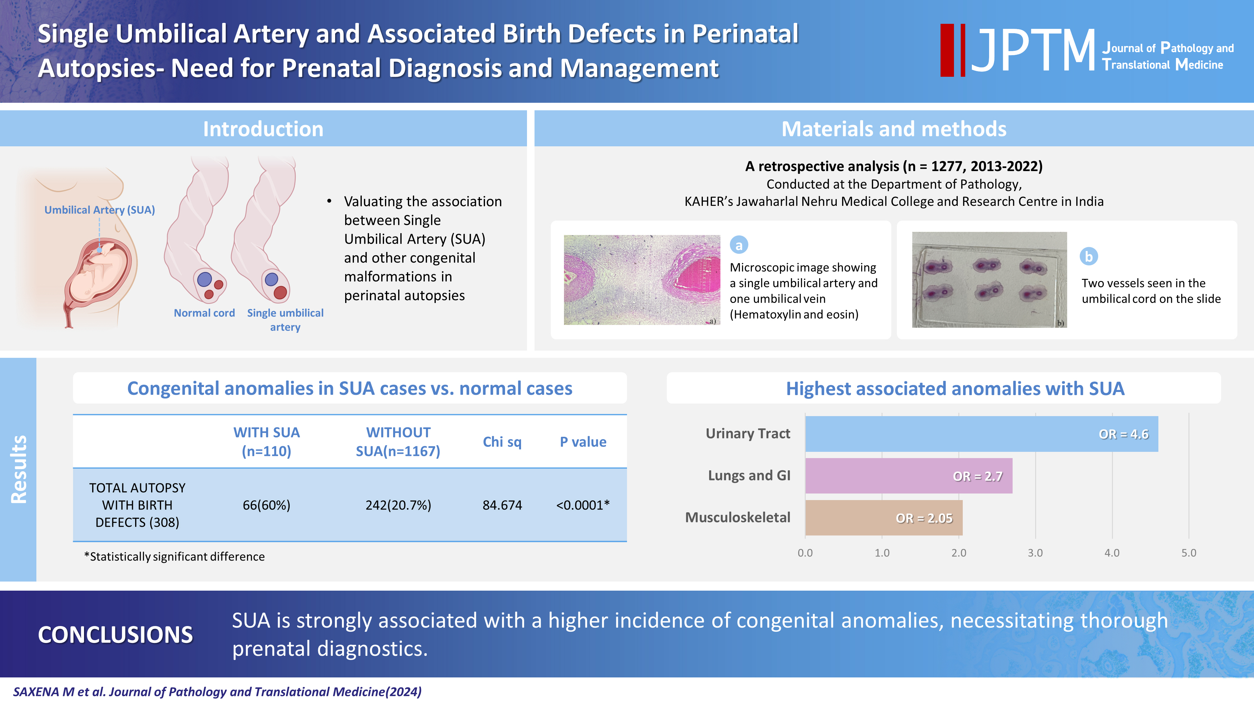
The umbilical cord forms the connection between the fetus and the placenta at the feto-maternal interface and normally comprises two umbilical arteries and one umbilical vein. In some cases, only a single umbilical artery (SUA) is present. This study was conducted to evaluate associations between SUA and other congenital malformations discovered in perinatal autopsies and to ascertain the existence of preferential associations between SUA and certain anomalies.
Methods
We evaluated records of all fetuses sent for autopsy to the Department of Pathology during the 10-year period from 2013 through 2022 (n = 1,277). The data were obtained from the hospital’s pathology laboratory records. The congenital anomalies were grouped by organ or system for analysis and included cardiovascular, urinary tract, nervous system, gastrointestinal tract, musculoskeletal, and lung anomalies.
Results
A SUA was present in 8.61% of the autopsies. The gestational age of the affected fetuses ranged between 13 to 40 weeks. An SUA presented as an isolated single anomaly in 44 cases (3.4%). Of the 110 SUA cases, 60% had other congenital anomalies. There was a significant association between birth defects and SUAs (p < .001). Strong associations between SUA and urinary tract, lung, and musculoskeletal anomalies were observed.
Conclusions
A SUA is usually seen in association with other congenital malformations rather than as an isolated defect. Therefore, examination for associated anomalies when an SUA is detected either antenatally or postnatally is imperative. The findings of this study should be helpful in counseling expectant mothers and their families in cases of SUA.
Figure
Reference
-
References
1. Vrabie SC, Novac L, Manolea MM, Dijmarescu LA, Novac M, Siminel MA. Abnormalities of the umbilical cord. In: Tudorache S, ed. Congenital anomalies: from the embryo to the neonate. London: IntechOpen;2017. p. 345–62.2. Heil JR, Bordoni B. Embryology, umbilical cord [Internet]. Treasure Island: StatPearls Publishing, 2023 [cited 2024 Apr 4]. Available from: https://pubmed.ncbi.nlm.nih.gov/32491422/.3. Csecsei K, Kovacs T, Hinchliffe SA, Papp Z. Incidence and associations of single umbilical artery in prenatally diagnosed malformed, midtrimester fetuses: a review of 62 cases. Am J Med Genet. 1992; 43:524–30.4. Thummala MR, Raju TN, Langenberg P. Isolated single umbilical artery anomaly and the risk for congenital malformations: a metaanalysis. J Pediatr Surg. 1998; 33:580–5.5. Persutte WH, Hobbins J. Single umbilical artery: a clinical enigma in modern prenatal diagnosis. Ultrasound Obstet Gynecol. 1995; 6:216–29.6. Tulek F, Kahraman A, Taskin S, Ozkavukcu E, Soylemez F. Determination of risk factors and perinatal outcomes of singleton pregnancies complicated by isolated single umbilical artery in Turkish population. J Turk Ger Gynecol Assoc. 2015; 16:21–4.7. Murphy-Kaulbeck L, Dodds L, Joseph KS, Van den Hof M. Single umbilical artery risk factors and pregnancy outcomes. Obstet Gynecol. 2010; 116:843–50.8. Lilja M. Infants with single umbilical artery studied in a national registry. 3: a case control study of risk factors. Paediatr Perinat Epidemiol. 1994; 8:325–33.9. Tasha I, Brook R, Frasure H, Lazebnik N. Prenatal detection of cardiac anomalies in fetuses with single umbilical artery: diagnostic accuracy comparison of maternal-fetal-medicine and pediatric cardiologist. J Pregnancy. 2014; 2014:265421.10. Burshtein S, Levy A, Holcberg G, Zlotnik A, Sheiner E. Is single umbilical artery an independent risk factor for perinatal mortality? Arch Gynecol Obstet. 2011; 283:191–4.11. Bombrys AE, Neiger R, Hawkins S, et al. Pregnancy outcome in isolated single umbilical artery. Am J Perinatol. 2008; 25:239–42.12. Dagklis T, Defigueiredo D, Staboulidou I, Casagrandi D, Nicolaides KH. Isolated single umbilical artery and fetal karyotype. Ultrasound Obstet Gynecol. 2010; 36:291–5.13. Rittler M, Mazzitelli N, Fuksman R, de Rosa LG, Grandi C. Single umbilical artery and associated malformations in over 5500 autopsies: relevance for perinatal management. Pediatr Dev Pathol. 2010; 13:465–70.14. Chow JS, Benson CB, Doubilet PM. Frequency and nature of structural anomalies in fetuses with single umbilical arteries. J Ultrasound Med. 1998; 17:765–8.15. Abuhamad AZ, Shaffer W, Mari G, Copel JA, Hobbins JC, Evans AT. Single umbilical artery: does it matter which artery is missing? Am J Obstet Gynecol. 1995; 173:728–32.16. Leung AK, Robson WL. Single umbilical artery: a report of 159 cases. Am J Dis Child. 1989; 143:108–11.17. Tortora M, Chervenak FA, Mayden K, Hobbins JC. Antenatal sonographic diagnosis of single umbilical artery. Obstet Gynecol. 1984; 63:693–6.18. Gossett DR, Lantz ME, Chisholm CA. Antenatal diagnosis of single umbilical artery: is fetal echocardiography warranted? Obstet Gynecol. 2002; 100:903–8.19. Nayak SS, Shukla A, Girisha KM. Anomalies associated with single umbilical artery at perinatal autopsy. Indian Pediatr. 2015; 52:73–4.20. Froehlich LA, Fujikura T. Follow-up of infants with single umbilical artery. Pediatrics. 1973; 52:6–13.21. Singh V, Patel R, Pradhan P. Single umbilical artery and associated hydronephrosis: a report of 2 cases. J Reprod Med. 2004; 49:136–8.22. Heifetz SA. Single umbilical artery: a statistical analysis of 237 autopsy cases and review of the literature. Perspect Pediatr Pathol. 1984; 8:345–78.23. Srinivasan R, Arora RS. Do well infants born with an isolated single umbilical artery need investigation? Arch Dis Child. 2005; 90:100–1.24. Martinez-Payo C, Gaitero A, Tamarit I, Garcia-Espantaleon M, Iglesias Goy E. Perinatal results following the prenatal ultrasound diagnosis of single umbilical artery. Acta Obstet Gynecol Scand. 2005; 84:1068–74.25. Ebbing C, Kessler J, Moster D, Rasmussen S. Single umbilical artery and risk of congenital malformation: population-based study in Norway. Ultrasound Obstet Gynecol. 2020; 55:510–5.26. Rodriguez AM, Downs KM. Visceral endoderm and the primitive streak interact to build the fetal-placental interface of the mouse gastrula. Dev Biol. 2017; 432:98–124.
- Full Text Links
- Actions
-
Cited
- CITED
-
- Close
- Share
- Similar articles
-
- Pseudo-Single Umbilical Artery by Spontaneous Intrauterine Umbilical Artery Thrombosis
- Perinatal Outcome, Prevalence and Clinical Significance of Pregnancies with Single Umbilical Artery
- Fetal Intra-abdominal Umbilical Vein Varix Complicated with Patent Ductus Venosus and Atrial Septal Defect
- A Case of Limb-Body Wall Complex Diagnosed by Prenatal Ultrasonography
- Clinical Analysis of Fetuses with Single Umbilical Artery


