Perinatology.
2019 Sep;30(3):183-185. 10.14734/PN.2019.30.3.183.
Pseudo-Single Umbilical Artery by Spontaneous Intrauterine Umbilical Artery Thrombosis
- Affiliations
-
- 1Department of Obstetrics and Gynecology, The Catholic University of Korea St. Vincent's Hospital, Suwon, Korea. leegsr@catholic.ac.kr
- 2Department of Pathology, The Catholic University of Korea St. Vincent's Hospital, Suwon, Korea.
- KMID: 2460034
- DOI: http://doi.org/10.14734/PN.2019.30.3.183
Abstract
- Umbilical artery thrombosis is rare event and few prenatally diagnosed cases have been reported. Antenatal diagnosis is very critical, as it is associated with high risk of perinatal motility and morbidity such as intrauterine growth restriction and intrauterine fetal death. This study presents a rare case of intrauterine umbilical artery thrombosis, diagnosed by ultrasonography, at 32 weeks and 5 days of gestation in a 34-year-old woman who had an uneventful pregnancy. Early diagnosis with color Doppler ultrasonography is critical for pregnancy outcomes when fetal movement has diminished or intrauterine growth restriction is diagnosed, even though two umbilical artery already been confirmed on the first or the second-trimester ultrasonography scans.
MeSH Terms
Figure
-
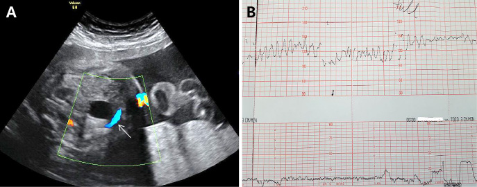
Fig. 1 (A) There was single paravesical color Doppler flow in the left umbilical artery (arrow). (B)Fetal heart rate monitoring showed “saw-tooth pattern” with bradycardia.
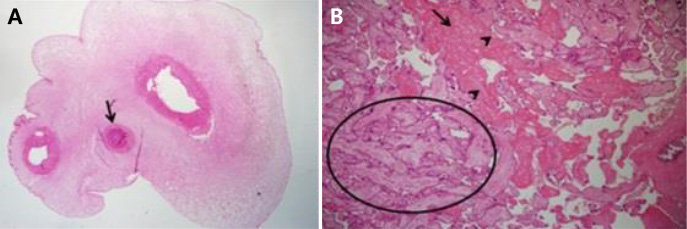
Fig. 2 (A) Three vessels were noted with thromboic occlusion of one of umbilical arteries (arrow; H&E, ×12). (B) The placenta microscopically presented necrotic and avasular villi (arrowheads) embedded in large fibrinoid (arrow), and the collapsed intervillous space (circle) which consistent with early ischemic change (H&E, ×40).
Reference
-
1. Shilling C, Walsh C, Downey P, Mooney E. Umbilical artery thrombosis is a rare but clinically important finding: a series of 7 cases with clinical outcomes. Pediatr Dev Pathol. 2014; 17:89–93.
Article2. Sato Y, Benirschke K. Umbilical arterial thrombosis with vascular wall necrosis: clinicopathologic findings of 11 cases. Placenta. 2006; 27:715–718.
Article3. Mittal A, Nanda S, Sen J. Antenatal umbilical coiling index as a predictor of perinatal outcome. Arch Gynecol Obstet. 2015; 291:763–768.
Article4. Thompson JM, Irgens LM, Skjaerven R, Rasmussen S. Placenta weight percentile curves for singleton deliveries. BJOG. 2007; 114:715–720.
Article5. Klaritsch P, Haeusler M, Karpf E, Schlembach D, Lang U. Spontaneous intrauterine umbilical artery thrombosis leading to severe fetal growth restriction. Placenta. 2008; 29:374–377.
Article6. Tanaka K, Tanigaki S, Matsushima M, Miyazaki N, Hashimoto R, Izawa T, et al. Prenatal diagnosis of umbilical artery thrombosis. Fetal Diagn Ther. 2014; 35:148–150.
Article7. Cook V, Weeks J, Brown J, Bendon R. Umbilical artery occlusion and fetoplacental thromboembolism. Obstet Gynecol. 1995; 85(5 Pt 2):870–872.
Article8. Lutfallah F, Oufkir N, Markou GA, Frimigacci D, Poncelet C. A case of umbilical artery thrombosis in the third trimester of pregnancy. Am J Case Rep. 2018; 19:72–75.
Article9. Oliveira GH, Dias Cde M, Vaz-Oliani DC, Oliani AH. Intrauterine thrombosis of umbilical artery - case report. Sao Paulo Med J. 2016; 134:355–358.
Article10. FreemanRF , Garite TG, NageotteMP , Miller LA. Fetal heart rate monitoring. 4th ed. Philadelphia: Lippincott Williams & Wilkins;2012. p. 149–151.11. Redline RW, Pappin A. Fetal thrombotic vasculopathy: the clinical significance of extensive avascular villi. Hum Pathol. 1995; 26:80–85.
Article12. Saleemuddin A, Tantbirojn P, Sirois K, Crum CP, Boyd TK, Tworoger S, et al. Obstetric and perinatal complications in placentas with fetal thrombotic vasculopathy. Pediatr Dev Pathol. 2010; 13:459–464.
Article
- Full Text Links
-
- Actions
-
Cited
- CITED
-
- Close
- Share
-
- Similar articles
-
- Ultrason ographic Diagnosis of Aortic Thrombosis after Umbilical Artery Catheterization in Neonate
- Perinatal Outcome, Prevalence and Clinical Significance of Pregnancies with Single Umbilical Artery
- A Case Report of Non-surgical Removal of Fragmented Remnant of Umbilical Vein Catheter Using an Intravascular Snare
- A case of one fetal demise of twin pregnancy by umbilical artery stricture
- Clinical Analysis of Fetuses with Single Umbilical Artery
