Ethmoid Sinus Mucocele Penetrating the Anterior Skull Base: A Case Report
- Affiliations
-
- 1Division of Rhinology, Department of Otorhinolaryngology-Head and Neck Surgery, Korea University Ansan Hospital, Korea University College of Medicine, Ansan, Republic of Korea
- KMID: 2558238
- DOI: http://doi.org/10.18787/jr.2024.00013
Abstract
- Sinus mucoceles are nonmalignant cystic tumors lined by non-neoplastic epithelium, typically involving the frontal and ethmoid sinuses. Although it is common for these mucoceles to cause destruction of surrounding bone tissue due to their growth, cerebrospinal fluid leaks resulting from skull base penetration by an ethmoid sinus mucocele have rarely been reported. A 24-year-old male patient presented with right proptosis and right periorbital pain, who underwent bilateral endoscopic sinus surgery 12 years ago. Endoscopic sinus surgery was performed to treat the right ethmoid sinus mucocele, and confirmed the presence of a basal skull defect during surgery. We reconstructed the skull base defect using septal cartialge and free mucosal graft. The symptoms were completely resolved after surgery and no cerebrospinal fluid leakage was noted during follow up period. This case report highlights a rare instance of direct mucocele extension to the skull base.
Keyword
Figure
-
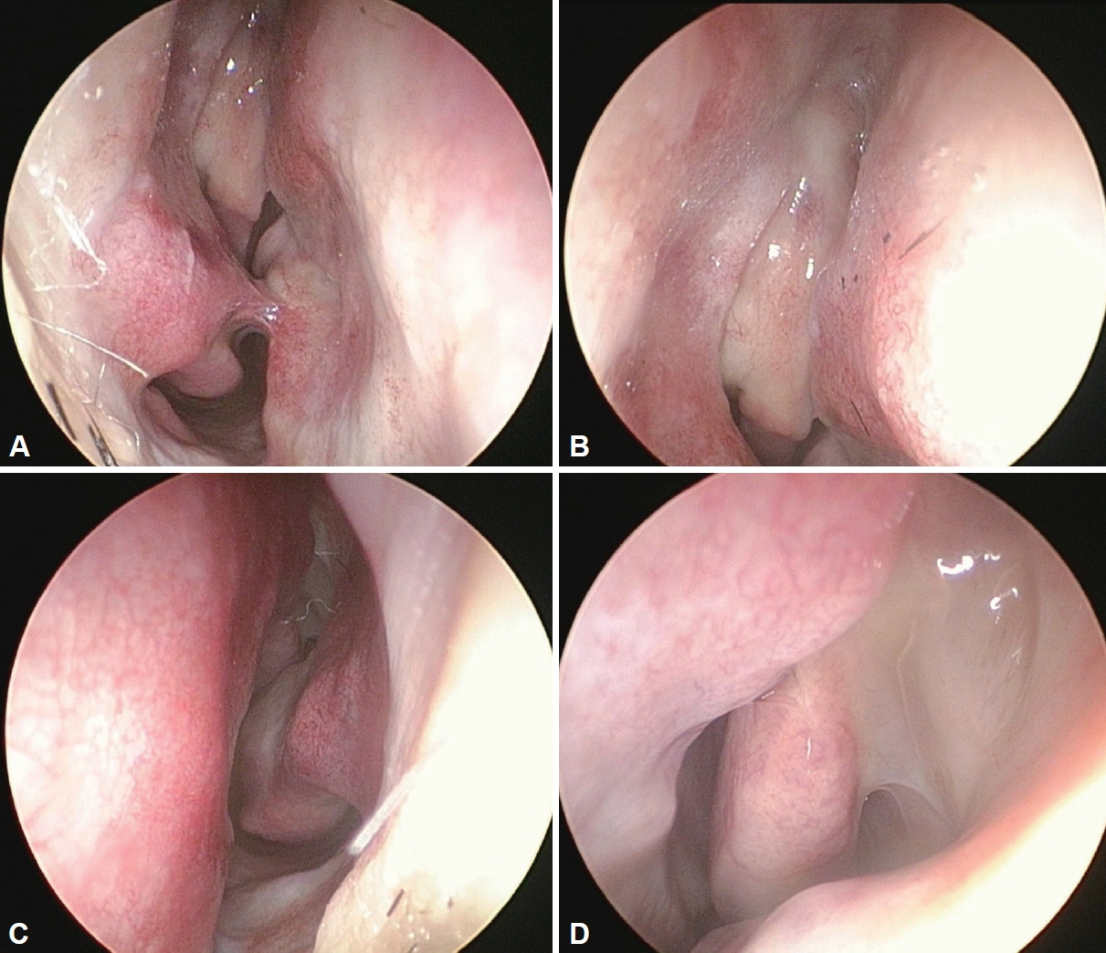
Fig. 1. Preoperative nasal endoscopic finding. A: Preoperative endoscopic findings of the right nasal cavity. The patient underwent endoscopic sinus surgery 12 years ago. There were no signs of mucosal swelling or discharge. Synechia was observed between the right inferior turbinate and the nasal septum, presumably caused by previous surgery. B: The right uncinated process was swollen. C: Preoperative endoscopic findings of the left nasal cavity showed no signs of mucosal swelling. D: There was purulent discharge around the left uncinate process.
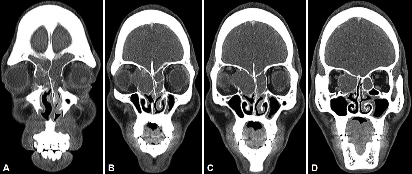
Fig. 2. Preoperative paranasal sinus computed tomography finding. A: Preoperative paranasal sinus computed tomography, showing soft tissue attenuation in both the frontal and ethmoid sinuses. The nasal septum is deviated to the left. B: The mucocele appears to originate from the right ethmoid sinus. C: The right eyeball is laterally displaced by an expansile mucocele. The boundary between the right eye and the tumor appears clear. A bone defect is suspected in the right skull base. D: The right medial rectus muscle is displaced by the mucocele. There is mild mucosal thickening in the left maxillary sinus.
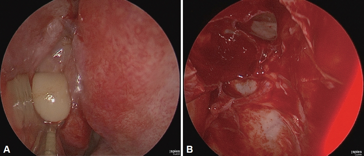
Fig. 3. Intraoperative endoscopic finding. A: Intraoperative endoscopic findings revealed massive pus drainage from a mucocele filling the right ethmoid and frontal sinuses. B: There was a bony defect in the lateral lamella of the right cribriform plate, measuring 1×0.5 cm. Cerebrospinal fluid leakage was observed.
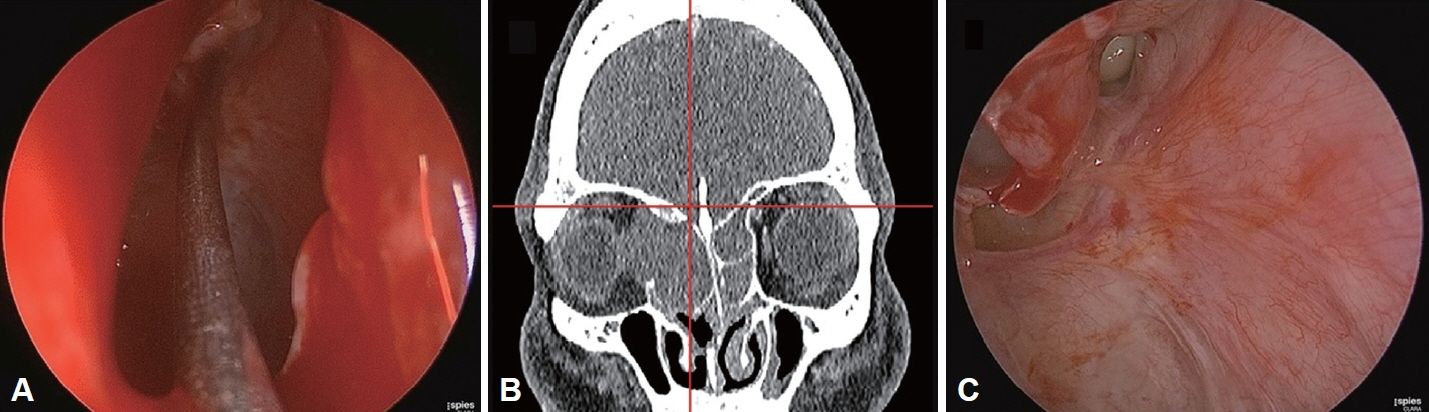
Fig. 4. Intraoperative finding of skullbase defect. A: A navigation pointer is partially inserted into the suspected site of a skull base defect. B: A skull base defect is present at the location indicated by the navigation pointer. C: The endoscopic view is utilized to confirm a defect at the skull base.
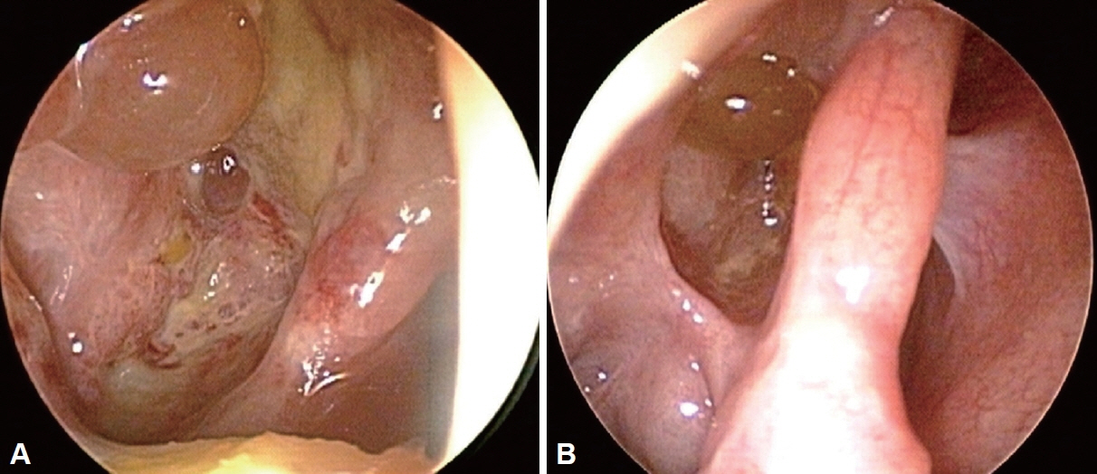
Fig. 5. Postoperative endoscopic view. The reconstruction of the skull base remains stable. A: One-month postoperative status. B: Two-month postoperative status.
Reference
-
References
1. Capra GG, Carbone PN, Mullin DP. Paranasal sinus mucocele. Head Neck Pathol. 2012; 6(3):369–72.
Article2. Natvig K, Larsen TE. Mucocele of the paranasal sinuses. A retrospective clinical and histological study. J Laryngol Otol. 1978; 92(12):1075–82.3. Moriyama H, Nakajima T, Honda Y. Studies on mucocoeles of the ethmoid and sphenoid sinuses: analysis of 47 cases. J Laryngol Otol. 1992; 106(1):23–7.
Article4. Devars du Mayne M, Moya-Plana A, Malinvaud D, Laccourreye O, Bonfils P. Sinus mucocele: natural history and long-term recurrence rate. Eur Ann Otorhinolaryngol Head Neck Dis. 2012; 129(3):125–30.
Article5. Busaba NY, Salman SD. Ethmoid mucocele as a late complication of endoscopic ethmoidectomy. Otolaryngol Head Neck Surg. 2003; 128(4):517–22.
Article6. Pizzo LJ, Mishler KE. Frontoethmoid mucocele with CSF leak. Ear Nose Throat J. 1984; 63(11):571–3.
- Full Text Links
-
- Actions
-
Cited
- CITED
-
- Close
- Share
-
- Similar articles
-
- A Case of Ethmoid Mucocele
- A Case of Brain Herniation and Cerebrospinal Fluid Leakage during Endoscopic Marsupialization of Ethmoid Sinus Mucocele
- Complex Anterior Skullbase Fracture Caused by a Bottle Cap: A Case Report and Review of the Literature
- A Case of Ethmoid Sinus Mucocele with Transient Intraocular Pressure Elevation
- A Case Report of 41-Year-Old Female with Fibrous Dysplasia Combined with Ethmoid Mucocele
