J Rhinol.
2016 May;23(1):49-54. 10.18787/jr.2016.23.1.49.
Complex Anterior Skullbase Fracture Caused by a Bottle Cap: A Case Report and Review of the Literature
- Affiliations
-
- 1Department of Otorhinolaryngology-Head and Neck Surgery, Samsung Medical Center, Sungkyunkwan University School of Medicine, Seoul, Korea. kkam97@gmail.com
- 2Department of Rhinology, Hana ENT Hospital, Seoul, Korea.
- KMID: 2165155
- DOI: http://doi.org/10.18787/jr.2016.23.1.49
Abstract
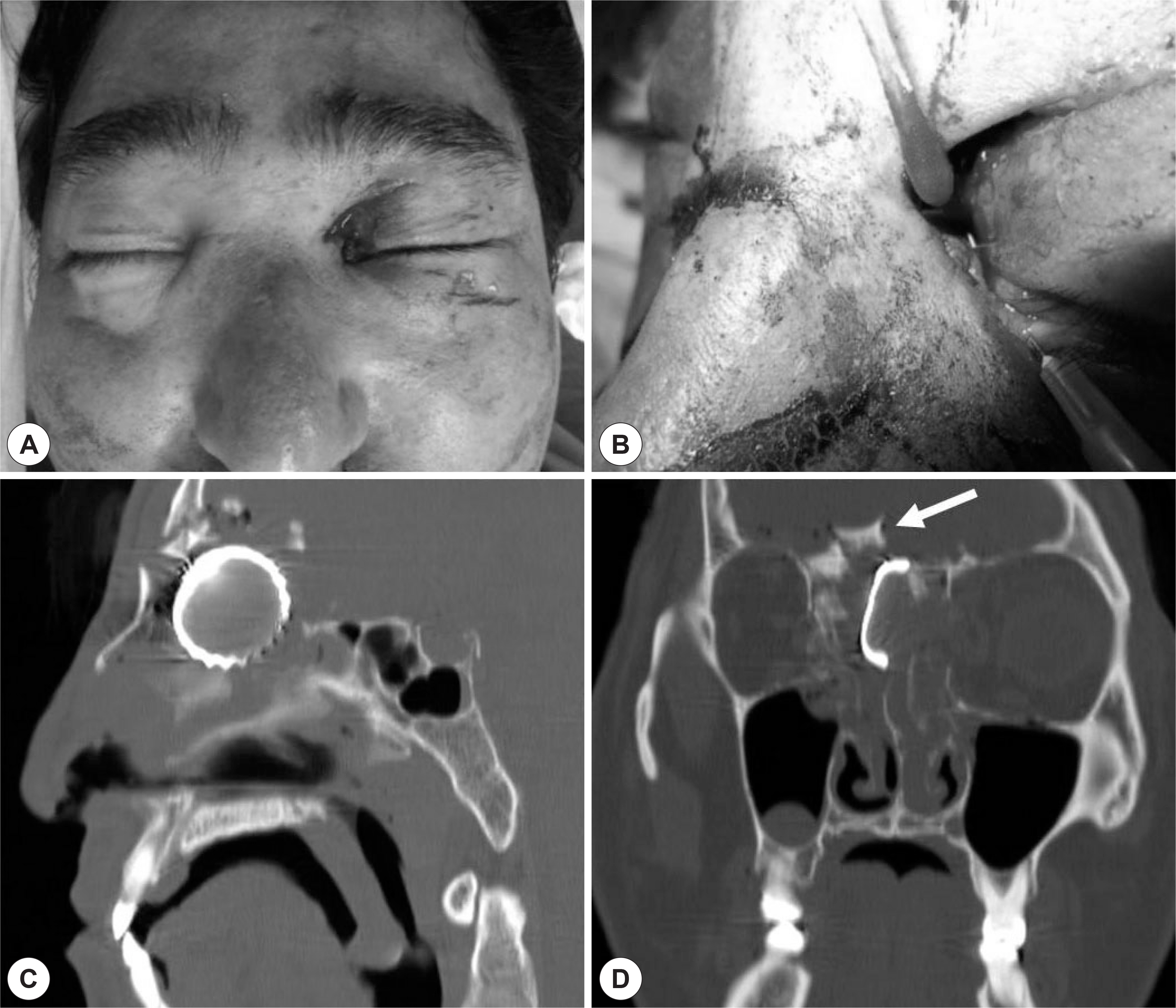
- We report a case of foreign body presence in the ethmoid sinus cavity with anterior skull base fracture and visual loss. A 42-year-old male had an uncertain history of trauma and a penetrating wound near the left medial canthus. Computed tomography imaging showed a 3.0-cm bottle cap penetrating into the anterior skull base. He underwent foreign body removal, canalicular repair, ethmoidectomy, and cerebrospinal fluid leakage repair using packing material. Six months after the initial surgery, a second-stage operation for blow-out fracture repair was performed. At the 18-month postoperative follow-up from the initial surgery, the patient had no complaints except anosmia. This is a very rare case of a large, blunt, foreign body penetrating into the anterior skull base without long-term complications after successful removal and skull base repair. Simultaneous repair of cerebrospinal fluid leakage, management of canaliculi injury, and traumatic optic nerve neuropathy should be considered in such cases.
Keyword
MeSH Terms
Figure
Reference
-
References
1). Brinson GM, Senior BA, Yarbrough WG. Endoscopic management of retained airgun projectiles in the paranasal sinuses. Otolaryngol Head Neck Surg. 2004; 130:25–30.
Article2). Udwadia RA, Maniar D, Acharya M. A transethmoid transorbital foreign body. J Laryngol Otol. 1994; 108:441–2.
Article3). Tsao YH, Kao CH, Wang HW, Chin SC, Moe KS. Transorbital penetrating injury of paranasal sinuses and anterior skull base by a plastic chair glide: management options of a foreign body in multiple anatomic compartments. Otolaryngol Head Neck Surg. 2006; 134:177–9.
Article4). Garces SM, Norris CW. Unusual frontal sinus foreign body. J Laryngol Otol. 1972; 86:1265–8.
Article5). Fallon MJ, Plante DM, Brown LW. Wooden transnasal intracranial penetration: an unusual presentation. J Emerg Med. 1992; 10:439–43.
Article6). Donald PJ, Gadre AK. Neuralgia-like symptoms in a patient with an airgun pellet in the ethmoid sinus: a case report. J Laryngol Otol. 1995; 109:646–9.
Article7). Asano T, Ohno K, Takada Y, Suzuki R, Hirakawa K, Monma S. Fractures of the floor of the anterior cranial fossa. J Trauma. 1995; 39:702–6.
Article8). Cetinkaya EA, Okan C, Pelin K. Transnasal, intracranial penetrating injury treated endoscopically. J Laryngol Otol. 2006; 120:325–6.
Article9). Dodson KM, Bridges MA, Reiter ER. Endoscopic transnasal management of intracranial foreign bodies. Arch Otolaryngol Head Neck Surg. 2004; 130:985–8.
Article10). Sharif S, Roberts G, Phillips J. Transnasal penetrating brain injury with a ball-pen. Br J Neurosurg. 2000; 14:159–60.11). Thomas S, Daudia A, Jones NS. Endoscopic removal of foreign body from the anterior cranial fossa. J Laryngol Otol. 2007; 121:794–5.
Article12). Gray ST, Wu AW. Pathophysiology of iatrogenic and traumatic skull base injury. Adv Otorhinolaryngol. 2013; 74:12–23.
Article13). Friedman JA, Ebersold MJ, Quast LM. Post-traumatic cerebrospinal fluid leakage. World J Surg. 2001; 25:1062–6.
Article14). Emanuelli E, Bignami M, Digilio E, Fusetti S, Volo T, Castelnuovo P. Post-traumatic optic neuropathy: our surgical and medical protocol. Eur Arch Otorhinolaryngol 2014. In press.15). Kumaran AM, Sundar G, Chye LT. Traumatic optic neuropathy: a review. Craniomaxillofac Trauma Reconstr. 2015; 8:31–41.
Article16). Levin LA, Joseph MP, Rizzo JF 3rd, Lessell S. Optic canal decompression in indirect optic nerve trauma. Ophthalmology. 1994; 101:566–9.
Article17). Mine S, Yamakami I, Yamaura A, Hanawa K, Ikejiri M, Mizota A, et al. Outcome of traumatic optic neuropathy. Comparison between surgical and nonsurgical treatment. Acta Neurochir (Wien). 1999; 141:27–30.
Article18). Yu-Wai-Man P, Griffiths PG. Steroids for traumatic optic neuropathy. Cochrane Database Syst Rev. 2013; 6:CD006032.
Article19). Beck RW, Cleary PA, Anderson MM Jr, Keltner JL, Shults WT, Kaufman DI, et al. A randomized, controlled trial of corticosteroids in the treatment of acute optic neuritis. The Optic Neuritis Study Group. N Engl J Med. 1992; 326:581–8.
- Full Text Links
- Actions
-
Cited
- CITED
-
- Close
- Share
- Similar articles
-
- Barotraumatic Perforation of Pharyngoesophagus by Explosion of a Bottle into the Mouth
- Anterior Dislocation of the Ankle without Malleolar Fractures: A Case Report
- Avulsion Fracture of Medial Cuneiform by Tibialis Anterior Tendon (A Case Report)
- The combined qudriple lesion : fracture of acromion, distal end of clavicle, distal coracoid and glenoid rim associated with anterior shoulder dislocation: A Case Report
- Avulsion of the Femoral Attachment of Anterior Cruciate Ligament Associated with Ipsilateral Femoral Shaft Fracture in Skeletally Mature Patient: A Case Report


