J Korean Assoc Oral Maxillofac Surg.
2024 Jun;50(3):140-145. 10.5125/jkaoms.2024.50.3.140.
Assessing the efficacy of apicoectomy without retrograde filling in treating periapical inflammatory cysts
- Affiliations
-
- 1Department of Oral and Maxillofacial Surgery, School of Dentistry and Institute of Oral Bioscience, Research Institute of Clinical Medicine of Jeonbuk National University-Biomedical Research Institute of Jeonbuk National University Hospital, Jeonbuk National University, Jeonju, Korea
- 2Department of Conservative Dentistry, Section of Dentistry, Seoul National University Bundang Hospital, Seongnam, Korea
- KMID: 2557630
- DOI: http://doi.org/10.5125/jkaoms.2024.50.3.140
Abstract
Objectives
The necessity of retrograde filling after apicoectomy is controversial in cases of non-inflammatory cysts as opposed to bacteria-related periapical abscesses. This study aims to investigate whether the presence or absence of retrograde filling during apicoectomy has differential long-term prognostic implications between inflammatory and non-inflammatory cysts.
Materials and Methods
This retrospective study included patients who underwent tooth apicoectomy during jaw cyst enucleation between 2013 and 2022, and who underwent follow-up cone-beam computed tomography for at least 6 months. The prognosis of the tooth was evaluated during the follow-up period according to the cyst type, the presence or absence of retrograde filling, mandible or maxilla, and location.
Results
A total of 147 teeth was included in this study. All the operated teeth underwent preoperative root canal treatment by an endodontic specialist. Apicoectomy was performed for 119 inflammatory cysts and 28 non-inflammatory cysts. Retrograde filling was performed on 22 teeth with inflammatory cysts and 3 teeth with non-inflammatory cysts. All teeth survived the 3.5-year follow-up (range, 1.0-9.1 years). However, 1 tooth with an inflammatory cyst developed complications 1 year after surgery that required re-endodontic treatment.
Conclusion
The prognosis of a tooth treated by apicoectomy without retrograde filling during cyst enucleation is favorable, regardless of the cyst type.
Figure
-
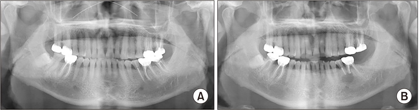
Fig. 1 Radiograph of a patient with a radicular cyst. A. Preoperative panoramic view. B. Postoperative panoramic view.
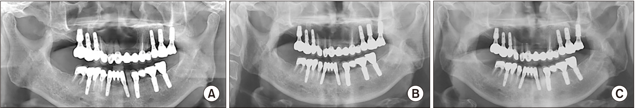
Fig. 2 Radiograph of a patient with an odontogenic keratocyst on the anterior maxilla. A. Initial panoramic view. B. Preoperative panoramic view after prior root canal therapy. C. Postoperative panoramic view.
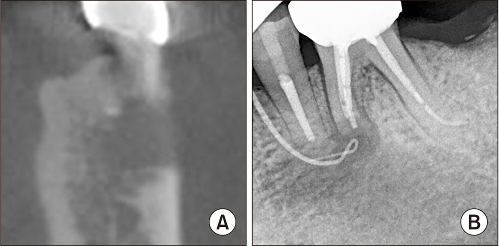
Fig. 3 Postoperative radiograph of the patient from Fig. 1. A. Incomplete bone healing demonstrated on cone-beam computed tomography one year after the surgery. B. Gutta percha tracing from the gingival fistula.
Reference
-
References
1. Ricucci D, Mannocci F, Ford TR. 2006; A study of periapical lesions correlating the presence of a radiopaque lamina with histological findings. Oral Surg Oral Med Oral Pathol Oral Radiol Endod. 101:389–94. https://doi.org/10.1016/j.tripleo.2005.08.026. DOI: 10.1016/j.tripleo.2005.08.026. PMID: 16504874.
Article2. Rios Osorio N, Caviedes-Bucheli J, Mosquera-Guevara L, Adames-Martinez JS, Gomez-Pinto D, Jimenez-Jimenez K, et al. 2023; The paradigm of the inflammatory radicular cyst: biological aspects to be considered. Eur Endod J. 8:20–36. https://doi.org/10.14744/eej.2022.26918. DOI: 10.14744/eej.2022.26918. PMID: 36748442. PMCID: PMC10098462.
Article3. Hwang MJ, Lee YP, Lang MJ, Wu YH, Chiang CP, Chueh LH. 2021; Clinicopathological study of radicular cysts with actinomycosis. J Dent Sci. 16:825–30. https://doi.org/10.1016/j.jds.2021.04.008. DOI: 10.1016/j.jds.2021.04.008. PMID: 34141095. PMCID: PMC8189886.
Article4. Elhakim A, Kim S, Kim E, Elshazli AH. 2021; Preserving the vitality of teeth adjacent to a large radicular cyst in periapical microsurgery: a case report with 4-year follow-up. BMC Oral Health. 21:382. https://doi.org/10.1186/s12903-021-01738-2. DOI: 10.1186/s12903-021-01738-2. PMID: 34344347. PMCID: PMC8336380.
Article5. Lieblich SE. 2020; Current concepts of periapical surgery: 2020 update. Oral Maxillofac Surg Clin North Am. 32:571–82. https://doi.org/10.1016/j.coms.2020.07.007. DOI: 10.1016/j.coms.2020.07.007. PMID: 32912776.
Article6. Ng YL, Gulabivala K. 2023; Factors that influence the outcomes of surgical endodontic treatment. Int Endod J. 56 Suppl 2:116–39. https://doi.org/10.1111/iej.13896. DOI: 10.1111/iej.13896. PMID: 36710526.
Article7. Huh JK, Yang DK, Jeon KJ, Shin SJ. 2016; Progression of periapical cystic lesion after incomplete endodontic treatment. Restor Dent Endod. 41:137–42. https://doi.org/10.5395/rde.2016.41.2.137. DOI: 10.5395/rde.2016.41.2.137. PMID: 27200282. PMCID: PMC4868877.
Article8. Zhao Y, Liu B, Cheng G, Wang SP, Wang YN. 2012; Recurrent keratocystic odontogenic tumours: report of 19 cases. Dentomaxillofac Radiol. 41:96–102. https://doi.org/10.1259/dmfr/22891281. DOI: 10.1259/dmfr/22891281. PMID: 22301637. PMCID: PMC3520371.
Article9. García CC, Sempere FV, Diago MP, Bowen EM. 2007; The post-endodontic periapical lesion: histologic and etiopathogenic aspects. Med Oral Patol Oral Cir Bucal. 12:E585–90.10. Angerame D, De Biasi M, Lenhardt M, Porrelli D, Bevilacqua L, Generali L, et al. 2022; Root-end resection with or without retrograde obturation after orthograde filling with two techniques: a micro-CT study. Aust Endod J. 48:423–30. https://doi.org/10.1111/aej.12634. DOI: 10.1111/aej.12634. PMID: 35665570.
Article11. Jung J, Kim S, Kim E, Shin SJ. 2020; Volume of voids in retrograde filling: comparison between calcium silicate cement alone and combined with a calcium silicate-based sealer. J Endod. 46:97–102. https://doi.org/10.1016/j.joen.2019.10.015. DOI: 10.1016/j.joen.2019.10.015. PMID: 31759678.
Article12. Villa-Machado PA, Botero-Ramírez X, Tobón-Arroyave SI. 2013; Retrospective follow-up assessment of prognostic variables associated with the outcome of periradicular surgery. Int Endod J. 46:1063–76. https://doi.org/10.1111/iej.12100. DOI: 10.1111/iej.12100. PMID: 23560363.
Article13. Kim E, Song JS, Jung IY, Lee SJ, Kim S. 2008; Prospective clinical study evaluating endodontic microsurgery outcomes for cases with lesions of endodontic origin compared with cases with lesions of combined periodontal-endodontic origin. J Endod. 34:546–51. https://doi.org/10.1016/j.joen.2008.01.023. DOI: 10.1016/j.joen.2008.01.023. PMID: 18436032.
Article14. Antunes HS, Gominho LF, Andrade-Junior CV, Dessaune-Neto N, Alves FR, Rôças IN, et al. 2016; Sealing ability of two root-end filling materials in a bacterial nutrient leakage model. Int Endod J. 49:960–5. https://doi.org/10.1111/iej.12543. DOI: 10.1111/iej.12543. PMID: 26334201.
Article15. Nair PNR, Pajarola G, Schroeder HE. 1996; Types and incidence of human periapical lesions obtained with extracted teeth. Oral Surg Oral Med Oral Pathol Oral Radiol Endod. 81:93–102. https://doi.org/10.1016/s1079-2104(96)80156-9. DOI: 10.1016/S1079-2104(96)80156-9. PMID: 8850492.
Article16. Nair PNR. 2006; On the causes of persistent apical periodontitis: a review. Int Endod J. 39:249–81. https://doi.org/10.1111/j.1365-2591.2006.01099.x. DOI: 10.1111/j.1365-2591.2006.01099.x. PMID: 16584489.
Article17. European Society of Endodontology. 2006; Quality guidelines for endodontic treatment: consensus report of the European Society of Endodontology. Int Endod J. 39:921–30. https://doi.org/10.1111/j.1365-2591.2006.01180.x. DOI: 10.1111/j.1365-2591.2006.01180.x. PMID: 17180780.18. Cohen S, Burns RC. 1998. Pathways of the pulp. 7th ed. Mosby.19. Ma X, Li C, Jia L, Wang Y, Liu W, Zhou X, et al. 2016; Materials for retrograde filling in root canal therapy. Cochrane Database Syst Rev. 12:CD005517. https://doi.org/10.1002/14651858.cd005517.pub2. DOI: 10.1002/14651858.CD005517.pub2.
Article20. Altan H, Tosun G. 2016; The setting mechanism of mineral trioxide aggregate. J Istanb Univ Fac Dent. 50:65–72. https://doi.org/10.17096/jiufd.50128. DOI: 10.17096/jiufd.50128. PMID: 28955558. PMCID: PMC5573456.
Article21. Peñarrocha-Oltra D, Soto-Peñaloza D, Peñarrocha-Diago M, Cervera-Ballester J, Cabanes-Gumbau G, Peñarrocha-Diago M. 2020; Hemostatic agents in endodontic surgery of maxillary molars: a randomized controlled pilot study of polytetrafluoroethylene (PTFE) strips as an adjunct to epinephrine impregnated gauze versus aluminum chloride. Med Oral Patol Oral Cir Bucal. 25:e634–43. https://doi.org/10.4317/medoral.23652. DOI: 10.4317/medoral.23652. PMID: 32683388. PMCID: PMC7473427.
Article22. Jang Y, Kim H, Roh BD, Kim E. 2014; Biologic response of local hemostatic agents used in endodontic microsurgery. Restor Dent Endod. 39:79–88. https://doi.org/10.5395/rde.2014.39.2.79. DOI: 10.5395/rde.2014.39.2.79. PMID: 24790919. PMCID: PMC3978108.
Article23. Amador-Cabezalí A, Pardal-Peláez B, Quispe-López N, Lobato-Carreño M, Sanz-Sánchez Á, Montero J. 2022; Influence of the retrograde filling material on the success of periapical surgery. Systematic review and meta-analysis by groups. Coatings. 12:1140. https://doi.org/10.3390/coatings12081140. DOI: 10.3390/coatings12081140.
Article24. Simon JH. 1980; Incidence of periapical cysts in relation to the root canal. J Endod. 6:845–8. https://doi.org/10.1016/s0099-2399(80)80039-2. DOI: 10.1016/S0099-2399(80)80039-2. PMID: 6935342.
Article25. Ricucci D, Rôças IN, Hernández S, Siqueira JF Jr. 2020; "True" versus "bay" apical cysts: clinical, radiographic, histopathologic, and histobacteriologic features. J Endod. 46:1217–27. https://doi.org/10.1016/j.joen.2020.05.025. DOI: 10.1016/j.joen.2020.05.025. PMID: 32544498.
Article26. Song M, Kim HC, Lee W, Kim E. 2011; Analysis of the cause of failure in nonsurgical endodontic treatment by microscopic inspection during endodontic microsurgery. J Endod. 37:1516–9. https://doi.org/10.1016/j.joen.2011.06.032. DOI: 10.1016/j.joen.2011.06.032. PMID: 22000454.
Article27. Furusawa M, Asai Y. 2002; SEM observations of resected root canal ends following apicoectomy. Bull Tokyo Dent Coll. 43:7–12. https://doi.org/10.2209/tdcpublication.43.7. DOI: 10.2209/tdcpublication.43.7. PMID: 12013825.
Article
- Full Text Links
-
- Actions
-
Cited
- CITED
-
- Close
- Share
-
- Similar articles
-
- Retrograde filling with Lid technique in periapical surgery: case report
- A RETROSPECTIVE CLINICAL STUDY OF PERIAPICALLY INFECTED TEETH TREATED WITH PERIAPICAL SURGERY
- A Clinical Study of Periapical Lesions
- Interplay of collagen and mast cells in periapical granulomas and periapical cysts: a comparative polarizing microscopic and immunohistochemical study
- Apicoectomy of maxillary anterior teeth through a piezoelectric bony-window osteotomy: two case reports introducing a new technique to preserve cortical bone
