Retrograde filling with Lid technique in periapical surgery: case report
- Affiliations
-
- 1Department of Conservative Dentistry, College of Dentistry, Gangneung-Wonju National University, Gangneung, Republic of Korea
- KMID: 2549251
- DOI: http://doi.org/10.14368/jdras.2023.39.1.45
Abstract
- Apical sealing with proper retrograde filling material and technique is the key factor for successful apical surgery. In order to provide impeccable apical sealing and healing, various mechanical and chemical characteristics of retrograde filling material is required. MTA has been used as the gold standard of retrograde filling due to its unprecedented advantages. As MTA has long setting time and difficult handling properties, premixed putty type bioceramic material has been newly developed. For efficient retrofilling with premixed putty type biocreamic material, ‘Lid technique’ was also proposed. The following cases present apical surgery using newly developed premixed putty type bioceramic material with Lid technique.
Figure
-
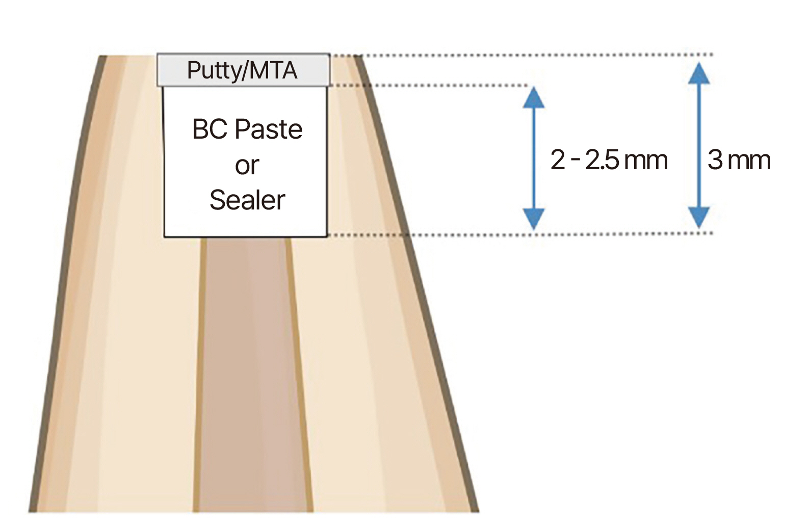
Fig. 1 Schematic diagram of the retrograde filling using “Lid technique”.
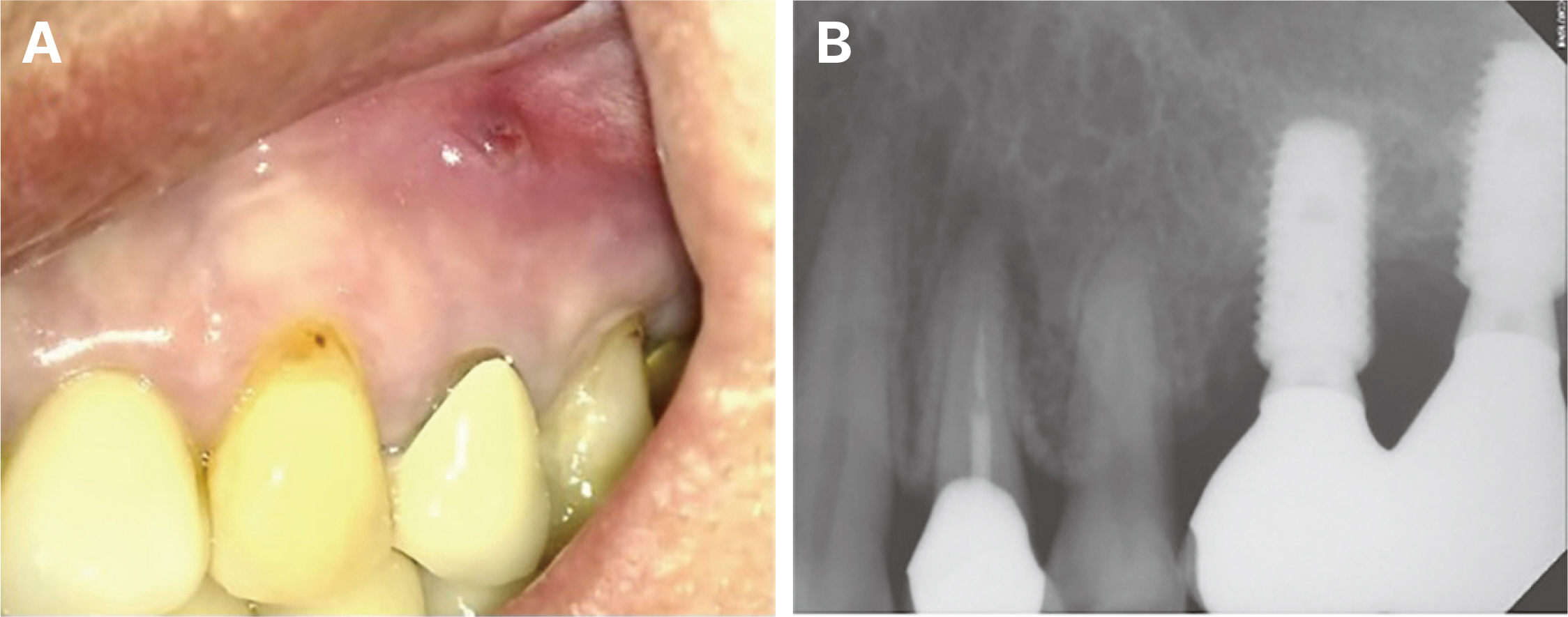
Fig. 2 (A) Pretreament clinical view of maxillary left first premolar, (B) Radiograph of maxillary left first premolar before treatment.
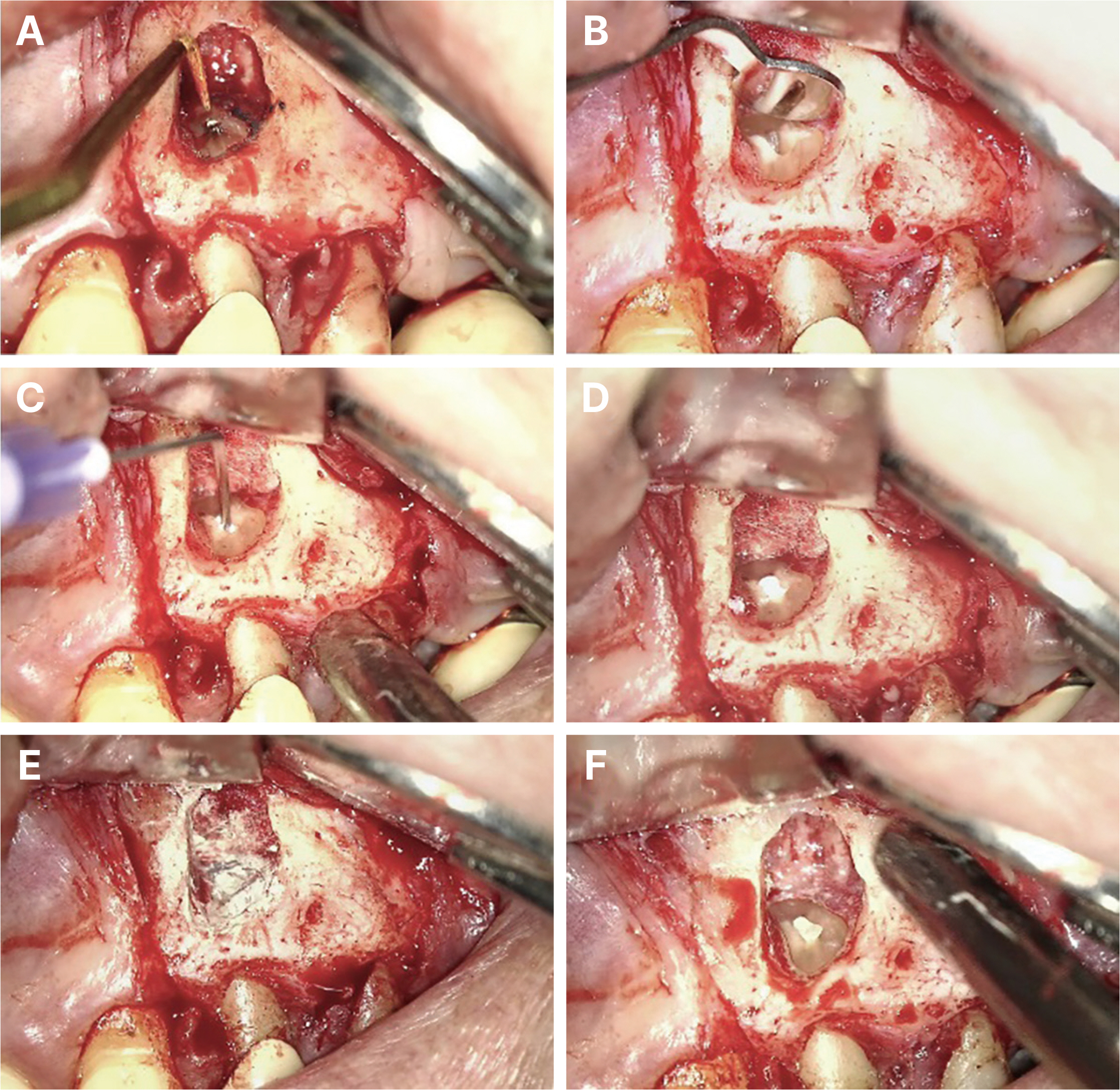
Fig. 3 (A, B) Retropreparation using ultrasonic instruments, (C, D) Application of the syringe type material, (E, F) Application of the putty type material.
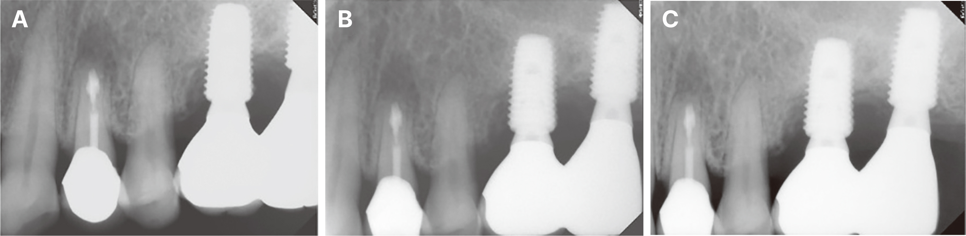
Fig. 4 (A) Radiograph of maxillary left first premolar after treatment, (B) 3 months later follow up, (C) 6 months later follow up.
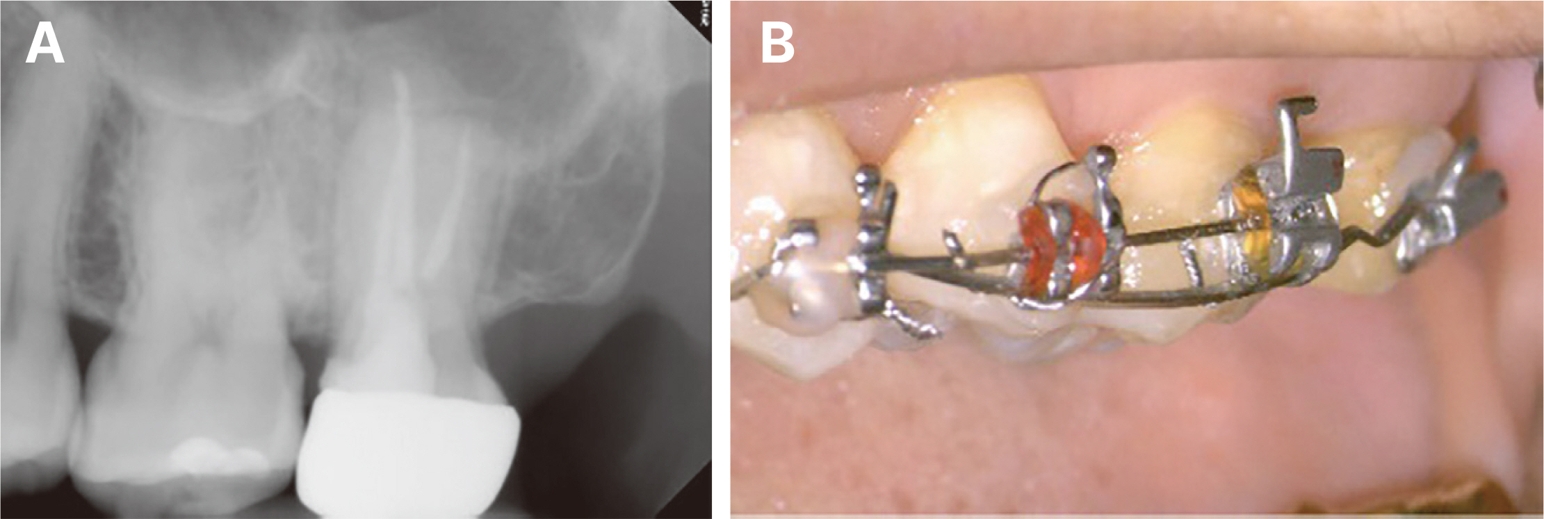
Fig. 5 (A) Radiograph of maxillary left second molar before treatment, (B) Pretreatment clinical view of maxillary left second molar after orthodontic extrusion.

Fig. 6 (A) Pretreatment clinical view of maxillary left second molar after removal of orthodontic wire, (B) Extraction of left maxillary second molar using forcep, (C) Clinical view of the apex before apical resection, (D) Apical resection, (E) Retropreparation, (F) Application of syringe type material followed by putty type material.

Fig. 7 (A) Radiograph of left maxillary second molar after treatment, (B) 3 months later follow up, (C) 6 months later follow up.
Reference
-
References
1. Chong BS, Rhodes JS. 2014; Endodontic surgery. Br Dent J. 216:281–90. DOI: 10.1038/sj.bdj.2014.220. PMID: 24651333.2. Antony JK, George L, Mathew J, Joy A. 2022; Sealing ability of mineral trioxide aggregate, Biodentine, and Endosequence RRM putty used as retrograde restorative material: An in vitro bacterial leakage model study. Endodontology. 34:16–21. DOI: 10.4103/endo.endo_176_21. PMID: 7f5247f643744764b99b0d59062a870c.
Article3. Li H, Guo Z, Li C, Ma X, Wang Y, Zhou X, Johnson TM, Huang D. 2021; Materials for retrograde filling in root canal therapy. Cochrane Database Syst Rev. 10:CD005517. DOI: 10.1002/14651858.CD005517.pub3. PMID: 34647617. PMCID: PMC8515509.4. Ali Nasseh A. 2022. Endodontic Practice Us. Available from: https://endopracticeus.com/premixed-nanoparticulate-bioceramics-endodontics-first-decade. updated 2022 July 18.5. Pereira IR, Carvalho C, Paulo S, Martinho JP, Coelho AS, Paula AB, Marto CM, Carrilho E, Botelho MF, Abrantes AM, Ferreira MM. 2021; Apical sealing ability of two calcium silicate-based sealers using a radioactive isotope method: An In Vitro Apexification Model. Materials. 14:6456. DOI: 10.3390/ma14216456. PMID: 34771981. PMCID: PMC8585189. PMID: 24f9f14b84c344478250d38dfb35f1fa.
Article6. Antunes HS, Gominho LF, Andrade-Junior CV, Dessaune-Neto N, Alves FRF, Rôças IN, Siqueira JF Jr. 2016; Sealing ability of two root-end filling materials in a bacterial nutrient leakage model. Int Endod J. 49:960–5. DOI: 10.1111/iej.12543. PMID: 26334201.7. Ali Nasseh A. 2022. Real World Endo. Available from: https://realworldendo.com/the-rwe-lid-technique-for-retrofilling-during-apicoectomy. updated 2022 July 18.
- Full Text Links
-
- Actions
-
Cited
- CITED
-
- Close
- Share
-
- Similar articles
-
- A RETROSPECTIVE CLINICAL STUDY OF PERIAPICALLY INFECTED TEETH TREATED WITH PERIAPICAL SURGERY
- Assessing the efficacy of apicoectomy without retrograde filling in treating periapical inflammatory cysts
- Modified Huches Procedure
- Bioblock technique to treat severe internal resorption with subsequent periapical pathology: a case report
- Plasmacytoma presented as a lid mass: A case report
