Potential Risk of Choline Alfoscerate on Isoflurane-Induced Toxicity in Primary Human Astrocytes
- Affiliations
-
- 1Department of Anesthesiology and Pain Medicine, College of Medicine, Ewha Womans University, Seoul, Korea
- 2Department of Neurosurgery, Korea University Medicine, Korea University College of Medicine, Seoul, Korea
- 3Photo-Theranosis and Bioinformatics for Tumor Laboratory, Research Institute for Future Medicine, Samsung Medical Center, Seoul, Korea
- 4Department of Neurosurgery, Samsung Medical Center, Sungkyunkwan University School of Medicine, Seoul, Korea
- KMID: 2556739
- DOI: http://doi.org/10.3340/jkns.2023.0208
Abstract
Objective
: Isoflurane, a widely used common inhalational anesthetic agent, can induce brain toxicity. The challenge lies in protecting neurologically compromised patients from neurotoxic anesthetics. Choline alfoscerate (L-α-Glycerophosphorylcholine, α-GPC) is recognized for its neuroprotective properties against oxidative stress and inflammation, but its optimal therapeutic window and indications are still under investigation. This study explores the impact of α-GPC on human astrocytes, the most abundant cells in the brain that protect against oxidative stress, under isoflurane exposure.
Methods
: This study was designed to examine changes in factors related to isoflurane-induced toxicity following α-GPC administration. Primary human astrocytes were pretreated with varying doses of α-GPC (ranging from 0.1 to 10.0 μM) for 24 hours prior to 2.5% isoflurane exposure. In vitro analysis of cell morphology, water-soluble tetrazolium salt-1 assay, quantitative real-time polymerase chain reaction, proteome profiler array, and transcriptome sequencing were conducted.
Results
: A significant morphological damage to human astrocytes was observed in the group that had been pretreated with 10.0 mM of α-GPC and exposed to 2.5% isoflurane. A decrease in cell viability was identified in the group pretreated with 10.0 μM of α-GPC and exposed to 2.5% isoflurane compared to the group exposed only to 2.5% isoflurane. Quantitative real-time polymerase chain reaction revealed that mRNA expression of heme-oxygenase 1 and hypoxia-inducible factor-1α, which were reduced by isoflurane, was further suppressed by 10.0 μM α-GPC pretreatment. The proteome profiler array demonstrated that α-GPC pretreatment influenced a variety of factors associated with apoptosis induced by oxidative stress. Additionally, transcriptome sequencing identified pathways significantly related to changes in isoflurane-induced toxicity caused by α-GPC pretreatment.
Conclusion
: The findings suggest that α-GPC pretreatment could potentially enhance the vulnerability of primary human astrocytes to isoflurane-induced toxicity by diminishing the expression of antioxidant factors, potentially leading to amplified cell damage.
Figure
-
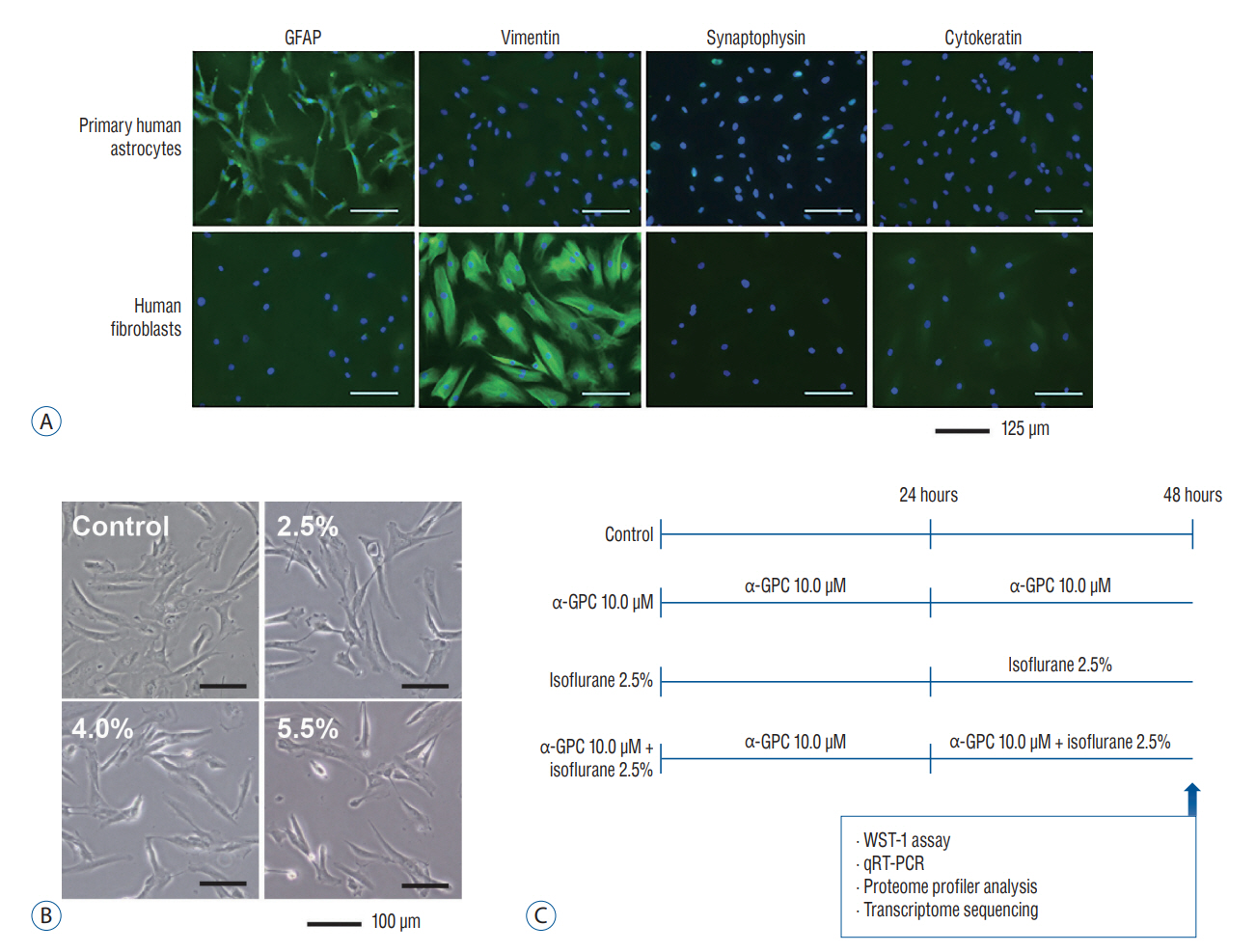
Fig. 1. A : Immunocytochemistry images of cultured primary human astrocytes (×100 magnification; green : Alexa Fluor 488-conjugated secondary antibody; blue : 4',6-diamidino-2-phenylindole). Human fibroblasts were used as a positive control for vimentin and a negative control for GFAP. The results indicate that the cultures were enriched with mature astrocytes. B : Morphological alteration of primary human astrocytes by isoflurane exposure in a dose-dependent manner (×100 magnification). The characteristics of morphological damage are manifested through cell shrinkage and diminished intercellular adherence as observed under a light microscope. The numbers depicted in the image denote the concentration of isoflurane. C : Schematic diagram of experimental protocol designed to evaluate the impact of α-GPC on isoflurane-induced toxicity. GFAP : glial fibrillary acidic protein, α-GPC : L-α-Glycerophosphorylcholine, WST : water-soluble tetrazolium salt, qRT-PCR : quantitative real-time polymerase chain reaction.
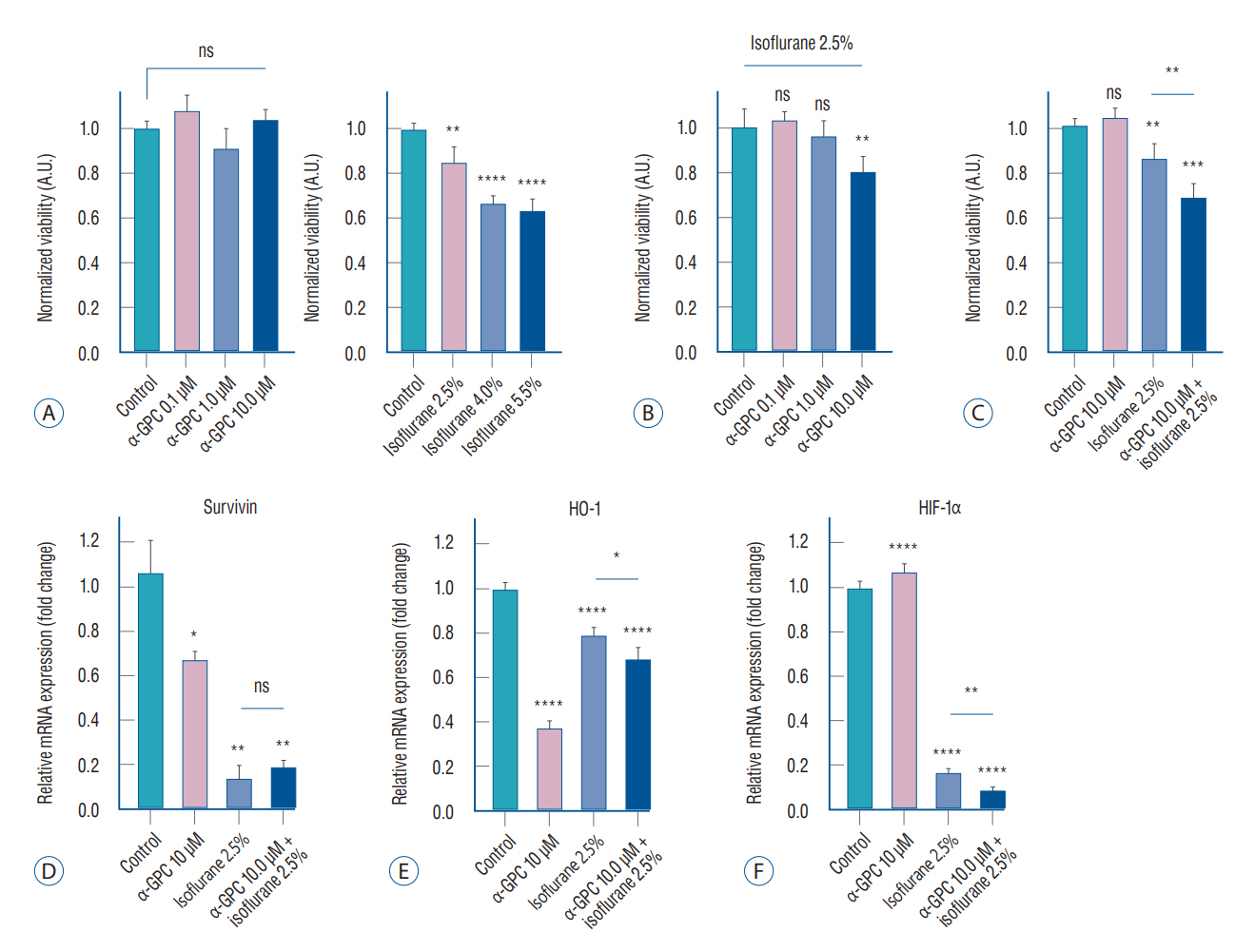
Fig. 2. A : WST-1 cell viability assay results of primary human astrocytes subjected to α-GPC and isoflurane. There was no significant difference in cell viability at any dose of treatment with α-GPC for 48 hours compared to control (n=6 for each, left), but isoflurane exposure reduced cell viability in a dose-dependent manner (n=6 for each, right). B : The effect on cell viability by 0.1, 1.0, 10.0 μM of α-GPC pretreatment and 2.5% isoflurane exposure (n=6 for each). After 24 hours of exposure to 2.5% isoflurane, pretreatment with 10.0 μM of α-GPC significantly reduced cell viability by approximately 20% compared to the group not treated with α-GPC (n=6). C : Reanalyzed results considering the individual and combined effects of 10.0 μM of α-GPC and 2.5% isoflurane. Primary human astrocytes, when pretreated with 10.0 μM of α-GPC and subsequently exposed to 2.5% isoflurane, exhibited a 32% reduction in cell viability compared to the non-treated control group. This decrease was notably more substantial than the 14% reduction observed when cells were only treated with 2.5% isoflurane, compared to the non-treated control group. D : Analysis of survivin mRNA expression in each treatment group using quantitative real-time polymerase chain reaction (qRT-PCR). In comparison to the non-treated control group, all groups treated with either 10 μM of α-GPC, 2.5% isoflurane, or a combination of both, demonstrated a statistically significant decrease in survivin expression (n=3 for each). E : Analysis of heme oxygenase-1 (HO-1) mRNA expression in each treatment group using qRT-PCR (n=3 for each). The groups pre-treated with α-GPC exhibited a significant reduction in HO-1 mRNA expression when compared to the control group. F : Analysis of hypoxia-inducible factor-1α (HIF-1α) mRNA expression in each treatment group using qRT-PCR (n=3 for each). In all groups treated with 2.5% isoflurane, the HIF-1α expression significantly decreased by more than 80%. The results are presented as the means±standard deviations. *p<0.05, **p<0.01, ***p<0.001, ****p<0.0001. A.U. : arbitrary unit, ns : not statistically significant, α-GPC : L-α-Glycerophosphorylcholine, WST-1 : water-soluble tetrazolium salt.
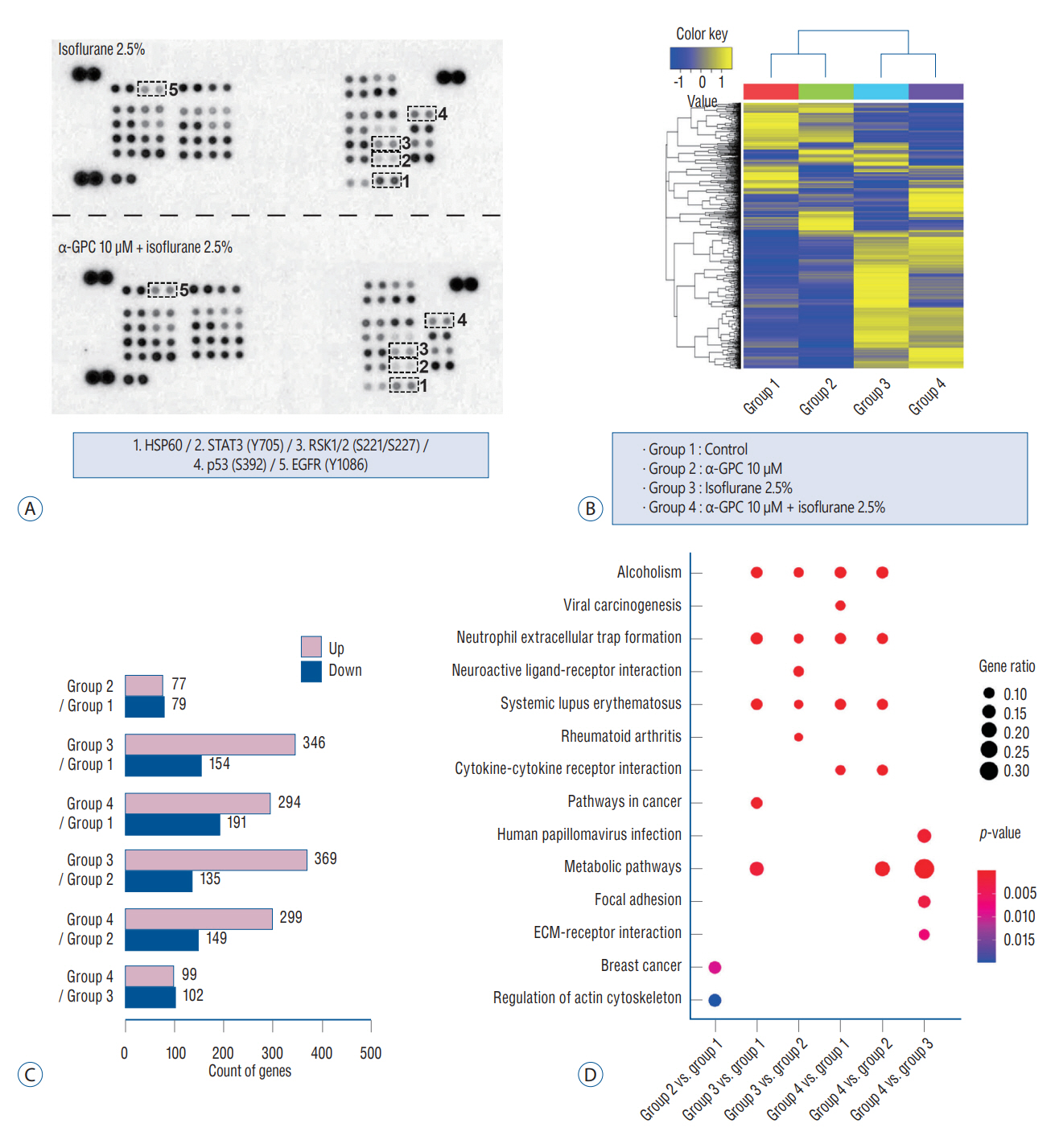
Fig. 3. A : Phosphorylation screening analysis using proteome profiler array. The α-GPC pretreatment was identified to decrease the phosphorylation of heat shock protein 60 (HSP60), signal transducer and activator of transcription3 (STAT3), 90 kDa ribosomal S6 kinase 1/2 (RSK1/2), and p53, while increasing the phosphorylation of epidermal growth factor receptor (EGFR). B : One-way hierarchical clustering heatmap that represents non-treated controls and various treated groups. The heatmap was created using the Z-Score of normalized values from 1321 genes that exhibited a fold change greater than 2 and a statistical significance of p<0.05. C : Count of up- and down-regulated genes exhibiting a fold change greater than 2 and statistical significance of p<0.05, according to group comparisons. The number of up- or down-regulated genes increased depending on the administration of 2.5% isoflurane, compared to the variation in gene expression with the administration of 10 μM of α-GPC. D : Gene-set enrichment analysis using the Kyoto Encyclopedia of Genes and Genomes database. The comparative analysis between the ‘isoflurane 2.5%’ group and the ‘α-GPC 10.0 μM + isoflurane 2.5%’ group revealed distinct patterns. The most notable finding was the further increase in the gene ratio for ‘metabolic pathways’, which exhibited the highest gene ratio with statistical significance. α-GPC : L-α-Glycerophosphorylcholine, ECM : extracellular matrix.
Reference
-
References
1. Bao F, Kang X, Xie Q, Wu J. HIF-α/PKM2 and PI3K-AKT pathways involved in the protection by dexmedetomidine against isoflurane or bupivacaine-induced apoptosis in hippocampal neuronal HT22 cells. Exp Ther Med. 17:63–70. 2019.
Article2. Barbagallo Sangiorgi G, Barbagallo M, Giordano M, Meli M, Panzarasa R. alpha-Glycerophosphocholine in the mental recovery of cerebral ischemic attacks. An Italian multicenter clinical trial. Ann N Y Acad Sci. 717:253–269. 1994.3. Bauer I, Raupach A. The role of heme oxygenase-1 in remote ischemic and anesthetic organ conditioning. Antioxidants (Basel). 8:403. 2019.
Article4. Burgaletto C, Di Benedetto G, Munafò A, Bernardini R, Cantarella G. Beneficial effects of choline alphoscerate on amyloid-β neurotoxicity in an in vitro model of Alzheimer's disease. Curr Alzheimer Res. 18:298–309. 2021.
Article5. Catanesi M, d'Angelo M, Antonosante A, Castelli V, Alfonsetti M, Benedetti E, et al. Neuroprotective potential of choline alfoscerate against β-amyloid injury: involvement of neurotrophic signals. Cell Biol Int. 44:1734–1744. 2020.
Article6. Chai D, Jiang H, Li Q. Isoflurane neurotoxicity involves activation of hypoxia inducible factor-1α via intracellular calcium in neonatal rodents. Brain Res. 1653:39–50. 2016.
Article7. Chen L, Chu C, Lu J, Kong X, Huang T, Cai YD. Gene ontology and KEGG pathway enrichment analysis of a drug target-based classification system. PLoS One. 10:e0126492. 2015.
Article8. Cui H, Xu Z, Qu C. Tetramethylpyrazine ameliorates isoflurane-induced cognitive dysfunction by inhibiting neuroinflammation via miR-150 in rats. Exp Ther Med. 20:3878–3887. 2020.
Article9. Culley DJ, Cotran EK, Karlsson E, Palanisamy A, Boyd JD, Xie Z, et al. Isoflurane affects the cytoskeleton but not survival, proliferation, or synaptogenic properties of rat astrocytes in vitro. Br J Anaesth 110 Suppl. 1:i19–i28. 2013.
Article10. De Jesus Moreno Moreno M. Cognitive improvement in mild to moderate Alzheimer's dementia after treatment with the acetylcholine precursor choline alfoscerate: a multicenter, double-blind, randomized, placebo-controlled trial. Clin Ther. 25:178–193. 2003.
Article11. Dringen R, Kussmaul L, Gutterer JM, Hirrlinger J, Hamprecht B. The glutathione system of peroxide detoxification is less efficient in neurons than in astroglial cells. J Neurochem. 72:2523–2530. 1999.
Article12. Evered LA, Silbert BS. Postoperative cognitive dysfunction and noncardiac surgery. Anesth Analg. 127:496–505. 2018.
Article13. Flecknell P, Lofgren JL, Dyson MC, Marini RR, Swindle MM, Wilson RP. Preanesthesia, anesthesia, analgesia, and euthanasia. In : Fox JG, Anderson LC, Otto GM, Pritchett-Corning KR, Whary MT, editors. Laboratory animal medicine. ed 3. Amsterdam: Elsevier;2015. p. 1135–1200.14. Guo H, Peng Z, Yang L, Liu X, Xie Y, Cai Y, et al. TREK-1 mediates isoflurane-induced cytotoxicity in astrocytes. BMC Anesthesiol. 17:124. 2017.
Article15. Hettiarachchi NT, Boyle JP, Dallas ML, Al-Owais MM, Scragg JL, Peers C. Heme oxygenase-1 derived carbon monoxide suppresses Aβ1-42 toxicity in astrocytes. Cell Death Dis. 8:e2884. 2017.
Article16. Kanehisa M, Furumichi M, Tanabe M, Sato Y, Morishima K. KEGG: new perspectives on genomes, pathways, diseases and drugs. Nucleic Acids Res. 45:D353–D361. 2017.
Article17. Kanehisa M, Sato Y, Kawashima M, Furumichi M, Tanabe M. KEGG as a reference resource for gene and protein annotation. Nucleic Acids Res. 44:D457–D462. 2016.
Article18. Kobayashi K, Hayashi M, Nakano H, Fukutani Y, Sasaki K, Shimazaki M, et al. Apoptosis of astrocytes with enhanced lysosomal activity and oligodendrocytes in white matter lesions in Alzheimer's disease. Neuropathol Appl Neurobiol. 28:238–251. 2002.
Article19. Lee G, Choi S, Chang J, Choi D, Son JS, Kim K, et al. Association of L-α glycerylphosphorylcholine with subsequent stroke risk after 10 years. JAMA Netw Open. 4:e2136008. 2021.
Article20. Lee SH, Choi BY, Kim JH, Kho AR, Sohn M, Song HK, et al. Late treatment with choline alfoscerate (l-alpha glycerylphosphorylcholine, α-GPC) increases hippocampal neurogenesis and provides protection against seizure-induced neuronal death and cognitive impairment. Brain Res. 1654(Pt A):66–76. 2017.
Article21. Lee YM, Song BC, Yeum KJ. Impact of volatile anesthetics on oxidative stress and inflammation. Biomed Res Int. 2015:242709. 2015.
Article22. Li HS, Zhou YN, Li L, Li SF, Long D, Chen XL, et al. HIF-1α protects against oxidative stress by directly targeting mitochondria. Redox Biol. 25:101109. 2019.
Article23. Li QF, Zhu YS, Jiang H, Xu H, Sun Y. Heme oxygenase-1 mediates the anti-inflammatory effect of isoflurane preconditioning in LPS-stimulated macrophages. Acta Pharmacol Sin. 30:228–234. 2009.
Article24. Longnecker DE, Murphy FL. Dripps/Eckenhoff/Vandam Introduction to Anesthesia. ed 9. Philadelphia: Saunders;1997. p. 75–87.25. Lunardi N, Hucklenbruch C, Latham JR, Scarpa J, Jevtovic-Todorovic V. Isoflurane impairs immature astroglia development in vitro: the role of actin cytoskeleton. J Neuropathol Exp Neurol. 70:281–291. 2011.
Article26. Malik JA, Lone R. Heat shock proteins with an emphasis on HSP 60. Mol Biol Rep. 48:6959–6969. 2021.
Article27. Min MH, Park JH, Hur JH, Shin HC, Cho Y, Kim DD. Formulation and bioequivalence studies of choline alfoscerate tablet comparing with soft gelatin capsule in healthy male volunteers. Drug Des Devel Ther. 13:1049–1058. 2019.28. Monk TG, Price CC. Postoperative cognitive disorders. Curr Opin Crit Care. 17:376–381. 2011.
Article29. Monk TG, Weldon BC, Garvan CW, Dede DE, van der Aa MT, Heilman KM, et al. Predictors of cognitive dysfunction after major noncardiac surgery. Anesthesiology. 108:18–30. 2008.
Article30. Needham MJ, Webb CE, Bryden DC. Postoperative cognitive dysfunction and dementia: what we need to know and do. Br J Anaesth. 119(suppl_1):i115–i125. 2017.
Article31. O'Driscoll L, Linehan R, Clynes M. Survivin: role in normal cells and in pathological conditions. Curr Cancer Drug Targets. 3:131–152. 2003.32. Parnetti L, Amenta F, Gallai V. Choline alphoscerate in cognitive decline and in acute cerebrovascular disease: an analysis of published clinical data. Mech Ageing Dev. 122:2041–2055. 2001.
Article33. Sarafian TA, Montes C, Imura T, Qi J, Coppola G, Geschwind DH, et al. Disruption of astrocyte STAT3 signaling decreases mitochondrial function and increases oxidative stress in vitro. PLoS One. 5:e9532. 2010.
Article34. Schnitzer J, Franke WW, Schachner M. Immunocytochemical demonstration of vimentin in astrocytes and ependymal cells of developing and adult mouse nervous system. J Cell Biol. 90:435–447. 1981.
Article35. Shi T, van Soest DMK, Polderman PE, Burgering BMT, Dansen TB. DNA damage and oxidant stress activate p53 through differential upstream signaling pathways. Free Radic Biol Med. 172:298–311. 2021.
Article36. Takata T, Araki S, Tsuchiya Y, Watanabe Y. Oxidative stress orchestrates MAPK and nitric-oxide synthase signal. Int J Mol Sci. 21:8750. 2020.
Article37. Tuboly E, Gáspár R, Ibor MO, Gömöri K, Kiss B, Strifler G, et al. L-alpha-glycerylphosphorylcholine can be cytoprotective or cytotoxic in neonatal rat cardiac myocytes: a double-edged sword phenomenon. Mol Cell Biochem. 460:195–203. 2019.
Article38. Vutskits L, Xie Z. Lasting impact of general anaesthesia on the brain: mechanisms and relevance. Nat Rev Neurosci. 17:705–717. 2016.
Article39. Wei H, Liang G, Yang H, Wang Q, Hawkins B, Madesh M, et al. The common inhalational anesthetic isoflurane induces apoptosis via activation of inositol 1,4,5-trisphosphate receptors. Anesthesiology. 108:251–260. 2008.
Article40. Weng MS, Chang JH, Hung WY, Yang YC, Chien MH. The interplay of reactive oxygen species and the epidermal growth factor receptor in tumor progression and drug resistance. J Exp Clin Cancer Res. 37:61. 2018.
Article41. Zhou CH, Zhang YH, Xue F, Xue SS, Chen YC, Gu T, et al. Isoflurane exposure regulates the cell viability and BDNF expression of astrocytes via upregulation of TREK-1. Mol Med Rep. 16:7305–7314. 2017.
Article
- Full Text Links
-
- Actions
-
Cited
- CITED
-
- Close
- Share
-
- Similar articles
-
- An Analysis on Prescribing Patterns of Alzheimer's Dementia Treatment and Choline Alfoscerate using HIRA Claims Data
- Effects of aluminum on choline uptake and activities of choline acetyltransferase and acetylcholinesterase in rat brain
- Human Brain Astrocytes Mediate TRAIL-mediated Apoptosis after Treatment with IFN-gamma
- Choline intake and its dietary reference values in Korea and other countries: a review
- Effect of Choline Alfoscerate on the Progression From Mild Cognitive Impairment to Dementia: Distributed Network Analysis of a Multicenter Korean Database Using a Common Data Model
