Ewha Med J.
2024 Apr;47(2):e28. 10.12771/emj.2024.e28.
Gastric adenocarcinoma with enteroblastic differentiation in a 67-year-old man in Korea: a case report
- Affiliations
-
- 1Division of Gastroenterology, Pusan National University Hospital, Busan, Korea
- 2Department of Internal Medicine, Pusan National University School of Medicine, Busan, Korea
- 3Biomedical Research Institute, Pusan National University Hospital, Busan, Korea
- 4Department of Pathology, Pusan National University Hospital, Busan, Korea
- KMID: 2556317
- DOI: http://doi.org/10.12771/emj.2024.e28
Abstract
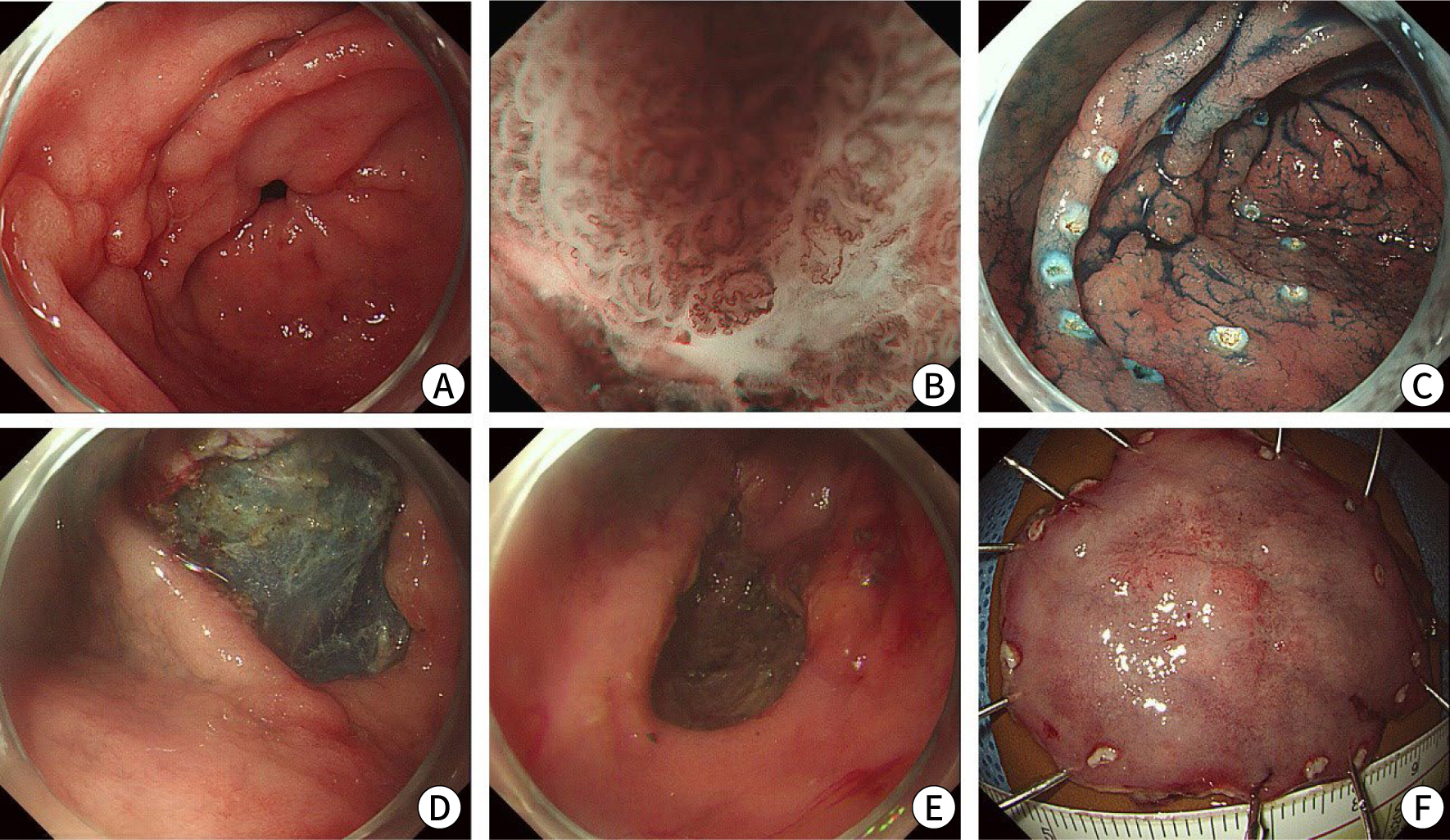
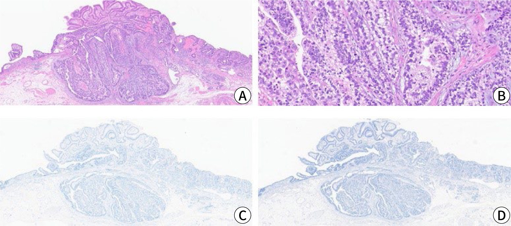
- We report a rare case of gastric adenocarcinoma with enteroblastic differentiation (GAED) that was treated with endoscopic submucosal dissection followed by additional distal gastrectomy with lymph node dissection. A 67-year-old man underwent endoscopic submucosal dissection for a gastric lesion, which was diagnosed as GAED with submucosal and lymphatic invasion. Histologically, GAED is characterized by a tubulopapillary growth pattern and clear cells that resemble those of the primitive fetal gut. Immunohistochemically, GAED variably expresses oncofetal proteins such as glypican-3, alpha-fetoprotein, and spalt-like transcription factor 4. Despite negative margins, additional gastrectomy with lymph node dissection was performed due to submucosal and lymphatic invasion. No residual tumor or metastasis was detected, and the patient remained disease-free for 2 years before dying from causes unrelated to GAED. Given its aggressive nature, frequent lymphovascular invasion, and high metastatic potential, clinicians should recognize the histopathological diagnosis of this rare tumor and its propensity for aggressiveness.
Keyword
Figure
Reference
-
References
1. Kumar S, Jabbar K. Gastric adenocarcinoma with enteroblastic differentiation: a rare find. Am J Clin Pathol. 2020; 154(Suppl 1):S65. DOI: 10.1093/ajcp/aqaa161.141.2. Murakami T, Yao T, Mitomi H, Morimoto T, Ueyama H, Matsumoto K, et al. Clinicopathologic and immunohistochemical characteristics of gastric adenocarcinoma with enteroblastic differentiation: a study of 29 cases. Gastric Cancer. 2016; 19(2):498–507. DOI: 10.1007/s10120-015-0497-9. PMID: 25893262.3. Dias E, Santos-Antunes J, Nunes AC, Rodrigues JA, Pinheiro J, Macedo G. Gastric adenocarcinoma with enteroblastic differentiation: an unexpected cause of upper gastrointestinal bleeding. Acta Gastroenterol Belg. 2021; 84(4):678–679. DOI: 10.51821/84.4.022. PMID: 34965054.4. Abada E, Anaya IC, Abada O, Lebbos A, Beydoun R. Colorectal adenocarcinoma with enteroblastic differentiation: diagnostic challenges of a rare case encountered in clinical practice. J Pathol Transl Med. 2022; 56(2):97–102. DOI: 10.4132/jptm.2021.10.28. PMID: 35051325. PMCID: PMC8935001.5. Afshar Ghotli Z, Serra S, Chetty R. Clear cell (glycogen rich) gastric adenocarcinoma: a distinct tubulo‐papillary variant with a predilection for the cardia/gastro‐oesophageal region. Pathology. 2007; 39(5):466–469. DOI: 10.1080/00313020701569972. PMID: 17886094.6. Govender D, Ramdial PK, Clarke B, Chetty R. Clear cell (glycogen-rich) gastric adenocarcinoma. Ann Diagn Pathol. 2004; 8(2):69–73. DOI: 10.1053/j.anndiagpath.2004.01.002. PMID: 15060883.7. Kwon MJ, Byeon S, Kang SY, Kim KM. Gastric adenocarcinoma with enteroblastic differentiation should be differentiated from hepatoid adenocarcinoma: a study with emphasis on clear cells and clinicopathologic spectrum. Pathol Res Pract. 2019; 215(9):152525. DOI: 10.1016/j.prp.2019.152525. PMID: 31301878.8. Ishikawa A, Nakamura K. Gastric adenocarcinoma with enteroblastic differentiation resected through endoscopic submucosal dissection: a case report. Case Rep Gastroenterol. 2024; 18(1):68–73. DOI: 10.1159/000535954. PMID: 38333765. PMCID: PMC10852983.9. Kato T, Hikichi T, Nakamura J, Takasumi M, Hashimoto M, Kobashi R, et al. Two cases of gastric adenocarcinoma with enteroblastic differentiation resected by endoscopic submucosal dissection. Clin J Gastroenterol. 2021; 14(3):736–744. DOI: 10.1007/s12328-021-01356-z. PMID: 33629257.10. Kinjo T, Taniguchi H, Kushima R, Sekine S, Oda I, Saka M, et al. Histologic and immunohistochemical analyses of α-fetoprotein: producing cancer of the stomach. Am J Surg Pathol. 2012; 36(1):56–65. DOI: 10.1097/PAS.0b013e31823aafec. PMID: 22173117.11. Ushiku T, Shinozaki A, Shibahara J, Iwasaki Y, Tateishi Y, Funata N, et al. SALL4 represents fetal gut differentiation of gastric cancer, and is diagnostically useful in distinguishing hepatoid gastric carcinoma from hepatocellular carcinoma. Am J Surg Pathol. 2010; 34(4):533–540. DOI: 10.1097/PAS.0b013e3181d1dcdd. PMID: 20182341.12. Yamauchi N, Watanabe A, Hishinuma M, Ohashi KI, Midorikawa Y, Morishita Y, et al. The glypican 3 oncofetal protein is a promising diagnostic marker for hepatocellular carcinoma. Mod Pathol. 2005; 18(12):1591–1598. DOI: 10.1038/modpathol.3800436. PMID: 15920546.13. Kono K, Amemiya H, Sekikawa T, Iizuka H, Takahashi A, Fujii H, et al. Clinicopathologic features of gastric cancers producing alpha-fetoprotein. Dig Surg. 2002; 19(5):359–365. DOI: 10.1159/000065838. PMID: 12435906.14. Kim JY, Park DY, Kim GH, Jeon TY, Lauwers GY. Does clear cell carcinoma of stomach exist? Clinicopathological and prognostic significance of clear cell changes in gastric adenocarcinomas. Histopathology. 2014; 65(1):90–99. DOI: 10.1111/his.12372. PMID: 25032253.
- Full Text Links
- Actions
-
Cited
- CITED
-
- Close
- Share
- Similar articles
-
- Gastric Cancer Showing Rapid Recurrence and Progression: A Case of Gastric Adenocarcinoma With Enteroblastic Differentiation
- Colorectal adenocarcinoma with enteroblastic differentiation: diagnostic challenges of a rare case encountered in clinical practice
- Gagtric Adenocarcinoma with Choriocarcinomatous and Hepatoid Differentiation: Report of a case
- Gastric-Type Extremely Well-Differentiated Adenocarcinoma of the Stomach: A Challenge for Preoperative Diagnosis
- Gastric Adenocarcinoma with Coexistent Hepatoid Adenocarcinoma and Neuroendocrine Carcinoma: A Case Report


