Cardioprotection via mitochondrial transplantation supports fatty acid metabolism in ischemia-reperfusion injured rat heart
- Affiliations
-
- 1Department of Physiology, College of Medicine, Chung-Ang University, Seoul 06974, Korea
- 2Divsion of Cardiology, Department of Internal Medicine, College of Medicine, Chung-Ang University Hospital, Seoul 06973, Korea
- 3Department of Medicine, College of Medicine, Chung-Ang University, Seoul 06974, Korea
- 4Cardiovascular and Metabolic Disease Center, SMART Marine Therapeutics Center, Inje University, Busan 47392, Korea
- 5Department of Physiology, School of Medicine, Jeju National University, Jeju 63243, Korea
- 6Department of Family Medicine, College of Medicine, Chung-Ang University Hospital, Seoul 06973, Korea
- KMID: 2555663
- DOI: http://doi.org/10.4196/kjpp.2024.28.3.209
Abstract
- In addition to cellular damage, ischemia-reperfusion (IR) injury induces substantial damage to the mitochondria and endoplasmic reticulum. In this study, we sought to determine whether impaired mitochondrial function owing to IR could be restored by transplanting mitochondria into the heart under ex vivo IR states. Additionally, we aimed to provide preliminary results to inform therapeutic options for ischemic heart disease (IHD). Healthy mitochondria isolated from autologous gluteus maximus muscle were transplanted into the hearts of Sprague–Dawley rats damaged by IR using the Langendorff system, and the heart rate and oxygen consumption capacity of the mitochondria were measured to confirm whether heart function was restored. In addition, relative expression levels were measured to identify the genes related to IR injury. Mitochondrial oxygen consumption capacity was found to be lower in the IR group than in the group that underwent mitochondrial transplantation after IR injury (p < 0.05), and the control group showed a tendency toward increased oxygen consumption capacity compared with the IR group. Among the genes related to fatty acid metabolism, Cpt1b (p < 0.05) and Fads1 (p < 0.01) showed significant expression in the following order: IR group, IR + transplantation group, and control group. These results suggest that mitochondrial transplantation protects the heart from IR damage and may be feasible as a therapeutic option for IHD.
Keyword
Figure
-
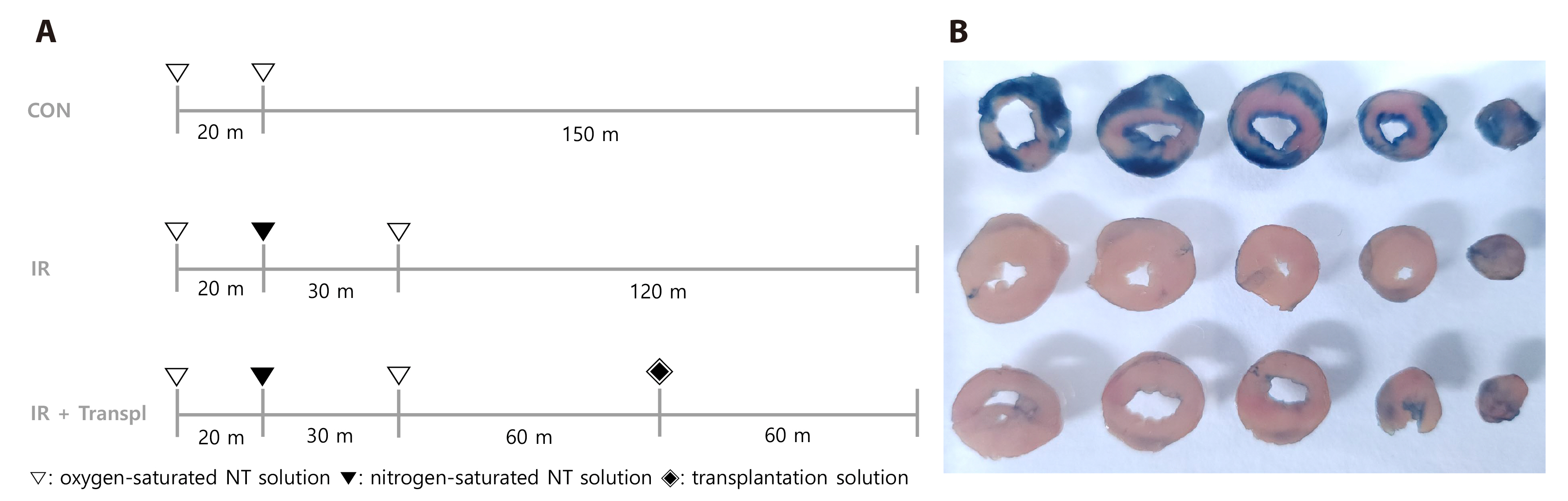
Fig. 1 Observation of non-ischemic area at endpoint of animal experiment. (A) Schematic illustration of experimental protocol. (B) Representative cardiac sections stained with 0.25% Evans blue. The upper panel shows control group cardiac section stained with 0.25% Evans blue. The middle panel shows IR group cardiac section stained with 0.25% Evans blue. The lower shows IR + transpl group cardiac section stained with 0.25% Evans blue. CON (control), group without ischemia or reperfusion injury; IR, group with damage induced by ischemia and reperfusion; IR + transpl, group transplanted with isolated mitochondria after inducing IR damage.
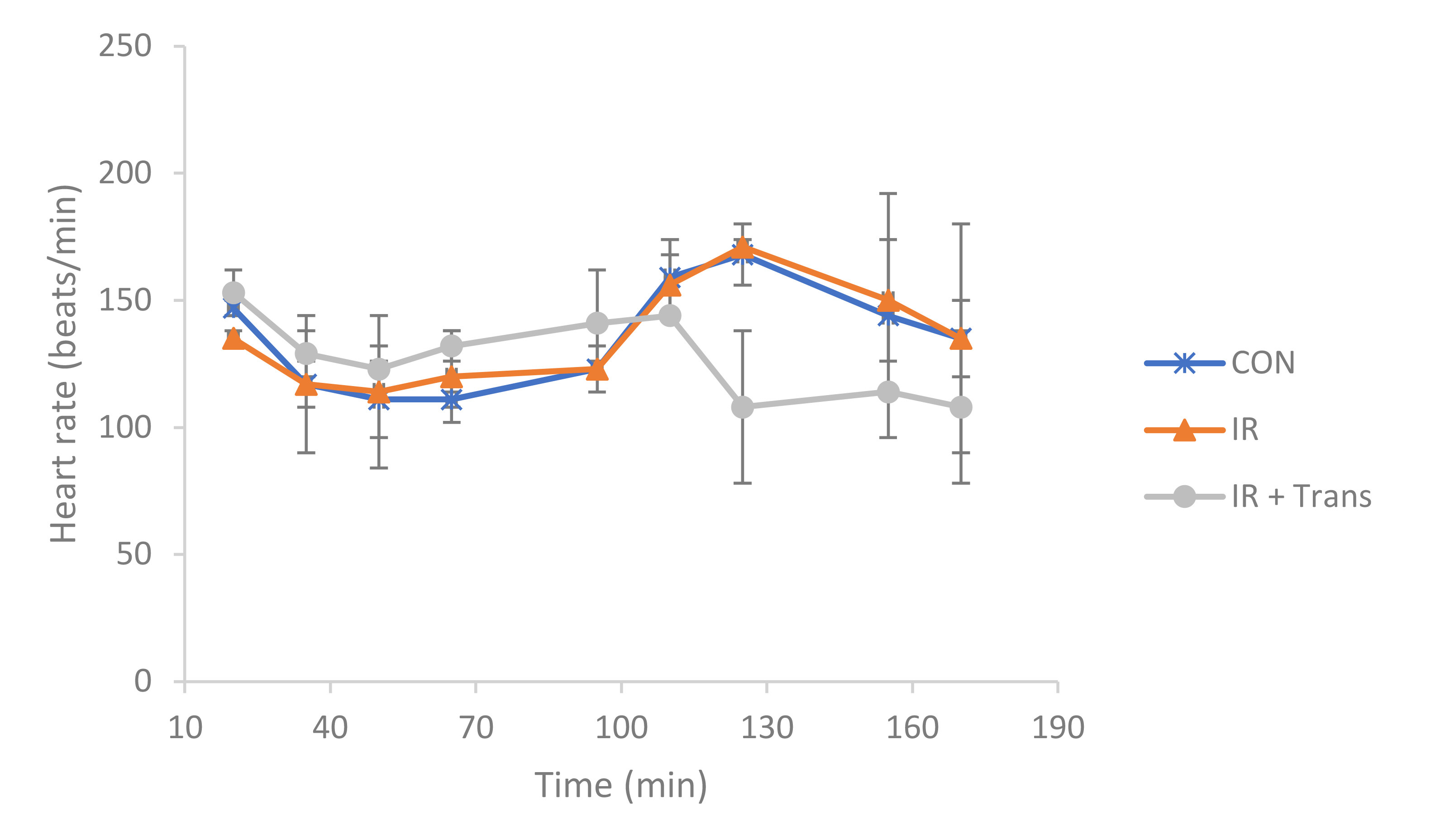
Fig. 2 Changes in heart rate over time by group. All isolated hearts showed normal ex vivo heart rates during the experiment. CON (control), group without ischemia or reperfusion injury; IR, group with damage induced by ischemia and reperfusion; IR + transpl, group transplanted with isolated mitochondria after inducing IR damage.
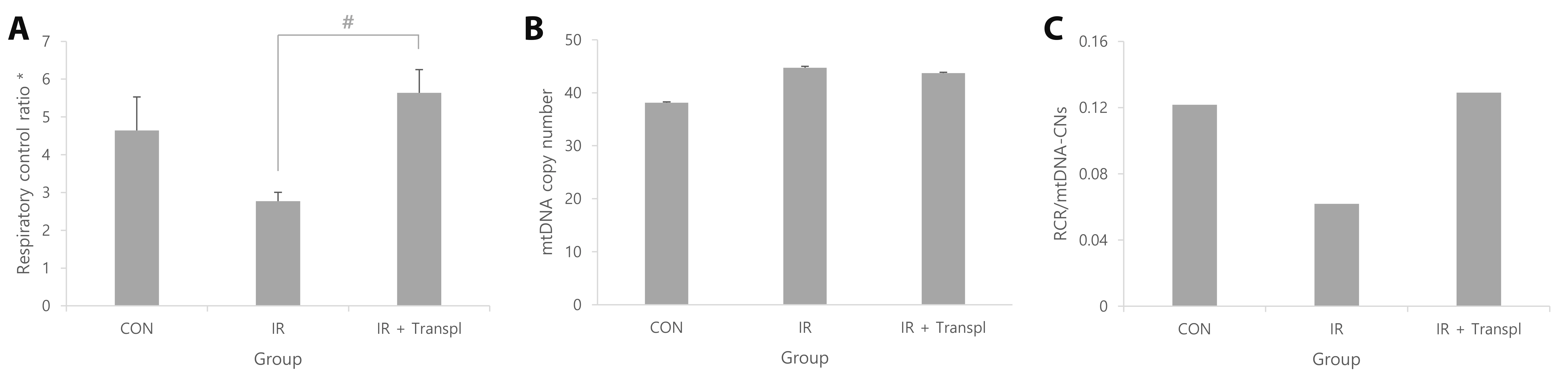
Fig. 3 Mitochondrial function analysis. (A) RCR tracked ATP production by assessing the oxygen consumption capacity of mitochondria. *p < 0.05 indicates that the group means are not all statistically equal. #p < 0.05 denoted a comparison between IR and IR + transpl. (B) mtDNA-DN assessed cellular ATP requirements. (C) Oxygen consumption per unit mitochondria was confirmed. CON (control), group without ischemia or reperfusion injury; IR, injury due to ischemia and reperfusion group; IR + transpl, group transplanted with isolated mitochondria after inducing IR injury; mtDNA-CN, mitochondrial DNA copy number; RCR, respiratory control ratio; ATP, adenosine triphosphate.
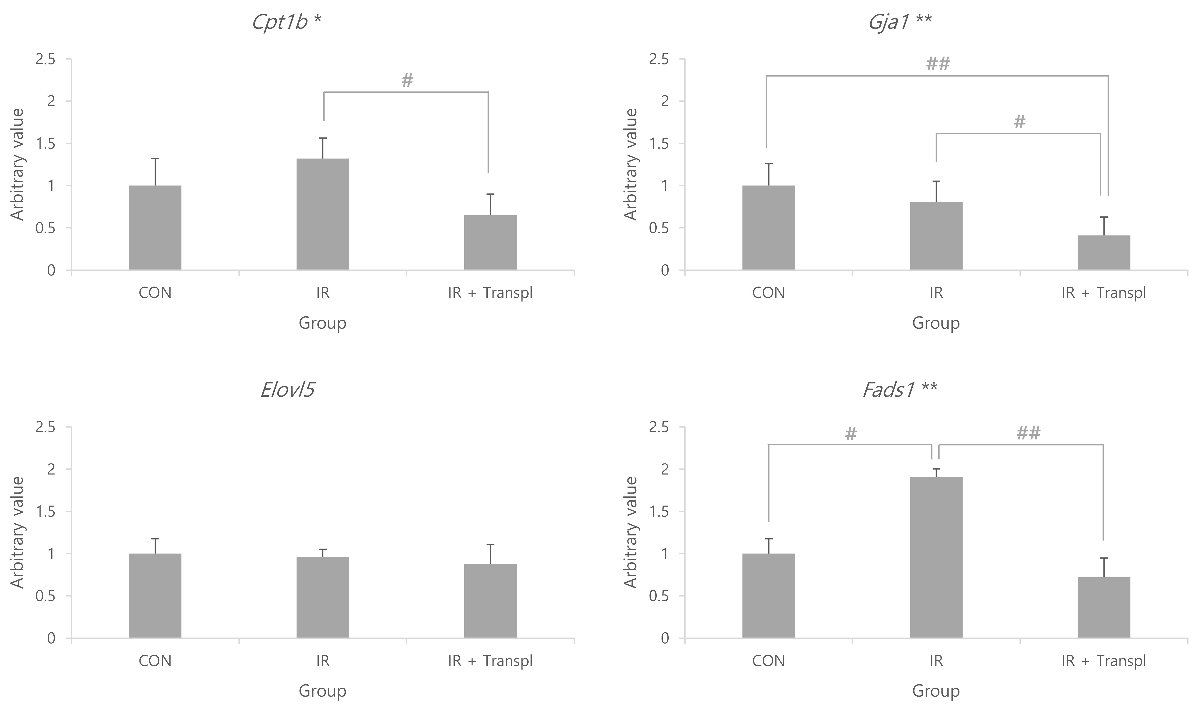
Fig. 4 Relative expression levels of target genes. Comparison of cardiac gene expression levels associated with fatty acid energy metabolism in the mitochondrial transplantation. Statistically significant results indicated that the group means are not equal, with significance levels of *p < 0.05 and **p < 0.01. The gene expression showed statistically significant differences, with #p < 0.05 and ##p < 0.01 indicating levels of significance. CON (control), group without ischemia or reperfusion injury; IR, group with damage induced by ischemia and reperfusion; IR + transpl, group transplanted with isolated mitochondria after inducing IR damage; Cpt1b, carnitine palmitoyltransferase 1B; Elovl5, ELOVL fatty acid elongase 5; Fads1, fatty acid desaturase 1; Gja1, gap function alpha 1.
Reference
-
1. Vedin O, Lam CSP, Koh AS, Benson L, Teng THK, Tay WT, Braun OÖ, Savarese G, Dahlström U, Lund LH. 2017; Significance of ischemic heart disease in patients with heart failure and preserved, midrange, and reduced ejection fraction: a nationwide cohort study. Circ Heart Fail. 10:e003875. DOI: 10.1161/CIRCHEARTFAILURE.117.003875. PMID: 28615366.
Article2. Benjamin EJ, Blaha MJ, Chiuve SE, Cushman M, Das SR, Deo R, de Ferranti SD, Floyd J, Fornage M, Gillespie C, Isasi CR, Jiménez MC, Jordan LC, Judd SE, Lackland D, Lichtman JH, Lisabeth L, Liu S, Longenecker CT, Mackey RH, et al. American Heart Association Statistics Committee and Stroke Statistics Subcommittee. 2017; Heart disease and stroke statistics-2017 update: a report from the American Heart Association. Circulation. 135:e146–e603. Erratum in: Circulation. 2017;135:e646. Erratum in: Circulation. 2017;136:e196.
Article3. Fuhrmann DC, Brüne B. 2017; Mitochondrial composition and function under the control of hypoxia. Redox Biol. 12:208–215. DOI: 10.1016/j.redox.2017.02.012. PMID: 28259101. PMCID: PMC5333533.
Article4. Leist M, Single B, Castoldi AF, Kühnle S, Nicotera P. 1997; Intracellular adenosine triphosphate (ATP) concentration: a switch in the decision between apoptosis and necrosis. J Exp Med. 185:1481–1486. DOI: 10.1084/jem.185.8.1481. PMID: 9126928. PMCID: PMC2196283.
Article5. Lesnefsky EJ, Chen Q, Slabe TJ, Stoll MS, Minkler PE, Hassan MO, Tandler B, Hoppel CL. 2004; Ischemia, rather than reperfusion, inhibits respiration through cytochrome oxidase in the isolated, perfused rabbit heart: role of cardiolipin. Am J Physiol Heart Circ Physiol. 287:H258–H267. DOI: 10.1152/ajpheart.00348.2003. PMID: 14988071.
Article6. Trankle C, Thurber CJ, Toldo S, Abbate A. 2016; Mitochondrial membrane permeability inhibitors in acute myocardial infarction: still awaiting translation. JACC Basic Transl Sci. 1:524–535. DOI: 10.1016/j.jacbts.2016.06.012. PMID: 30167535. PMCID: PMC6113419.
Article7. Zoratti M, Szabò I. 1995; The mitochondrial permeability transition. Biochim Biophys Acta. 1241:139–176. DOI: 10.1016/0304-4157(95)00003-A. PMID: 7640294.
Article8. Nisoli E, Falcone S, Tonello C, Cozzi V, Palomba L, Fiorani M, Pisconti A, Brunelli S, Cardile A, Francolini M, Cantoni O, Carruba MO, Moncada S, Clementi E. 2004; Mitochondrial biogenesis by NO yields functionally active mitochondria in mammals. Proc Natl Acad Sci U S A. 101:16507–16512. Erratum in: Proc Natl Acad Sci U S A. 2005;102:5635. DOI: 10.1073/pnas.0405432101. PMID: 15545607. PMCID: PMC534517.
Article9. Marin W, Marin D, Ao X, Liu Y. 2021; Mitochondria as a therapeutic target for cardiac ischemiareperfusion injury (Review). Int J Mol Med. 47:485–499. DOI: 10.3892/ijmm.2020.4823. PMID: 33416090. PMCID: PMC7797474.
Article10. D'Amato M, Morra F, Di Meo I, Tiranti V. 2023; Mitochondrial transplantation in mitochondrial medicine: current challenges and future perspectives. Int J Mol Sci. 24:1969. DOI: 10.3390/ijms24031969. PMID: 36768312. PMCID: PMC9916997.11. Pacak CA, Preble JM, Kondo H, Seibel P, Levitsky S, Del Nido PJ, Cowan DB, McCully JD. 2015; Actin-dependent mitochondrial internalization in cardiomyocytes: evidence for rescue of mitochondrial function. Biol Open. 4:622–626. DOI: 10.1242/bio.201511478. PMID: 25862247. PMCID: PMC4434813.
Article12. Zimmer HG. 1998; The isolated perfused heart and its pioneers. News Physiol Sci. 13:203–210. DOI: 10.1152/physiologyonline.1998.13.4.203. PMID: 11390791.
Article13. Bell RM, Mocanu MM, Yellon DM. 2011; Retrograde heart perfusion: the Langendorff technique of isolated heart perfusion. J Mol Cell Cardiol. 50:940–950. DOI: 10.1016/j.yjmcc.2011.02.018. PMID: 21385587.
Article14. Lau C, Ranasinghe MG, Shiels I, Keates H, Pasloske K, Bellingham MC. 2013; Plasma pharmacokinetics of alfaxalone after a single intraperitoneal or intravenous injection of Alfaxan(®) in rats. J Vet Pharmacol Ther. 36:516–520. DOI: 10.1111/jvp.12055. PMID: 23600373.
Article15. Kim HK, Kang SW, Jeong SH, Kim N, Ko JH, Bang H, Park WS, Choi TH, Ha YR, Lee YS, Youm JB, Ko KS, Rhee BD, Han J. 2012; Identification of potential target genes of cardioprotection against ischemia-reperfusion injury by express sequence tags analysis in rat hearts. J Cardiol. 60:98–110. DOI: 10.1016/j.jjcc.2012.02.004. PMID: 22512836.
Article16. Frezza C, Cipolat S, Scorrano L. 2007; Organelle isolation: functional mitochondria from mouse liver, muscle and cultured fibroblasts. Nat Protoc. 2:287–295. DOI: 10.1038/nprot.2006.478. PMID: 17406588.
Article17. Suzuki Y, Lyons JK, Yeung AC, Ikeno F. 2008; In vivo porcine model of reperfused myocardial infarction: in situ double staining to measure precise infarct area/area at risk. Catheter Cardiovasc Interv. 71:100–107. DOI: 10.1002/ccd.21329. PMID: 17985383.
Article18. Liu J, Liu C, Chen H, Cen H, Yang H, Liu P, Liu F, Ma L, Chen Q, Wang L. 2023; Tongguan capsule for treating myocardial ischemia-reperfusion injury: integrating network pharmacology and mechanism study. Pharm Biol. 61:437–448. DOI: 10.1080/13880209.2023.2175877. PMID: 36789620. PMCID: PMC9937005.
Article19. Kuznetsov AV, Veksler V, Gellerich FN, Saks V, Margreiter R, Kunz WS. 2008; Analysis of mitochondrial function in situ in permeabilized muscle fibers, tissues and cells. Nat Protoc. 3:965–976. DOI: 10.1038/nprot.2008.61. PMID: 18536644.
Article20. Gilmer LK, Roberts KN, Joy K, Sullivan PG, Scheff SW. 2009; Early mitochondrial dysfunction after cortical contusion injury. J Neurotrauma. 26:1271–1280. DOI: 10.1089/neu.2008.0857. PMID: 19637966. PMCID: PMC2850255.
Article21. Odinokova IV, Shalbuyeva N, Gukovskaya AS, Mareninova OA. 2011; Isolation of pancreatic mitochondria and measurement of their functional parameters. Pancreapedia: Exocrine Pancreas Knowledge Base. doi: 10.3998/panc.2011.25. DOI: 10.3998/panc.2011.25.
Article22. Salin K, Villasevil EM, Anderson GJ, Selman C, Chinopoulos C, Metcalfe NB. 2018; The RCR and ATP/O indices can give contradictory messages about mitochondrial efficiency. Integr Comp Biol. 58:486–494. DOI: 10.1093/icb/icy085. PMID: 29982616.
Article23. Lee D, Kim YW, Kim JH, Yang M, Bae H, Lim I, Bang H, Go KC, Yang GW, Rho YH, Park HS, Park EH, Ko JH. 2015; Improvement characteristics of bio-active materials coated fabric on rat muscular mitochondria. Korean J Physiol Pharmacol. 19:283–289. DOI: 10.4196/kjpp.2015.19.3.283. PMID: 25954135. PMCID: PMC4422970.
Article24. Kong HL, Hou AJ, Liu NN, Chen BH, Dai SN, Huang HT. 2018; The effects of ginsenoside Rb1 on fatty acid β-oxidation, mediated by AMPK, in the failing heart. Iran J Basic Med Sci. 21:731–737.25. Rooney JP, Ryde IT, Sanders LH, Howlett EH, Colton MD, Germ KE, Mayer GD, Greenamyre JT, Meyer JN. 2015; PCR based determination of mitochondrial DNA copy number in multiple species. Methods Mol Biol. 1241:23–38. DOI: 10.1007/978-1-4939-1875-1_3. PMID: 25308485. PMCID: PMC4312664.
Article26. Rao X, Huang X, Zhou Z, Lin X. 2013; An improvement of the 2ˆ(-delta delta CT) method for quantitative real-time polymerase chain reaction data analysis. Biostat Bioinforma Biomath. 3:71–85.27. R Core Team. 2023. R: a language and environment for statistical computing. R Foundation for Statistical Computing.28. Butova XA, Myachina TA, Khokhlova AD. 2020; A combined Langendorff-injection technique for simultaneous isolation of single cardiomyocytes from atria and ventricles of the rat heart. MethodsX. 8:101189. DOI: 10.1016/j.mex.2020.101189. PMID: 33376680. PMCID: PMC7758550.
Article29. Lee D, Seo Y, Kim YW, Kim S, Bae H, Choi J, Lim I, Bang H, Kim JH, Ko JH. 2019; Far-infrared radiation stimulates platelet-derived growth factor mediated skeletal muscle cell migration through extracellular matrix-integrin signaling. Korean J Physiol Pharmacol. 23:141–150. DOI: 10.4196/kjpp.2019.23.2.141. PMID: 30820158. PMCID: PMC6384197.
Article30. Kanehisa M, Goto S. 2000; KEGG: kyoto encyclopedia of genes and genomes. Nucleic Acids Res. 28:27–30. DOI: 10.1093/nar/28.1.27. PMID: 10592173. PMCID: PMC102409.
Article31. Kaza AK, Wamala I, Friehs I, Kuebler JD, Rathod RH, Berra I, Ericsson M, Yao R, Thedsanamoorthy JK, Zurakowski D, Levitsky S, Del Nido PJ, Cowan DB, McCully JD. 2017; Myocardial rescue with autologous mitochondrial transplantation in a porcine model of ischemia/reperfusion. J Thorac Cardiovasc Surg. 153:934–943. DOI: 10.1016/j.jtcvs.2016.10.077. PMID: 27938904.
Article32. Cowan DB, Yao R, Akurathi V, Snay ER, Thedsanamoorthy JK, Zurakowski D, Ericsson M, Friehs I, Wu Y, Levitsky S, Del Nido PJ, Packard AB, McCully JD. 2016; Intracoronary delivery of mitochondria to the ischemic heart for cardioprotection. PLoS One. 11:e0160889. DOI: 10.1371/journal.pone.0160889. PMID: 27500955. PMCID: PMC4976938.
Article33. Masuzawa A, Black KM, Pacak CA, Ericsson M, Barnett RJ, Drumm C, Seth P, Bloch DB, Levitsky S, Cowan DB, McCully JD. 2013; Transplantation of autologously derived mitochondria protects the heart from ischemia-reperfusion injury. Am J Physiol Heart Circ Physiol. 304:H966–H982. DOI: 10.1152/ajpheart.00883.2012. PMID: 23355340. PMCID: PMC3625892.
Article34. Cowan DB, Yao R, Thedsanamoorthy JK, Zurakowski D, Del Nido PJ, McCully JD. 2017; Transit and integration of extracellular mitochondria in human heart cells. Sci Rep. 7:17450. DOI: 10.1038/s41598-017-17813-0. PMID: 29234096. PMCID: PMC5727261.
Article35. Gambardella J, Lombardi A, Santulli G. 2020; Metabolic flexibility of mitochondria plays a key role in balancing glucose and fatty acid metabolism in the diabetic heart. Diabetes. 69:2054–2057. DOI: 10.2337/dbi20-0024. PMID: 32958606. PMCID: PMC7506829.
Article36. Warfel JD, Bermudez EM, Mendoza TM, Ghosh S, Zhang J, Elks CM, Mynatt R, Vandanmagsar B. 2016; Mitochondrial fat oxidation is essential for lipid-induced inflammation in skeletal muscle in mice. Sci Rep. 6:37941. DOI: 10.1038/srep37941. PMID: 27892502. PMCID: PMC5124994.
Article37. Tang L, Li J, Fu W, Wu W, Xu J. 2019; Suppression of FADS1 induces ROS generation, cell cycle arrest, and apoptosis in melanocytes: implications for vitiligo. Aging (Albany NY). 11:11829–11843. DOI: 10.18632/aging.102452. PMID: 31866583. PMCID: PMC6949104.
Article38. Basheer WA, Fu Y, Shimura D, Xiao S, Agvanian S, Hernandez DM, Hitzeman TC, Hong T, Shaw RM. 2018; Stress response protein GJA1-20k promotes mitochondrial biogenesis, metabolic quiescence, and cardioprotection against ischemia/reperfusion injury. JCI Insight. 3:e121900. DOI: 10.1172/jci.insight.121900. PMID: 30333316. PMCID: PMC6237442.
Article
- Full Text Links
-
- Actions
-
Cited
- CITED
-
- Close
- Share
-
- Similar articles
-
- Mitochondrial Dynamics in the Heart as a Novel Therapeutic Target for Cardioprotection
- Cardioprotection and ageing
- Evaluation of bioenergetic and mitochondrial function in liver transplantation
- Polyphenol (-)-Epigallocatechin Gallate during Ischemia Limits Infarct Size Via Mitochondrial K(ATP) Channel Activation in Isolated Rat Hearts
- Morphine and remifentanil-induced cardioprotection: its experimental and clinical outcomes
