Bisphosphonate’s effect on the tongue in adult male albino rats and the possible protective role of rutin: light and scanning electron microscopic study
- Affiliations
-
- 1Department of Histology and Cell Biology, Faculty of Medicine, Menoufia University, Al Menoufia, Egypt
- KMID: 2554247
- DOI: http://doi.org/10.5115/acb.23.230
Abstract
- Alendronate sodium (ALS) is a nitrogen-containing bisphosphonate used for the treatment of different bone disorders. However, its adverse effect on oral soft tissue has been detected. Rutin (RUT) is natural flavonoid with antioxidant and anti-inflammatory properties. This work aimed to investigate the possible effect of ALS on the tongue of adult male albino rats and to evaluate the possible protective role of RUT. Forty adult male albino rats were equally divided into four groups: group I (control), group II (RUT): Received RUT 50 mg/kg, group III (ALS): Received ALS 1 mg/kg, group IV (ALS+RUT): Received ALS and RUT with the same doses as pervious groups. The drugs were given once daily for 5 weeks. Tongue specimens were taken and processed for light and scanning electron microscopic inspection. ALS treated group revealed structural changes in the tongue in the form of decrease in the height of the filiform papillae with blunt ends, marked atrophy in some papillae with areas of focal loss, loss of some epithelial cells, pyknotic nuclei and cytoplasmic vacuoles in some epithelial cells. The lamina propria showed inflammatory cellular infiltration with congested blood vessels. Statistically, there were highly significant decrease in the number of proliferating cell nuclear antigen immunopositive cells, area percentage of Bcl-2 immunoexpression and highly significant increase in the collagen content compared to control group. Administration of RUT with ALS minimizes these changes. RUT protected the rat tongue against the histological and immunohistochemical changes induced by ALS through its antioxidant and anti-inflammatory properties.
Keyword
Figure
-

Fig. 1 A photomicrograph of H&E-stained sections of the anterior 2/3 of rat’s tongue from control group (I). (A) The dorsal surface showing regular orientation of numerous filiform papillae with tapering ends (arrows). Skeletal muscle fibers appear in different directions; some of them appear longitudinal (L), others appear polygonal (P). (B) A higher magnification of (panel A) showing each papilla (arrows) formed of a connective tissue (CT) core (*) with small blood vessels (Bv, blue arrow) and is covered by a keratinized stratified squamous epithelium (E) resting on a basement membrane with many epithelial ridges (arrowhead). The CT cores are continuous with the lamina propria (LP). Muscle fibers appear polygonal (P) with acidophilic cytoplasm and peripherally positioned nuclei. (C) A higher magnification of (panel B) showing the keratinized stratified squamous epithelium (E) with its basal cell layer (B), spinous cell layer (S), granulosum cell layer (G) and superficial corneum layer formed of keratin (K). (D) The dorsal surface showing fungiform papillae (arrowheads) in between the filiform ones (arrows). It has CT core (*) with small Bv and an E cover. (E) The ventral surface showing a smooth surface without papillae. It shows a thin keratin layer (arrows) over the stratified squamous E cover and the underlying LP (*). H&E stained, (A) ×100, (B, E) ×200, (C, D) ×400.

Fig. 2 A photomicrograph of H&E-stained sections of the anterior 2/3 of rat’s tongue from alendronate sodium group (III). (A) The dorsal surface showing an apparent decrease in the height of the filiform papillae with blunt ends (arrows). Focal loss of epithelial cells, connective tissue (CT) core and lamina propria is observed (*). Disorganized epithelial cells (arrowhead) are observed. Separated parts of keratin layer (K). (B) A higher magnification of (panel A) showing disorganized epithelial cells (arrowheads). Focal loss of epithelial cells is observed (*). Some epithelial cells appear with deeply stained pyknotic nuclei (arrows). (C) The dorsal surface showing an apparent decrease in the height of some filiform papillae with blunt ends (black arrow). Focal loss of epithelial cells is observed (*). The underlying CT core and lamina propria show inflammatory cellular infiltration (I, blue arrows). Disrupted muscle fibers (arrowheads). (D) The dorsal surface showing focal loss of epithelial cells (*), pyknotic nuclei and vacuoles of some epithelial cells (arrowheads). Mitotic activity is obvious in some epithelial cells (black arrow). Many basophilic granules were seen in the superficial cells (blue arrows). Congested blood vessel (Bv) in CT core. (E) The dorsal surface showing a fungiform papilla with pyknotic nuclei and vacuoles of some of its epithelial cell cover (arrows). The taste bud shows pyknotic nuclei in some cells (arrowhead). The CT core shows dilated congested Bv. (F) The ventral surface showing separation of the K layer from the underlying epithelium (E, arrow). The lamina propria shows dilated congested Bv. Disrupted muscles fibers (arrowheads). (G) The ventral surface showing areas of thinned and thickened stratified E. Vacuolated epithelial cells with pyknotic nuclei are observed (arrows). The lamina propria shows inflammatory cell (I) with elongated dilated congested Bv. (H) Showing disruption in the muscle fibers (arrows). Some muscle fibers appear hypereosinophilic (*). Wide spaces (S) between the muscles. H&E stained, (F) ×100, (A, C, G, H) ×200, (B, D, E) ×400.

Fig. 3 A photomicrograph of H&E-stained sections of the anterior 2/3 of rat’s tongue from alendronate sodium+rutin group (IV). (A) The dorsal surface showing regular orientation of the lingual papillae. Most of filiform papillae are long with pointed tips and covered by stratified squamous keratinized epithelium (arrows), few papillae appear disfigured (arrowheads). Dilated congested blood vessels (Bv) are seen in the connective tissue (CT) core and lamina propria. Normal appearance of muscles (M) that run in different directions. (B) A higher magnification of (panel A) showing the keratinized stratified squamous epithelium with most of the cells appearing normal with vesicular nuclei (black arrows), few ones appear with pyknotic nuclei (orange arrows). One papilla appears disfigured (arrowhead). Dilated congested Bv in the CT core and lamina propria. (C) The dorsal surface showing normal appearance of the fungiform papilla (arrow). Some pyknotic nuclei are seen (arrowhead). (D) The ventral surface showing a thin keratin layer (arrow) over the stratified squamous epithelial cover (E). Some pyknotic nuclei are seen (arrowhead). Most of the muscle (M) fibers show normal appearance. H&E stained, (A, D) ×200, (B, C) ×400.

Fig. 4 A photomicrograph of Masson’s trichrome-stained sections of the dorsal surface of the anterior 2/3 of the rat’s tongue from control and treated groups. (A) Control group (I) showing normal distribution of regularly arranged collagen fibers (green color) in the connective tissue (CT) core, lamina propria (*) and thin rims in between the muscle fibers (arrows). (B) Alendronate sodium (ALS) group (III) showing marked increase in the amount of the collagen fibers which appear disorganized and wavy in the CT core, lamina propria (*) and in between the muscle fibers (arrows). (C) ALS+rutin group (IV) showing moderate increase in the amount of the collagen fibers in the CT core, lamina propria (*) with normal distribution of collagen fibers appearing as thin rims in between the muscle fibers (arrows). Masson’s trichrome, (A–C) ×100.

Fig. 5 A photomicrograph of proliferating cell nuclear antigen (PCNA)-stained sections of the dorsal surface of the anterior 2/3 of the rat’s tongue from control and treated groups. (A) Control group (I) showing strong positive brown nuclear immunoreaction for PCNA in numerous epithelial cells in the basal and suprabasal layers of the epithelium (arrows). (B) Alendronate sodium (ALS) group (III) showing weak positive nuclear immunoreaction for PCNA in few epithelial cells mainly in the basal layer of the epithelium (arrows). (C) ALS+rutin group (IV) showing strong positive nuclear immunoreaction for PCNA in many epithelial cells in the basal and suprabasal layers of the epithelium (arrows). PCNA stained, (A–C) ×400.

Fig. 6 A photomicrograph of Bcl-2-stained sections of the dorsal surface of the anterior 2/3 of the rat’s tongue from control and treated groups. (A) Control group (I) showing strong positive cytoplasmic immunoexpression to Bcl-2 in numerous epithelial cells (arrows). (B) Alendronate sodium (ALS) group (III) showing negative cytoplasmic immunoexpression to Bcl-2 in the epithelial cells. (C) ALS+rutin group (IV) showing moderate positive cytoplasmic immunoexpression to Bcl-2 in many epithelial cells (arrows). Bcl2 stained, (A–C) ×400.
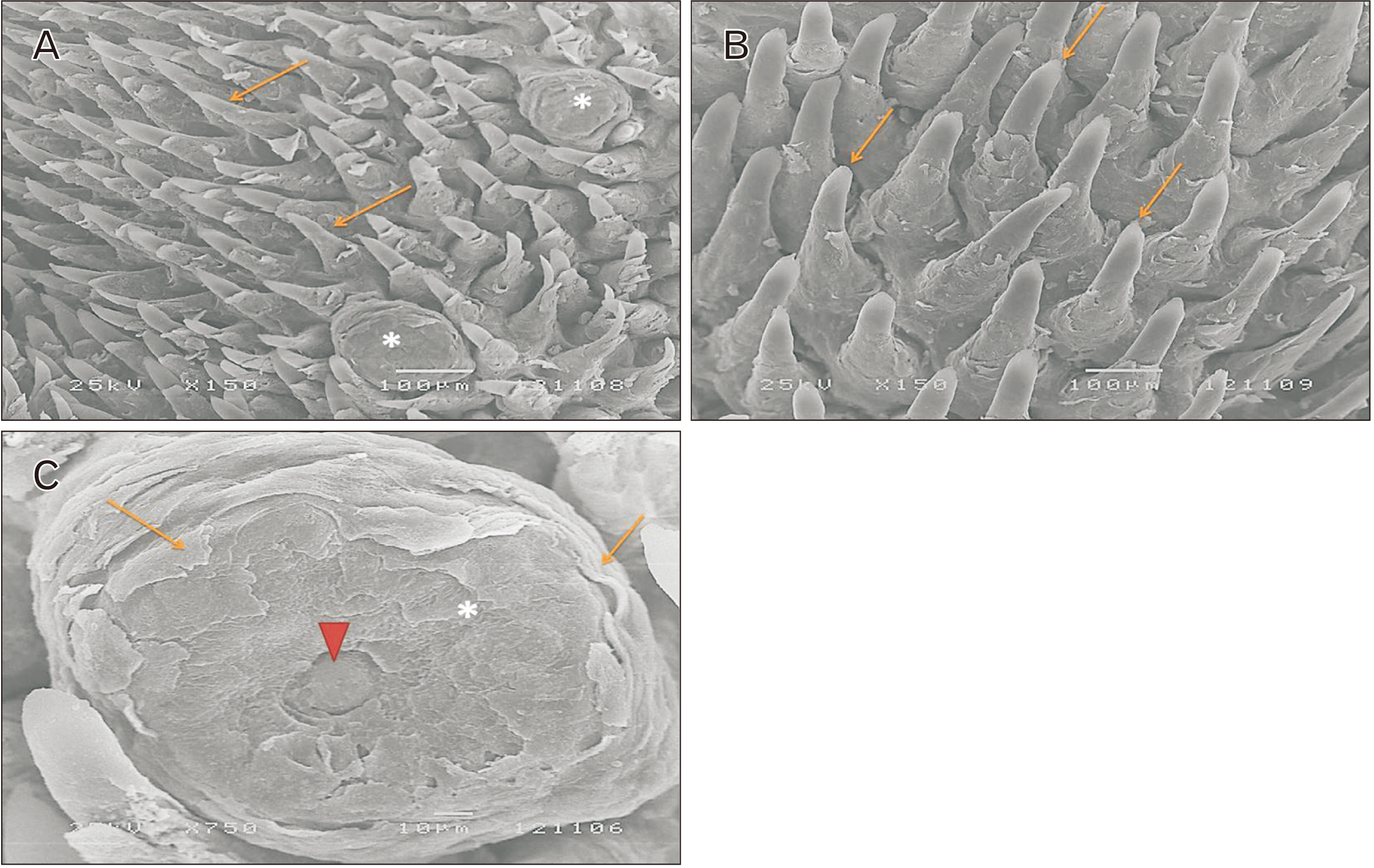
Fig. 7 A scanning electron micrograph of the dorsal surface of the anterior 2/3 of the rat’s tongue from control group (I). (A) Showing numerous filiform papillae (arrows) with scattered fungiform ones in between (*). (B) Showing numerous regularly arranged conical shaped filiform papillae with tapering tips that pointed into the same direction (arrows). (C) Showing a broad dome shaped fungiform papilla (*) with circular keratin packs (arrows) and centrally located well-defined taste pore (arrowhead). (A, B) ×150, (C) ×750.
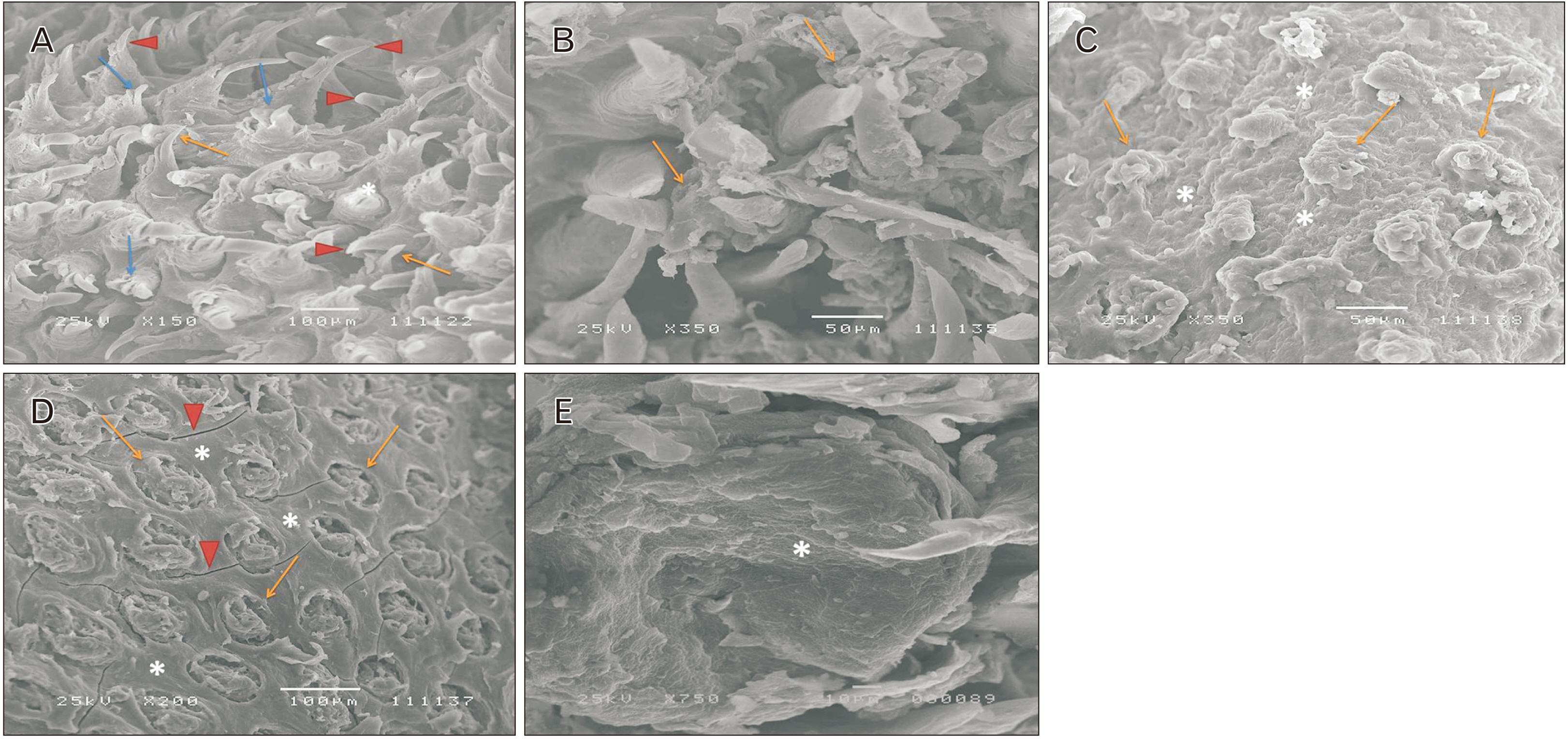
Fig. 8 A scanning electron micrograph of the dorsal surface of the anterior 2/3 of the rat’s tongue from alendronate sodium group (III). (A) Showing filiform papillae oriented in different directions (arrowheads). Some of papillae appear thin and atrophied (orange arrows). Others appear short and destructed (blue arrows). Less prominent and distorted fungiform papilla (*) in between filiform papillae. (B) Showing markedly destructed filiform papillae (arrows). (C) Showing disfigured filiform papillae which appear short with blunt ends (arrows). The surface of papillae appears irregular. Filiform papillae are widely separated with areas of focal loss (*). (D) Showing markedly atrophied filiform papillae (arrows) which appear widely separated with areas of focal loss (*). Fissures (arrowheads) on the dorsal mucosal surface in between the atrophied papillae. (E) Showing distorted fungiform papilla (*) with ill-defined taste pore. (A) ×150, (B, C) ×350, (D) ×200, (E) ×750.
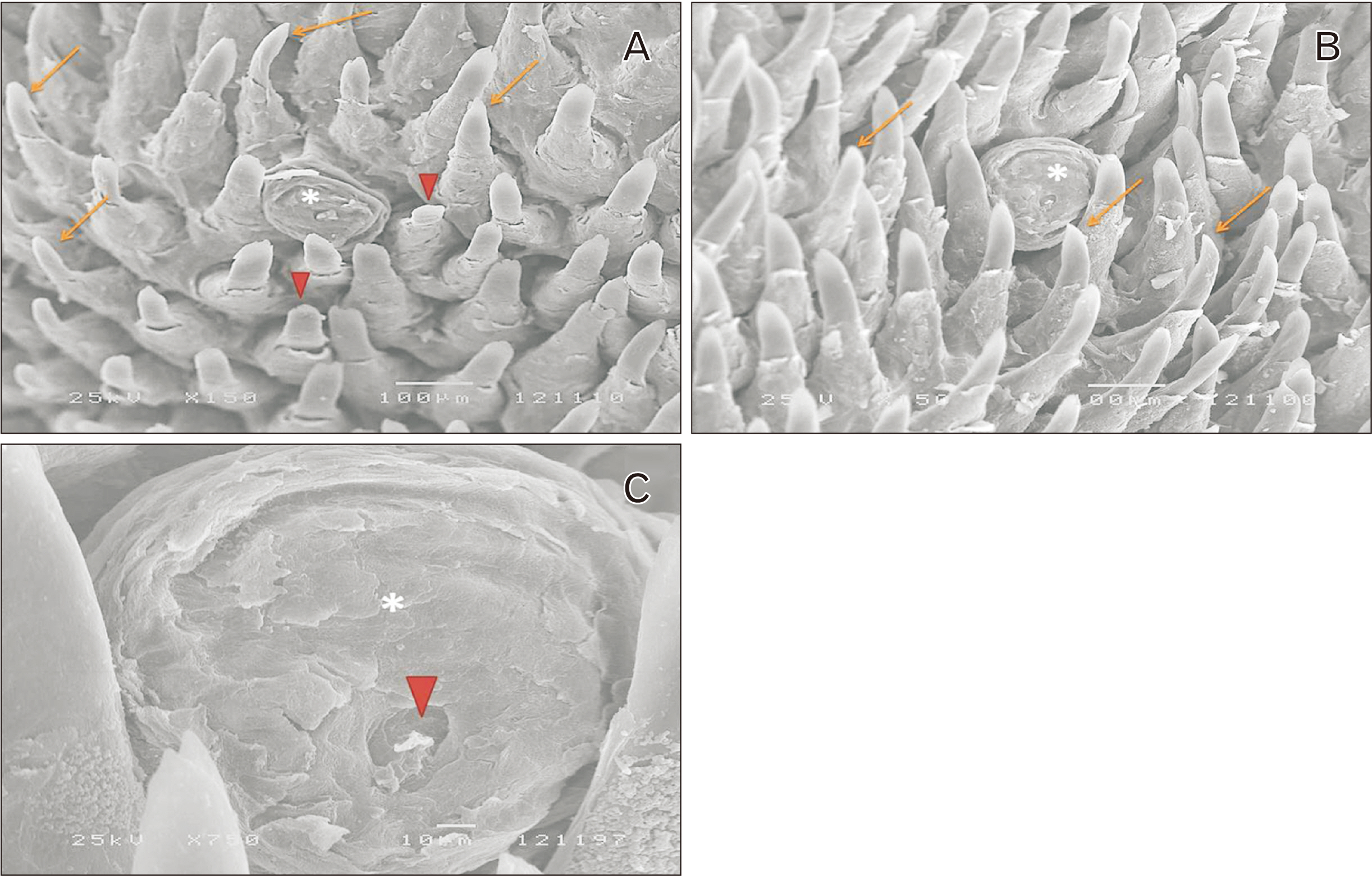
Fig. 9 A scanning electron micrograph of the dorsal surface of the anterior 2/3 of the rat’s tongue from alendronate sodium+rutin group (IV). (A) Showing conical shaped filiform papillae with tapering tips (arrows). Few of them appear short with blunt ends (arrowheads). Fungiform papilla (*) interposed in between filiform papillae. (B) Showing long conical shaped filiform papillae with tapering tips that pointed into the same direction (arrows). Fungiform papilla (*) interposed in between filiform papillae. (C) Showing normal fungiform papilla (*) with centrally located well-defined taste pore (arrowhead). (A, B) ×150, (C) ×750.
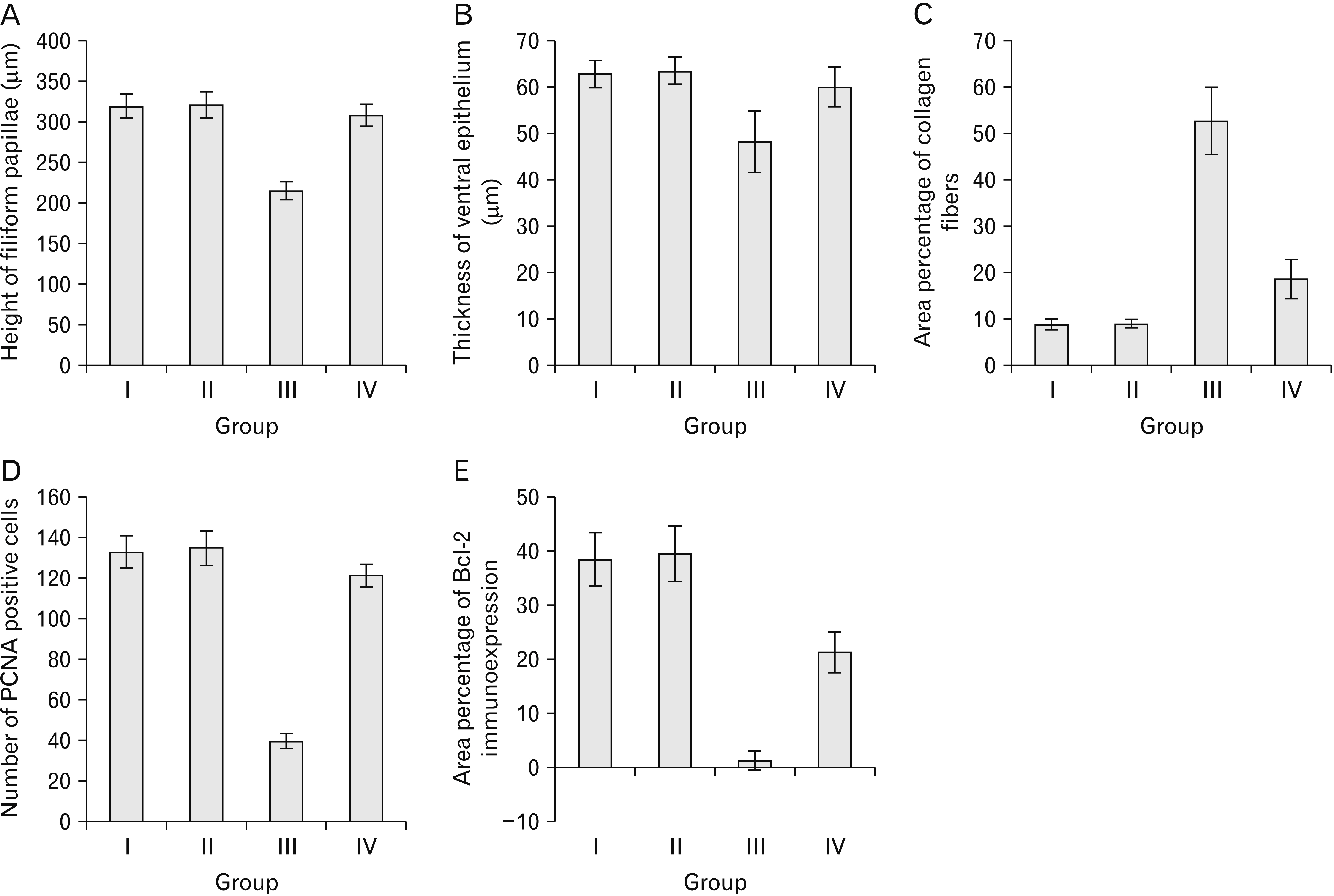
Fig. 10 Morphometric and statistical results in the control and experimental groups showing. (A) The mean height of filiform papillae (μm). (B) The mean thickness of ventral epithelium (μm). (C) The mean area percentage of collagen fibers. (D) The mean number of proliferating cell nuclear antigen positive cells. (E) The mean area percentage of Bcl-2 immunoexpression.
Reference
-
References
1. Zandi M, Dehghan A, Talimkhani I, Rezaeian L, Mohammad Gholi Mezerji N. 2019; Histological evaluation of the healing process of autografted mandibular bone defects in rats under treatment with zoledronate. J Craniomaxillofac Surg. 47:1779–86. DOI: 10.1016/j.jcms.2019.08.003. PMID: 31635981.
Article2. Oyhanart SR, Escudero ND, Mandalunis PM. 2015; Effect of alendronate on the mandible and long bones: an experimental study in vivo. Pediatr Res. 78:618–25. DOI: 10.1038/pr.2015.163. PMID: 26331769.
Article3. Psimma C, Psimma Z, Willems HC, Klüter WJ, van der Maarel-Wierink CD. 2022; Oral bisphosphonates: adverse effects on the oral mucosa not related to the jaw bones. A scoping review. Gerodontology. 39:330–8. DOI: 10.1111/ger.12590. PMID: 34725854. PMCID: PMC9787882.
Article4. Farag DB, Mehanny SS. 2020; Histopathological alterations of the intrinsic tongue muscles following zoledronic acid treatment in a rat model. Dent Med Probl. 57:131–6. DOI: 10.17219/dmp/115368. PMID: 32463600.
Article5. Badae NM, Ghazala RA, Zaki EI, Abdel-Ghani SA. 2019; Evaluating the therapeutic effect of Diallyl disulfide compared to that of Alderonate on Glucocorticoids induced osteoporosis in rats: Biochemical and histomorphometric analysis. Bull Egypt Soc Physiol Sci. 39:129–42. DOI: 10.21608/besps.2019.6704.1011.
Article6. Tokmak Özşahin ET, Çam B, Dere F, Kürkçü M, Evrüke C, Soames R, Oğuz Ö. 2017; The effect of alendronate sodium on trabecular bone structure in an osteoporotic rat model. Turk J Phys Med Rehabil. 63:165–73. DOI: 10.5606/tftrd.2017.164. PMID: 31453446. PMCID: PMC6648129.
Article7. Papamitsou T, Morsi-Yeroyannis A, Papanastasiou A, Bakalopoulos N, Dietrich EM, Karachrysafi S, Toskas A, Mareti E, Morsi-Yeroyanni A, Sioga A. 2020; Bisphosphonate's effect on tongue mucosa: an experimental electron microscopy study. Medicina (Kaunas). 56:51. DOI: 10.3390/medicina56020051. PMID: 31991568. PMCID: PMC7073723.
Article8. Donetti E, Gualerzi A, Sardella A, Lodi G, Carrassi A, Sforza C. 2014; Alendronate impairs epithelial adhesion, differentiation and proliferation in human oral mucosa. Oral Dis. 20:466–72. DOI: 10.1111/odi.12154. PMID: 23837876.
Article9. Theodora P, Stella F, Angeliki P, Eva-Maria D, Dimitris K, Alexandros T, Sofia K, Antonia S. 2018; Effect of alendronic acid on buccal mucosa. J Dent Oral Health. 4:1–6.10. Erdogan E, Ilgaz Y, Gurgor PN, Oztas Y, Topal T, Oztas E. 2015; Rutin ameliorates methotrexate induced hepatic injury in rats. Acta Cir Bras. 30:778–84. DOI: 10.1590/S0102-865020150110000009. PMID: 26647798.
Article11. Al-Hamadawi HA, Al-Ankoshy AAM, Alqershi KA. 2022; Studying the protective and therapeutic role of Rutin on the histological structure of the liver and some physiological parameters in Rats treated with ciprofloxacin. BNIHS. 140:1113–22.12. Hussani MF, Sabour AN. 2022; Effect of rutin on the histological structure of the heart and some biochemical indicators of oxidative stress in male Albino rats. PJMHS. 16:653–7. DOI: 10.53350/pjmhs22167653.
Article13. Thandavamoorthy P, Balan R, Subramaniyan J, Arumugam M, John B, Krishnan G, Ramasamy E, Mani GK, Rajendran R, Thiruvengadam D. 2014; Alleviative role of rutin against 4-Nitroquinoline-1-oxide (4-NQO) provoked oral squamous cell carcinoma in experimental animal model. J Pharm Res. 8:899–906.14. Motamedshariaty VS, Amel Farzad S, Nassiri-Asl M, Hosseinzadeh H. 2014; Effects of rutin on acrylamide-induced neurotoxicity. Daru. 22:27. DOI: 10.1186/2008-2231-22-27. PMID: 24524427. PMCID: PMC3927829.
Article15. Gaertner D, Hallman T, Claire Hankenson F, Batchelder MA. Fish RE, Brown MJ, Danneman PJ, Karas AZ, editors. Anesthesia and analgesia for laboratory rodents. Anesthesia and Analgesia in Laboratory Animals. 2nd ed. Academic Press;2008. p. 239–97. DOI: 10.1016/B978-012373898-1.50014-0.
Article16. Kiernan JA. Histological and histochemical methods: theory and practice. 5th ed. Scion;2015. p. 238–310.17. Ramos-Vara JA, Kiupel M, Baszler T, Bliven L, Brodersen B, Chelack B, Czub S, Del Piero F, Dial S, Ehrhart EJ, Graham T, Manning L, Paulsen D, Valli VE, West K. 2008; Suggested guidelines for immunohistochemical techniques in veterinary diagnostic laboratories. J Vet Diagn Invest. 20:393–413. DOI: 10.1177/104063870802000401. PMID: 18599844.
Article18. Ozer H, Yenicesu G, Arici S, Cetin M, Tuncer E, Cetin A. 2012; Immunohistochemistry with apoptotic-antiapoptotic proteins (p53, p21, bax, bcl-2), c-kit, telomerase, and metallothionein as a diagnostic aid in benign, borderline, and malignant serous and mucinous ovarian tumors. Diagn Pathol. 7:124. DOI: 10.1186/1746-1596-7-124. PMID: 22995373. PMCID: PMC3523067.
Article19. Piroeva I, Atanassova-Vladimirova S, Dimowa L, Sbirkova H, Radoslavov G, Hristov P, Shivachev BL. 2013; A simple and rapid scanning electron microscope preparative technique for observation of biological samples: application on bacteria and DNA samples. Bulgarian Chem Commun. 45:510–5.20. White SE, Malley J, Carton L, Dawson B. Basic and clinical biostatistics. 5th ed. McGraw-Hill Companies;2020.21. Fideles LS, de Miranda JAL, Martins CDS, Barbosa MLL, Pimenta HB, Pimentel PVS, Teixeira CS, Scafuri MAS, Façanha SO, Barreto JEF, Carvalho PMM, Scafuri AG, Araújo JL, Rocha JA, Vieira IGP, Ricardo NMPS, da Silva Campelo M, Ribeiro MENP, de Castro Brito GA, Cerqueira GS. 2020; Role of Rutin in 5-fluorouracil-induced intestinal mucositis: prevention of histological damage and reduction of inflammation and oxidative stress. Molecules. 25:2786. DOI: 10.3390/molecules25122786. PMID: 32560278. PMCID: PMC7356626.
Article22. Kharazmi M, Sjöqvist K, Warfvinge G. 2012; Oral ulcers, a little known adverse effect of alendronate: review of the literature. J Oral Maxillofac Surg. 70:830–6. DOI: 10.1016/j.joms.2011.03.046. PMID: 21816532.
Article23. Carvalho NS, Silva MM, Silva RO, Nicolau LA, Araújo TS, Costa DS, Sousa NA, Souza LK, Soares PM, Medeiros JV. 2016; Protective effects of simvastatin against alendronate-induced gastric mucosal injury in rats. Dig Dis Sci. 61:400–9. DOI: 10.1007/s10620-015-3890-7. PMID: 26403426.
Article24. Kassab AA. 2019; Effect of Fosamax on the duodenal mucosa in adult male albino rats and the possible protection by nigella sativa oil: a histological and immunohistochemical study. Egypt J Histol. 42:900–14. DOI: 10.21608/ejh.2019.10902.1105.
Article25. Kassab AA, Moustafa KA, Abd-El-Hafez AA. 2020; The possible protective role of pumpkin seed oil in ameliorating tongue mucosal damage induced by orlistat in adult male albino rats: a light and scanning electron microscopic study. Egypt J Histol. 43:975–87. DOI: 10.21608/ejh.2020.25027.1258.
Article26. Pourgonabadi S, Ghorbani A, Tayarani Najarn Z, Mousavi SH. 2018; In vitro assessment of alendronate toxic and apoptotic effects on human dental pulp stem cells. Iran J Basic Med Sci. 21:905–10. DOI: 10.22038/IJBMS.2018.22877.5816. PMID: 30524690. PMCID: PMC6272077.27. Saso L, Suzen S, Borges F, Csont T. 2020; Chemistry and pharmacology of modulators of oxidative stress. Curr Med Chem. 27:2038–9. DOI: 10.2174/092986732713200425202016. PMID: 32368965.
Article28. Thoma A, Lightfoot AP. 2018; NF-kB and inflammatory cytokine signalling: role in skeletal muscle atrophy. Adv Exp Med Biol. 1088:267–79. DOI: 10.1007/978-981-13-1435-3_12. PMID: 30390256.
Article29. Tian C, Liu X, Chang Y, Wang R, Yang M, Liu M. 2021; Rutin prevents inflammation induced by lipopolysaccharide in RAW 264.7 cells via conquering the TLR4-MyD88-TRAF6-NF-κB signalling pathway. J Pharm Pharmacol. 73:110–7. DOI: 10.1093/jpp/rgaa015. PMID: 33791807.
Article30. Li Y, Qin L, Ying L, Dong H, Wang D. 2019; Rutin prevents retinal ganglion cell death and exerts protective effects by regulating transforming growth factor-β2/Smad2/3Akt/PTEN signaling in experimental rat glaucoma. Trop J Pharm Res. 18:985–93. DOI: 10.4314/tjpr.v18i5.11.
Article31. Bai L, Li A, Gong C, Ning X, Wang Z. 2020; Protective effect of rutin against bleomycin induced lung fibrosis: Involvement of TGF-β1/α-SMA/Col I and III pathway. Biofactors. 46:637–44. DOI: 10.1002/biof.1629. PMID: 32233122.
Article
- Full Text Links
-
- Actions
-
Cited
- CITED
-
- Close
- Share
-
- Similar articles
-
- Studies on intestinal trematodes in Korea XIX. Light and scanning electron microscopy of Fibricola seoulensis collected from albino rats treated with praziquantel
- Scanning electron microscopic study of capillary change in bleomycin-induced pulmonary fibrosis
- Does oral ciprofloxacin affect the structure of thoracic aorta in adult and senile male albino rats? A clue to fluoroquinolones-induced risk of aortic dissection
- Morphological, ultrastructural, and biochemical changes induced by sodium fluoride in the tongue of adult male albino rat and the ameliorative effect of resveratrol
- Morpholgical Study of Korean Pubic Louse , Phthirus pubis ( Linnaeus , 1758 ) by Light and Scanning Electron Microscopy
