Association between occurrence of multiple white and flat elevated gastric lesions and oral proton pump inhibitor intake
- Hasegawa R
 1
1 - Yao K1
- Kanemitsu T1
- Arima H4
- Hirase T2
- Hiratsuka Y1
- Takeda K1
- Imamura K2
- Ohtsu K2
- Ono Y2
- Miyaoka M1
- Hisabe T2
- Ueki T2
- Tanabe H3
- Ohta A3
- Nimura S3
- Affiliations
-
- 1Department of Endoscopy, Fukuoka University Chikushi Hospital, Fukuoka, Japan
- 2Department of Gastroenterology, Fukuoka University Chikushi Hospital, Fukuoka, Japan
- 3Department of Pathology, Fukuoka University Chikushi Hospital, Fukuoka, Japan
- 4Department of Preventive Medicine and Public Health, Faculty of Medicine, Fukuoka University, Fukuoka, Japan
- KMID: 2551194
- DOI: http://doi.org/10.5946/ce.2022.257
Abstract
- Background/Aims
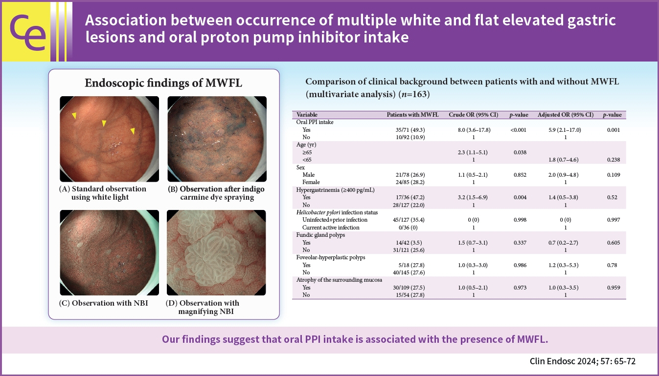
Multiple white and flat elevated lesions (MWFL) that develop from the gastric corpus to the fornix may be strongly associated with oral antacid intake. Therefore, this study aimed to determine the association between the occurrence of MWFL and oral proton pump inhibitor (PPI) intake and clarify the endoscopic and clinicopathological characteristics of MWFL.
Methods
The study included 163 patients. The history of oral drug intake was collected, and serum gastrin levels and anti-Helicobacter pylori immunoglobulin G antibody titers were measured. Upper gastrointestinal endoscopy was performed. The primary study endpoint was the association between MWFL and oral PPI intake.
Results
In the univariate analyses, MWFL were observed in 35 (49.3%) of 71 patients who received oral PPIs and 10 (10.9%) of 92 patients who did not receive oral PPIs. The occurrence of MWFL was significantly higher among patients who received PPIs than in those who did not (p<0.001). Moreover, the occurrence of MWFL was significantly higher in patients with hypergastrinemia (p=0.005). In the multivariate analyses, oral PPI intake was the only significant independent factor associated with the presence of MWFL (p=0.001; odds ratio, 5.78; 95% confidence interval, 2.06–16.2).
Conclusions
Our findings suggest that oral PPI intake is associated with the presence of MWFL (UMINCTR 000030144).
Figure
-

Fig. 1. Endoscopic features of multiple white and flat elevated lesions. (A) Standard observation using white light. The gastric fundus and corpus have numerous flat elevated lesions of various sizes and heights in white tones (arrowheads). (B) Observation following the indigo carmine dye application. Flat protrusions repelling the dye are observed on the surface. (C) Observation without magnification using NBI. The surrounding mucosa is brownish in color with increased contrast to the whitish flat elevated lesions, and the lesions are readily observed compared to the standard observation using white light. (D) Observation with magnification using narrow-band imaging. Demarcation lines are clearly observed at the base, and a microvascular pattern is absent. The microsurface pattern indicates a wider and oval-shaped marginal crypt epithelium, and the intervening parts between the crypts are wider with a brownish center compared to the surrounding mucosa.
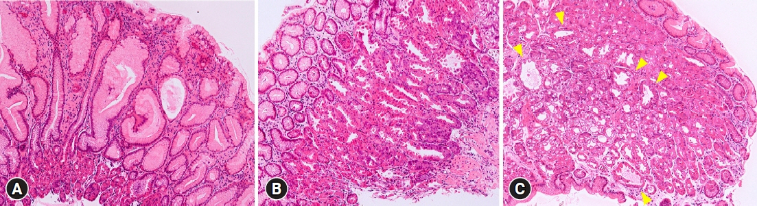
Fig. 2. Histological characteristics of patients with multiple white and flat elevated lesions (MWFL). (A) Biopsy specimens from MWFL show foveolar hyperplasia. (B) Parietal cell protrusions are observed in the surrounding gastric mucosa in patients with MWFL. (C) Oxyntic gland dilatations (arrowheads) are observed in the surrounding gastric mucosa of patients with MWFL. (A–C) Hematoxylin and eosin staining, ×20.
Reference
-
1. Noffsinger A. Fenoglio-Preiser's gastrointestinal pathology. 4th ed. Philadelphia: Wolters Kluwer;2017.2. Kawaguchi M, Arai E, Nozawa H. An investigation of white flat elevations in the gastric body. Gastroenterol Endosc. 2007; 49(Suppl 1):958.3. Kamada T, Kawaguchi M, Maruyama Y, et al. New gastric lesion in the cardia induced by proton pump inhibitor treatment. Gastroenterology. 2011; 140(5 Suppl 1):S–719.4. Hasegawa R, Yao K, Ihara S, et al. Magnified endoscopic findings of multiple white flat lesions: a new subtype of gastric hyperplastic polyps in the stomach. Clin Endosc. 2018; 51:558–562.5. Haruma K, Shiotani A, Kamada T, et al. Problems of prolonged administration of PPI-occurrence of gastric polyps. Shokakinaika. 2013; 56:190–193.6. Yamaoka R, Yao K, Chuman K, et al. Novel endoscopic findings of multiple white flat lesions: a new subtype of hyperplastic polyps in the stomach. United European Gastroenterol J. 2016; 3(Suppl 1):A386.7. Ally MR, Veerappan GR, Maydonovitch CL, et al. Chronic proton pump inhibitor therapy associated with increased development of fundic gland polyps. Dig Dis Sci. 2009; 54:2617–2622.8. Kim GH. Proton pump inhibitor-related gastric mucosal changes. Gut Liver. 2021; 15:646–652.9. Sugawara K, Imai Y, Saito E. Cases of gastric fundic gland polyps increasing in the size during the long-term therapy with proton pump inhibitor. Gastroenterol Endosc. 2009; 51:1686–1691.10. Hongo M, Fujimoto K; Gastric Polyps Study Group. Incidence and risk factor of fundic gland polyp and hyperplastic polyp in long-term proton pump inhibitor therapy: a prospective study in Japan. J Gastroenterol. 2010; 45:618–624.11. Takeda T, Asaoka D, Tajima Y, et al. Hemorrhagic polyps formed like fundic gland polyps during long-term proton pump inhibitor administration. Clin J Gastroenterol. 2017; 10:478–484.12. Aoi K, Yasunaga Y, Matsuura N, et al. Fundic gland polyp with dysplasia developed during long-term proton pump inhibitor therapy, report of a case. Stomach Intest. 2012; 47:1270–1274.13. Yao K. The endoscopic diagnosis of early gastric cancer. Ann Gastroenterol. 2013; 26:11–22.14. Kimura K, Takemoto T. An endoscopic recognition of the atrophic border and its significance in chronic gastritis. Endoscopy. 1969; 3:87–97.15. Shinozaki S, Osawa H, Miura Y, et al. The effect of proton pump inhibitors and vonoprazan on the development of 'gastric mucosal redness'. Biomed Rep. 2022; 16:51.16. Pimentel-Nunes P, Libânio D, Lage J, et al. A multicenter prospective study of the real-time use of narrow-band imaging in the diagnosis of premalignant gastric conditions and lesions. Endoscopy. 2016; 48:723–730.17. Pimentel-Nunes P, Dobru D, Libânio D, et al. White flat lesions in the gastric corpus may be intestinal metaplasia. Endoscopy. 2017; 49:617–618.18. Uedo N, Yamaoka R, Yao K. Multiple white flat lesions in the gastric corpus are not intestinal metaplasia. Endoscopy. 2017; 49:615–616.19. Kumar KR, Iqbal R, Coss E, et al. Helicobacter gastritis induces changes in the oxyntic mucosa indistinguishable from the effects of proton pump inhibitors. Hum Pathol. 2013; 44:2706–2710.20. Declich P, Ambrosiani L, Bellone S, et al. Parietal cell hyperplasia with deep cystic dilations: a lesion closely mimicking fundic gland polyps. Am J Gastroenterol. 2000; 95:566–568.
- Full Text Links
-
- Actions
-
Cited
- CITED
-
- Close
- Share
-
- Similar articles
-
- Proton Pump Inhibitor-Related Gastric Mucosal Changes
- Do Histamine-2 Receptor Antagonists and Proton Pump Inhibitors Really Have No Effect on the Gastric Emptying Rate?
- Magnified Endoscopic Findings of Multiple White Flat Lesions: A New Subtype of Gastric Hyperplastic Polyps in the Stomach
- Proton Pump Inhibitors Reduce the Size and Acidity of the Gastric Acid Pocket
- Long Term Proton Pump Inhibitor Use and Gastric Cancer
