Regeneration of total tissue using alveolar ridge augmentation with soft tissue substitute on periodontally compromised extraction sites: case report
- Affiliations
-
- 1Department of Periodontology and Research Institute of Oral Sciences, Gangneung-Wonju National University College of Dentistry, Gangneung, Republic of Korea
- KMID: 2550437
- DOI: http://doi.org/10.14368/jdras.2023.39.4.276
Abstract
- After tooth extraction, alveolar bone is resorbed over time. Loss of alveolar bone and reduction of upper soft tissue poses difficulties in future implant placement and long-term survival of the implant. This case report focuses on increasing the soft and hard tissues at the implant placement site by using alveolar ridge augmentation and a xenogeneic collagen matrix as a soft tissue substitute in an extraction socket affected by periodontal disease. In each case, the width of the alveolar bone increased to 6 mm, 8 mm, and 4 mm, and regeneration of the interdental papilla around the implant was shown, as well as buccal keratinized gingiva of 4 mm, 6 mm, and 4 mm, respectively. Enlarged alveolar bone facilitates implant surgery, and interdental papillae and keratinized gingiva enable aes-thetic prosthesis. This study performed alveolar ridge augmentation on patients with extraction sockets affected by periodontal dis-ease and additionally used soft tissue substitutes to provide a better environment for implant placement and have positive effects for aesthetic and predictive implant surgery.
Keyword
Figure
-
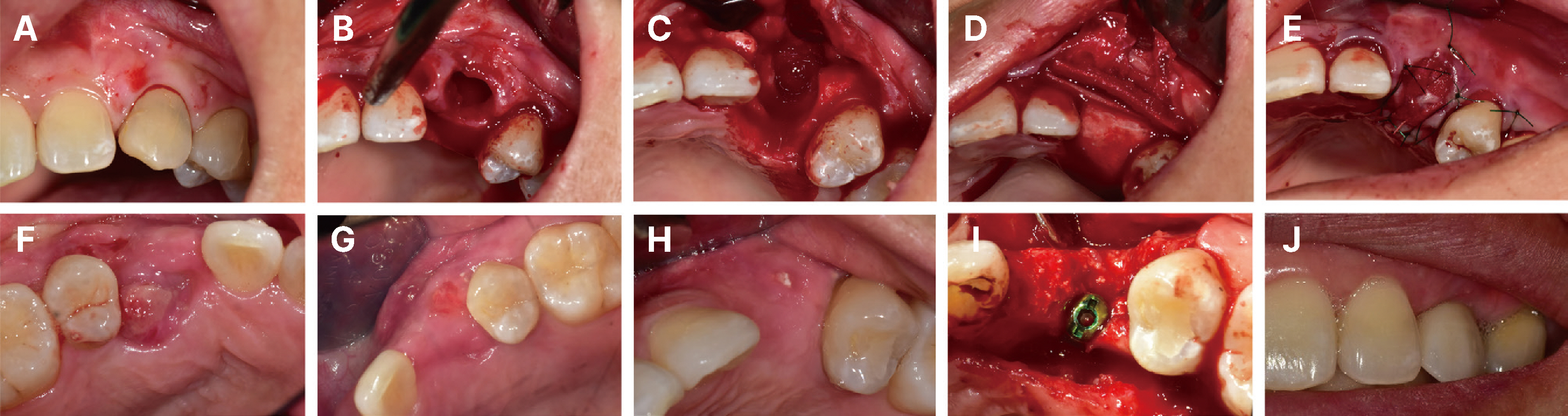
Fig. 1 Clinical photograph of case 1. (A) Preoperative. (B) After tooth #23 was extracted, a horizontal incision was made on the palatal side, and the full-thickness flap including the interdental papilla was lifted labially. (C) Buccal bone defects were observed after mucoperiosteal flap elevation. (D) Biphasic calcium phosphate with collagen was placed on the extraction socket. A soft stiffness type of resorbable collagen membrane was inserted into the crestal area and another medium stiffness type of resorbable collage membrane was overlaid on the labial side. A soft tissue substitute was inserted into the outermost part of the labial side. (E) The operative area was intentionally left open with hidden X suture and simple interrupted suture. (F) Post-operative 1 month, 2ndary healing stage was seen. (G) Post-operative 2 month, epithelization was completed. (H) Post-operative 9 month, 4 mm width of keratinized tissue and smooth surface was made. (I) About 6mm width of augmented bone was observed. (J) Clinical photograph after connection of the definitive prosthesis. Compared to before surgery, interdental papilla and 4 mm width of labial keratinized gingiva was regenerated
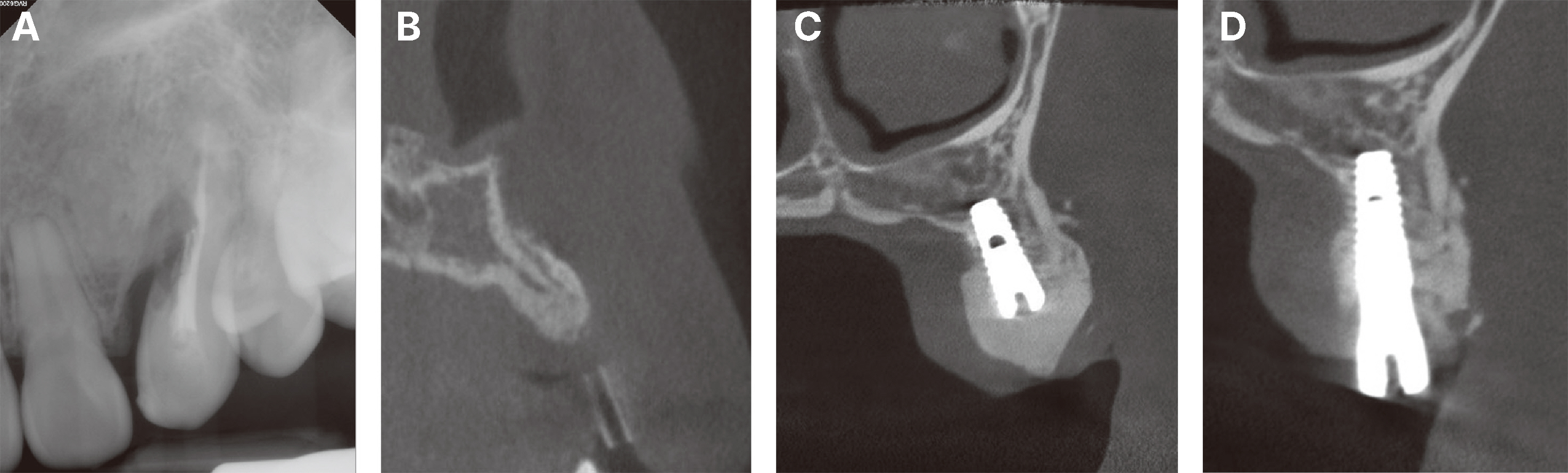
Fig. 2 Radiograph of case 1. (A) The initial periapical view of the left maxillary canine. External root resorption is seen in the middle third of the root. (B) Pre-implant site in cone-beam computed tomography image. About 6 mm width of augmented bone is observed. (C) After implant placement with guided bone regeneration, bone material around implant fixture is observed. (D) After implant second stage operation, the contour of bone graft is well maintained.

Fig. 3 Clinical photograph of case 2. (A) After full-thickness flap elevation, buccal plate of #16 extraction socket was absent. (B) Deproteinized bovine bone mineral with collagen was placed on the extraction socket. (C) A resorbable collagen membrane was laid on the bone graft material, and a soft tissue substitute was overlaid on the outermost part of the crestal area. (D) The operative area was intentionally left open with hidden X suture. (E) Post-operation 10 day, secondary intention healing was observed. (F) Post-operation 1 month, epithelization was completed. (G) Post-operative 10 month, about 8mm width augmented bone was seen. (H) Implants were placed on the augmented bone of #16,17 area. (I) Concerned about buccal and palatal plate perforation, additional guided bone regeneration with deproteinized bovine bone mineral and a resorbable collagen membrane was performed. (J) Clinical photograph after connection of the definitive prosthesis. Compared to before surgery, sufficient amount of augmented bone and 6mm width of buccal keratinized gingiva was seen on the buccal side.

Fig. 4 Radiograph of case 2. (A) Pre-operative panoramic view at the first visit. (B) Soft tissue substitute and underlying bone graft material are seen on #16,17 area after alveolar ridge augmentation. (C, D) Implants are placed on #16,17 area, respectively. (E) Panoramic view after connection of the definitive prosthesis.

Fig. 5 Clinical photograph of case 3. (A) Pre-operative. Crowding on lower anterior teeth is observed. (B) A horizontal incision is made on lingual side, and full thickness flap including interdental papilla was elevated labially. Horizontal bone resorption was observed on #31 buccal plate. (C) Biphasic calcium phosphate with collagen was inserted on the extraction socket. (D) A resorbable collagen membrane was placed on the bone graft material and soft tissue substitute was overlaid on the outermost part of crestal area. The operative area was left open with simple interrupted suture. (E) Post-operation 2 week, secondary intention healing was observed. (F) Post-operation 1 month, epithelization was completed and a conical shape of interdental papilla was maintained. (G) Post-operation 3 month, implants were placed on #32,42 area, respectively. About 4mm width of augmented bone was seen. (H),(I) Post-implant placement 2 month and 3 month, blunted shape of interdental papilla was maintained, respectively. (J) Clinical photograph after connection of the definitive prosthesis. Interdental papilla and 4 mm width of labial keratinized gingiva was observed.
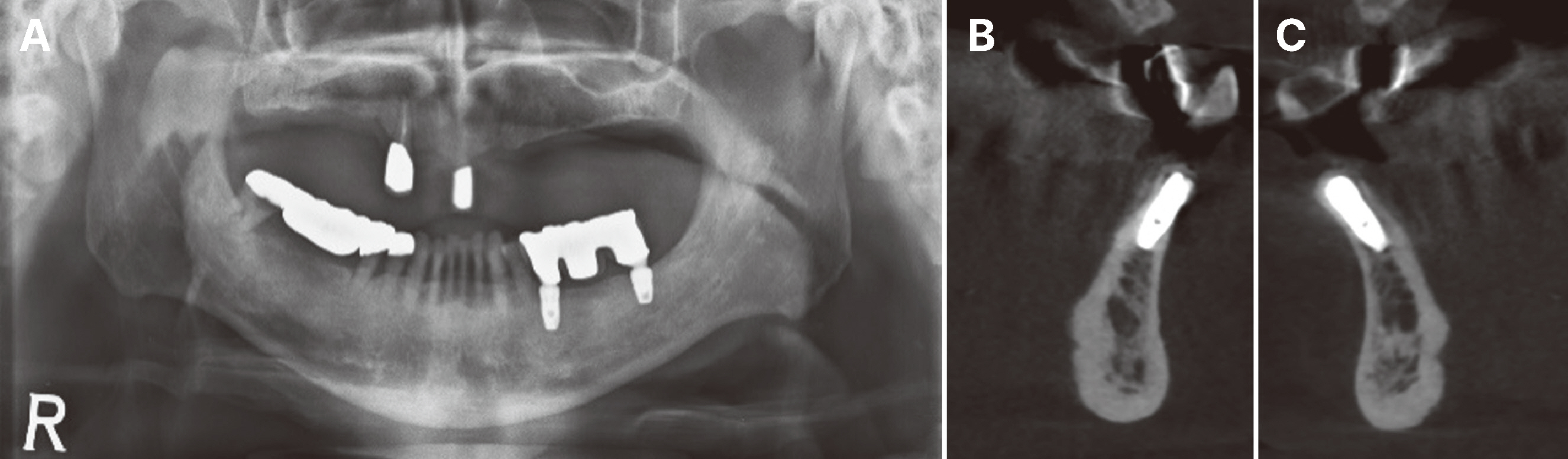
Fig. 6 Radiograph of Case 3. (A) Pre-operative panoramic view at the first visit. Crowding and alveolar bone loss are seen on lower anterior teeth. (B, C) CBCT image acquisition after implant first stage operation on #32,42, respectively. Implant fixture is surrounded with augmented bone and placed directly into bone.
Reference
-
References
1. Johnson K. 1969; A study of the dimensional changes occurring in the maxilla following closed face immediate denture treatment. Aust Dent J. 14:370–6. DOI: 10.1111/j.1834-7819.1969.tb02290.x. PMID: 5264550.2. Araújo MG, Silva CO, Misawa M, Sukekava F. 2015; Alveolar socket healing: what can we learn? Periodontol 2000. 68:122–34. DOI: 10.1111/prd.12082. PMID: 25867983.3. Ashman A, Bruins P. 1985; Prevention of alveolar bone loss postextraction with HTR grafting material. Oral Surg Oral Med Oral Pathol. 60:146–53. DOI: 10.1016/0030-4220(85)90282-8. PMID: 3862021.4. Darby I, Chen ST, Buser D. 2009; Ridge preservation techniques for implant therapy. Int J Oral Maxillofac Implants. 24 Suppl:260–71.5. Lang NP, Löe H. 1972; The relationship between the width of keratinized gingiva and gingival health. J Periodontol. 43:623–7. DOI: 10.1902/jop.1972.43.10.623. PMID: 4507712.6. Lin GH, Chan HL, Wang HL. 2013; The significance of keratinized mucosa on implant health: a systematic review. J Periodontol. 84:1755–67. DOI: 10.1902/jop.2013.120688. PMID: 23451989.7. Berglundh T, Lindhe J. 1996; Dimension of the periimplant mucosa. Biological width revisited. J Clin Periodontol. 23:971–3. DOI: 10.1111/j.1600-051X.1996.tb00520.x. PMID: 8915028.8. MacBeth N, Trullenque-Eriksson A, Donos N, Mardas N. 2017; Hard and soft tissue changes following alveolar ridge preservation: a systematic review. Clin Oral Implants Res. 28:982–1004. DOI: 10.1111/clr.12911. PMID: 27458031.9. Becker W, Becker BE. 1990; Guided tissue regeneration for implants placed into extraction sockets and for implant dehiscences; surgical techniques and case report. Int J Periodontics Restorative Dent. 10:376–91.10. Rosenquist B. 1997; A comparison of various methods of soft tissue management following the immediate placement of implants into extraction sockets. Int J Oral Maxillofac Implants. 12:43–51. PMID: 9048453.11. Schmitt CM, Moest T, Lutz R, Wehrhan F, Neukam FW, Schlegel KA. 2016; Long-term outcomes after vestibuloplasty with a porcine collagen matrix (Mucograft®) versus the free gingival graft: a comparative prospective clinical trial. Clin Oral Implants Res. 27:e125–33. DOI: 10.1111/clr.12575.12. Basegmez C, Karabuda ZC, Demirel K, Yalcin S. 2013; The comparison of acellular dermal matrix allografts with free gingival grafts in the augmentation of peri-implant attached mucosa: a randomised controlled trial. Eur J Oral Implantol. 6:145–52. PMID: 23926586.13. Sanz M, Lorenzo R, Aranda JJ, Martin C, Orsini M. 2009; Clinical evaluation of a new collagen matrix (Mucograft prototype) to enhance the width of keratinized tissue in patients with fixed prosthetic restorations: a randomized prospective clinical trial. J Clin Periodontol. 36:868–76. DOI: 10.1111/j.1600-051X.2009.01460.x. PMID: 19678861.14. Schropp L, Wenzel A, Kostopoulos L, Karring T. 2003; Bone healing and soft tissue contour changes following single-tooth extraction: a clinical and radiographic 12-month prospective study. Int J Periodontics Restorative Dent. 23:313–23. PMID: 12956475.15. Iasella JM, Greenwell H, Miller RL, Hill M, Drisko C, Bohra AA, Scheetz JP. 2003; Ridge preservation with freeze-dried bone allograft and a collagen membrane compared to extraction alone for implant site development: A clinical and histologic study in humans. J Periodontol. 74:990–9. DOI: 10.1902/jop.2003.74.7.990. PMID: 12931761.16. Araújo MG, Lindhe J. 2009; Ridge alterations following tooth extraction with and without flap elevation: an experimental study in the dog. Clin Oral Implants Res. 20:545–9. DOI: 10.1111/j.1600-0501.2008.01703.x. PMID: 19515033.17. Thalmair T, Fickl S, Schneider D, Hinze M, Wachtel H. 2013; Dimensional alterations of extraction sites after different alveolar ridge preservation techniques - a volumetric study. J Clin Periodontol. 40:721–7. DOI: 10.1111/jcpe.12111. PMID: 23647007.18. Ghanaati S, Schlee M, Webber MJ, Willershausen I, Barbeck M, Balic E, Görlach C, Stupp SI, Sader RA, Kirkpatrick CJ. 2011; Evaluation of the tissue reaction to a new bilayered collagen matrix in vivo and its translation to the clinic. Biomed Mater. 6:015010. DOI: 10.1088/1748-6041/6/1/015010. PMID: 21239849.19. Jung RE, Philipp A, Annen BM, Signorelli L, Thoma DS, Hämmerle CH, Attin T, Schmidlin P. 2013; Radiographic evaluation of different techniques for ridge preservation after tooth extraction: a randomized controlled clinical trial. J Clin Periodontol. 40:90–8. DOI: 10.1111/jcpe.12027. PMID: 23163915.20. De Risi V, Clementini M, Vittorini G, Mannocci A, De Sanctis M. 2015; Alveolar ridge preservation techniques: A systematic review and meta-analysis of histological and histomorphometrical data. Clin Oral Implants Res. 26:50–68. DOI: 10.1111/clr.12288. PMID: 27007188.
- Full Text Links
-
- Actions
-
Cited
- CITED
-
- Close
- Share
-
- Similar articles
-
- Guided bone regeneration using K-incision technique
- Compromised extraction sockets: a new classification and prevalence involving both soft and hard tissue loss
- Ridge augmentation using of hard tissue replacement(htrtm): a case report
- Anterior maxillary defect reconstruction with a staged bilateral rotated palatal graft
- Ridge Augmentation Using Vascularized Interpositional Periosteal- Connective Tissue (VIP-CT) in Conjunction with Anterior Implant Placement in Maxilla: Report of Three Cases
