Clinical and radiographic outcomes of regenerative endodontic treatment performed by endodontic postgraduate students: a retrospective study
- Affiliations
-
- 1College of Dentistry, Riyadh Elm University, Riyadh, Saudi Arabia
- 2Department of Restorative Dentistry – Endodontics, College of Dentistry, Riyadh Elm University, Riyadh, Saudi Arabia
- 3Periodontitis, Ministry of Health, Riyadh, Saudi Arabia
- 4Department of Preventive Dental Sciences – Biostatistics, College of Dentistry, King Saud University, Riyadh, Saudi Arabia
- KMID: 2548128
- DOI: http://doi.org/10.5395/rde.2022.47.e24
Abstract
Objectives
Regenerative endodontic treatment is a clinical procedure aimed at biologically regenerating damaged root canal tissue of immature permanent teeth. This study aimed to report the outcomes of regenerative endodontic treatment performed by endodontic postgraduate students.
Materials and Methods
Clinical and radiographic data of 27 patients, aged 10–22 years, who underwent regenerative treatment of immature permanent teeth from 2015 to 2019 were followed up, wherein clinical and radiographic examinations were performed for each patient. Postoperative success rate and tooth survival were analyzed, and the postoperative radiographic root area changes were quantified.
Results
A total of 23 patients attended the dental appointments, showing that all teeth survived and were asymptomatic. Specifically, 7 periapical pathosis cases were completely healed, 12 were incompletely healed, and 4 cases failed. Moreover, significant differences were found between discolored and non-discolored teeth, and between the presence or absence of periapical radiolucency. Additionally, 3 anterior teeth showed complete closure of the apical foramen, while the apical foramen width was reduced in 17 teeth and failed in 3 teeth. Root length was also found to have been increased in 7 anterior and 4 posterior teeth, and the average length ranged from 4.00–0.63 mm in the anterior teeth, 2.85–1.48 mm of the mesial root, and 2.73–2.16 mm of the molar teeth distal root. Furthermore, calcified tissue deposition was observed in 7 teeth.
Conclusions
A favorable outcome of regenerative endodontic treatment of immature permanent teeth with necrotic pulp was achieved with a high survival rate.
Figure
-
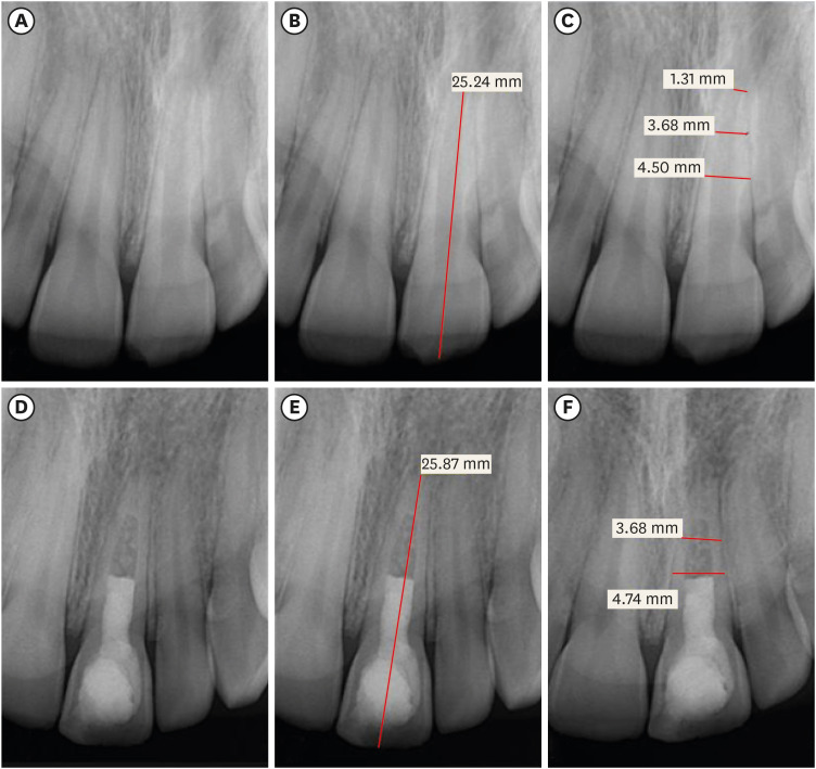
Figure 1 Maxillary left central incisor with open apex and apical radiolucency due to trauma (A-C). Calcium hydroxide was used as an intracanal medicament and the access was filled with mineral trioxide aggregate. 31-month postoperative radiographic follow-up shows complete apical closure, an increase in the tooth length, and healed (complete) apical radiolucency (D-F).
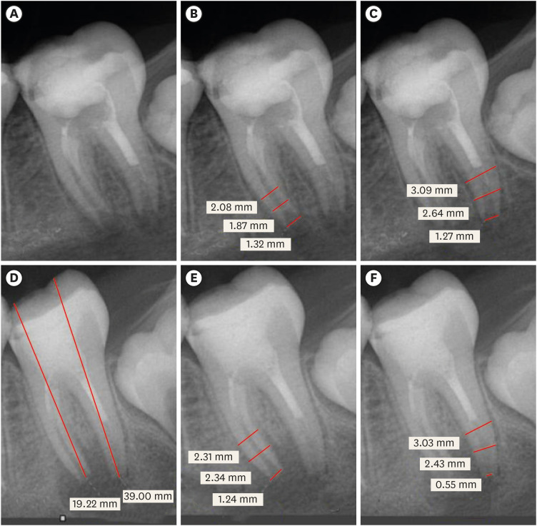
Figure 2 Mandibular left the second molar of 13-years old female patient with the open apex of both mesial and distal root and apical radiolucency due to caries. Calcium hydroxide was used as an intracanal medicament and the access and part of the canals were filled with mineral trioxide aggregate (A-C). 9 months post-operative radiograph shows complete apical closure of the distal root and reduction of the apical foramen width of the mesial root regardless of the increased apical radiolucency (D-F).
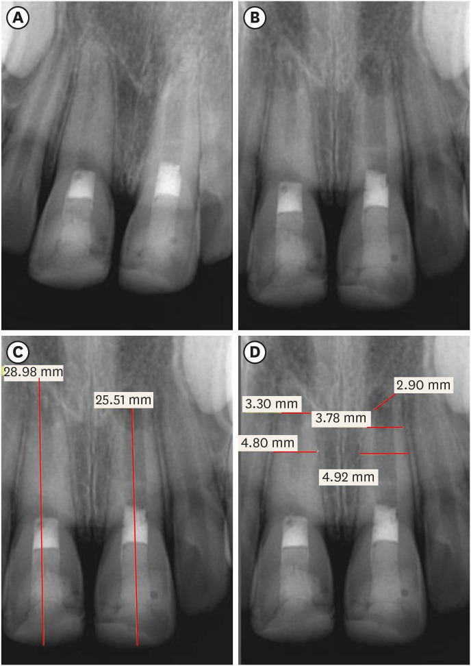
Figure 3 Maxillary left central incisor of 11-years old male patient with open apex and no apical radiolucency due to trauma. Calcium hydroxide was used as an intracanal medicament and the access and part of the canals were filled with mineral trioxide aggregate (A). The 6-months (B) and 12-months (C, D) post-operative radiograph shows the development of periapical radiolucency and the apical foramen failed to close.
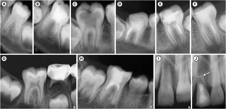
Figure 4 Root developmental patterns in response to regenerative endodontic therapy of immature permanent teeth with infected necrotic pulp described by Chen et al. [8] observed in the current study. Type 1 (A, B); Type 2 (C, D); Type 3 (E, F); Type 4 (G, H); and Type 5 (I, J).
Reference
-
1. Huang GT. Apexification: the beginning of its end. Int Endod J. 2009; 42:855–866. PMID: 19549154.
Article2. Hargreaves KM, Diogenes A, Teixeira FB. Treatment options: biological basis of regenerative endodontic procedures. J Endod. 2013; 39(3 Supplement):S30–S43. PMID: 23439043.
Article3. Thibodeau B, Teixeira F, Yamauchi M, Caplan DJ, Trope M. Pulp revascularization of immature dog teeth with apical periodontitis. J Endod. 2007; 33:680–689. PMID: 17509406.
Article4. Al-Ghamdi NS, Al-Nazhan S. Pulp revascularization of immature maxillary first premolar. J Conserv Dent. 2015; 18:496–499. PMID: 26752847.
Article5. Estefan BS, El Batouty KM, Nagy MM, Diogenes A. Influence of age and apical diameter on the success of endodontic regeneration procedures. J Endod. 2016; 42:1620–1625. PMID: 27623497.
Article6. Murray PE, Garcia-Godoy F, Hargreaves KM. Regenerative endodontics: a review of current status and a call for action. J Endod. 2007; 33:377–390. PMID: 17368324.
Article7. Wigler R, Kaufman AY, Lin S, Steinbock N, Hazan-Molina H, Torneck CD. Revascularization: a treatment for permanent teeth with necrotic pulp and incomplete root development. J Endod. 2013; 39:319–326. PMID: 23402501.
Article8. Alobaid AS, Cortes LM, Lo J, Nguyen TT, Albert J, Abu-Melha AS, Lin LM, Gibbs JL. Radiographic and clinical outcomes of the treatment of immature permanent teeth by revascularization or apexification: a pilot retrospective cohort study. J Endod. 2014; 40:1063–1070. PMID: 25069909.
Article9. Chen MY, Chen KL, Chen CA, Tayebaty F, Rosenberg PA, Lin LM. Responses of immature permanent teeth with infected necrotic pulp tissue and apical periodontitis/abscess to revascularization procedures. Int Endod J. 2012; 45:294–305. PMID: 22077958.
Article10. Kontakiotis EG, Filippatos CG, Agrafioti A. Levels of evidence for the outcome of regenerative endodontic therapy. J Endod. 2014; 40:1045–1053. PMID: 25069906.
Article11. Kahler B, Mistry S, Moule A, Ringsmuth AK, Case P, Thomson A, Holcombe T. Revascularization outcomes: a prospective analysis of 16 consecutive cases. J Endod. 2014; 40:333–338. PMID: 24565648.
Article12. Saoud TM, Zaazou A, Nabil A, Moussa S, Lin LM, Gibbs JL. Clinical and radiographic outcomes of traumatized immature permanent necrotic teeth after revascularization/revitalization therapy. J Endod. 2014; 40:1946–1952. PMID: 25443280.
Article13. Bukhari S, Kohli MR, Setzer F, Karabucak B. Outcome of revascularization procedure: a retrospective case series. J Endod. 2016; 42:1752–1759. PMID: 27726882.
Article14. Chatha NR. Regenerative endodontics: chart review of treated cases [master thesis]. Boston, MA: Boston University;2017.15. Sedwick R. Observations of trends and successes of revascularization therapy at Virginia Commonwealth University: a retrospective study [master thesis]. Richmond, VA: Virginia Commonwealth University;2018.16. American Association of Endodontics. Clinical considerations for regenerative procedures [Internet]. Chicago, IL: American Association of Endodontics;2013. updated 2016 Aug 6. cited 2021 Aug 20. Available from: www.aae.org/regenerativeendo.17. Jeeruphan T, Jantarat J, Yanpiset K, Suwannapan L, Khewsawai P, Hargreaves KM. Mahidol study 1: comparison of radiographic and survival outcomes of immature teeth treated with either regenerative endodontic or apexification methods: a retrospective study. J Endod. 2012; 38:1330–1336. PMID: 22980172.
Article18. Nygaard-Østby B, Hjørtdal O. Tissue formation in the root canal following pulp removal. Scand J Dent Res. 1971; 79:333–349. PMID: 5315973.
Article19. Landis JR, Koch GG. The measurement of observer agreement for categorical data. Biometrics. 1977; 33:159–174. PMID: 843571.
Article20. Cehreli ZC, Isbitiren B, Sara S, Erbas G. Regenerative endodontic treatment (revascularization) of immature necrotic molars medicated with calcium hydroxide: a case series. J Endod. 2011; 37:1327–1330. PMID: 21846559.
Article21. Friedman S, Mor C. The success of endodontic therapy--healing and functionality. J Calif Dent Assoc. 2004; 32:493–503. PMID: 15344440.22. Strindberg LZ. The dependence of the results on pulp therapy on certain factors: an analytical study based on radiographic and clinical follow-up examinations. Acta Odontol Scand. 1956; 14(Supplement 21):1–175.23. Xu R, Guo D, Zhou X, Sun J, Zhou Y, Fan Y. Disturbed bone remodeling activity varies in different stages of experimental, gradually progressive apical periodontitis in rats. Int J Oral Sci. 2019; 11:27. PMID: 31451690.24. Anantula K, Annareddy A. Platelet-rich fibrin (PRF) as an autologous biomaterial after an endodontic surgery: case reports. JNTR Univ Health Sci. 2016; 5:49–54.
Article25. Nair PN, Sjögren U, Figdor D, Sundqvist G. Persistent periapical radiolucencies of root-filled human teeth, failed endodontic treatments, and periapical scars. Oral Surg Oral Med Oral Pathol Oral Radiol Endod. 1999; 87:617–627. PMID: 10348524.
Article26. Shah N, Logani A, Bhaskar U, Aggarwal V. Efficacy of revascularization to induce apexification/apexogensis in infected, nonvital, immature teeth: a pilot clinical study. J Endod. 2008; 34:919–925. PMID: 18634921.
Article27. Torabinejad M, Nosrat A, Verma P, Udochukwu O. Regenerative endodontic treatment or mineral trioxide aggregate apical plug in teeth with necrotic pulps and open apices: a systematic review and meta-analysis. J Endod. 2017; 43:1806–1820. PMID: 28822564.
Article28. Almutairi W, Yassen GH, Aminoshariae A, Williams KA, Mickel A. Regenerative endodontics: a systematic analysis of the failed cases. J Endod. 2019; 45:567–577. PMID: 30905573.
Article29. Bose R, Nummikoski P, Hargreaves K. A retrospective evaluation of radiographic outcomes in immature teeth with necrotic root canal systems treated with regenerative endodontic procedures. J Endod. 2009; 35:1343–1349. PMID: 19801227.
Article30. Lee JY, Kersten DD, Mines P, Beltran TA. Regenerative endodontic procedures among endodontists: a web-based survey. J Endod. 2018; 44:250–255. PMID: 29229459.
Article31. Sonoyama W, Liu Y, Yamaza T, Tuan RS, Wang S, Shi S, Huang GT. Characterization of the apical papilla and its residing stem cells from human immature permanent teeth: a pilot study. J Endod. 2008; 34:166–171. PMID: 18215674.
Article32. Laureys WG, Cuvelier CA, Dermaut LR, De Pauw GA. The critical apical diameter to obtain regeneration of the pulp tissue after tooth transplantation, replantation, or regenerative endodontic treatment. J Endod. 2013; 39:759–763. PMID: 23683275.
Article33. Chueh LH, Ho YC, Kuo TC, Lai WH, Chen YH, Chiang CP. Regenerative endodontic treatment for necrotic immature permanent teeth. J Endod. 2009; 35:160–164. PMID: 19166764.
Article34. Ding RY, Cheung GS, Chen J, Yin XZ, Wang QQ, Zhang CF. Pulp revascularization of immature teeth with apical periodontitis: a clinical study. J Endod. 2009; 35:745–749. PMID: 19410097.
Article35. da Silva LA, Nelson-Filho P, da Silva RA, Flores DS, Heilborn C, Johnson JD, Cohenca N. Revascularization and periapical repair after endodontic treatment using apical negative pressure irrigation versus conventional irrigation plus triantibiotic intracanal dressing in dogs’ teeth with apical periodontitis. Oral Surg Oral Med Oral Pathol Oral Radiol Endod. 2010; 109:779–787. PMID: 20416538.
Article36. Wang X, Thibodeau B, Trope M, Lin LM, Huang GT. Histologic characterization of regenerated tissues in canal space after the revitalization/revascularization procedure of immature dog teeth with apical periodontitis. J Endod. 2010; 36:56–63. PMID: 20003936.
Article37. Yamauchi N, Yamauchi S, Nagaoka H, Duggan D, Zhong S, Lee SM, Teixeira FB, Yamauchi M. Tissue engineering strategies for immature teeth with apical periodontitis. J Endod. 2011; 37:390–397. PMID: 21329828.38. Nosrat A, Homayounfar N, Oloomi K. Drawbacks and unfavorable outcomes of regenerative endodontic treatments of necrotic immature teeth: a literature review and report of a case. J Endod. 2012; 38:1428–1434. PMID: 22980193.39. Shimizu E, Ricucci D, Albert J, Alobaid AS, Gibbs JL, Huang GT, Lin LM. Clinical, radiographic, and histological observation of a human immature permanent tooth with chronic apical abscess after revitalization treatment. J Endod. 2013; 39:1078–1083. PMID: 23880282.
Article40. Alenazy M, Al-Nazhan S, Mosadomi H. Histologic, radiographic, and micro-computed tomography evaluation of experimentally enlarged root apices in dog teeth with apical periodontitis after regenerative treatment. Curr Ther Res Clin Exp. 2020; 94:100620. PMID: 34306261.
Article41. Marciano MA, Costa RM, Camilleri J, Mondelli RF, Guimarães BM, Duarte MA. Assessment of color stability of white mineral trioxide aggregate angelus and bismuth oxide in contact with tooth structure. J Endod. 2014; 40:1235–1240. PMID: 25069940.
Article42. Petrino JA, Boda KK, Shambarger S, Bowles WR, McClanahan SB. Challenges in regenerative endodontics: a case series. J Endod. 2010; 36:536–541. PMID: 20171379.
Article43. Ruparel NB, Teixeira FB, Ferraz CC, Diogenes A. Direct effect of intracanal medicaments on survival of stem cells of the apical papilla. J Endod. 2012; 38:1372–1375. PMID: 22980180.
Article
- Full Text Links
-
- Actions
-
Cited
- CITED
-
- Close
- Share
-
- Similar articles
-
- Outcome of endodontic treatments performed by Brazilian undergraduate students: 3- to 8-year follow up
- A review of the regenerative endodontic treatment procedure
- Outcome of Regenerative Endodontic Treatment for an Avulsed Immature Permanent Tooth: A Case Report
- Endodontic treatment enhances the regenerative potential of teeth with advanced periodontal disease with secondary endodontic involvement
- Prevalence of referral reasons and clinical symptoms for endodontic referrals
