Effects of dentin surface preparations on bonding of self-etching adhesives under simulated pulpal pressure
- Affiliations
-
- 1Department of Operative Dentistry and Endodontics, Faculty of Dentistry, Mahidol University, Bangkok, Thailand
- 2Department of Anatomy, Faculty of Dentistry, Mahidol University, Bangkok, Thailand
- 3Department of Restorative Dentistry, Graduate School of Dental Medicine, Hokkaido University, Sapporo, Japan
- KMID: 2548113
- DOI: http://doi.org/10.5395/rde.2022.47.e4
Abstract
Objectives
This study evaluated the effects of different smear layer preparations on the dentin permeability and microtensile bond strength (µTBS) of 2 self-etching adhesives (Clearfil SE Bond [CSE] and Clearfil Tri-S Bond Universal [CTS]) under dynamic pulpal pressure.
Materials and Methods
Human third molars were cut into crown segments. The dentin surfaces were prepared using 4 armamentaria: 600-grit SiC paper, coarse diamond burs, superfine diamond burs, and carbide burs. The pulp chamber of each crown segment was connected to a dynamic intra-pulpal pressure simulation apparatus, and the permeability test was done under a pressure of 15 cmH 2 O. The relative permeability (%P) was evaluated on the smear layer-covered and bonded dentin surfaces. The teeth were bonded to either of the adhesives under pulpal pressure simulation, and cut into sticks after 24 hours water storage for the µTBS test. The resin-dentin interface and nanoleakage observations were performed using a scanning electron microscope. Statistical comparisons were done using analysis of variance and post hoc tests.
Results
Only the method of surface preparation had a significant effect on permeability (p < 0.05). The smear layers created by the carbide and superfine diamond burs yielded the lowest permeability. CSE demonstrated a higher µTBS, with these values in the superfine diamond and carbide bur groups being the highest. Microscopic evaluation of the resin-dentin interface revealed nanoleakage in the coarse diamond bur and SiC paper groups for both adhesives.
Conclusions
Superfine diamond and carbide burs can be recommended for dentin preparation with the use of 2-step CSE.
Keyword
Figure
-
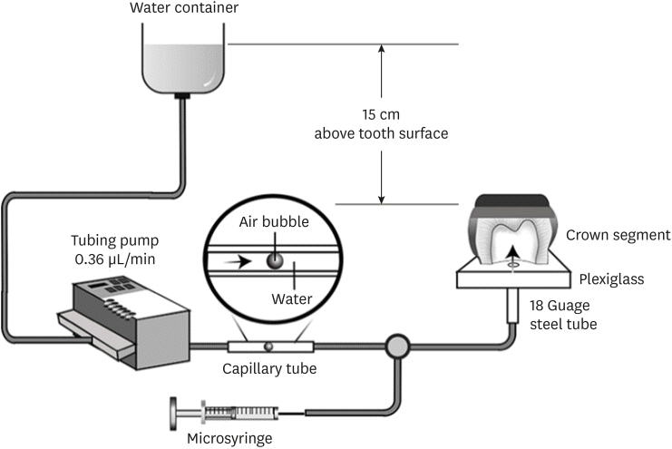
Figure 1 Schematic illustration of the simulated pulpal pressure model.

Figure 2 Mean and standard deviation of dentin permeability (%P) after surface preparation and after application of each tested adhesive. The %P of each tooth is expressed as a percentage of its own maximum permeability. The same uppercase letters indicate no significant difference in permeability after dentin surface preparation (p > 0.05). The same lowercase letters indicate no significant difference in dentin permeability after applying each adhesive (p > 0.05).SiC, silicon carbide.
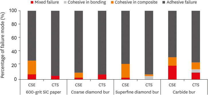
Figure 3 Percentage of failure mode distribution of the 2 self-etching adhesives with different dentin surface preparations (n = 40/group).CSE, Clearfil SE Bond; CTS, Clearfil Tri-S Bond Universal.

Figure 4 Representative scanning electron microscopy images of the resin-dentin interface of Clearfil SE Bond (upper) and Clearfil Tri-S Bond Universal (lower) at 3,000× magnification. (A, E) Dentin surface prepared with 600-grit SiC abrasive paper. (B, F) Dentin surface prepared with a coarse diamond bur. (C, G) Dentin surface prepared with a superfine diamond bur. (D, H) Dentin surface prepared with a carbide bur.
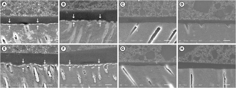
Figure 5 Representative scanning electron microscopy images of the resin-dentin interface with silver staining of Clearfil SE Bond (upper) and Clearfil Tri-S Bond Universal (lower) at 3,000× magnification. (A, E) Dentin surface prepared with 600-grit silicon carbide (SiC) abrasive paper. (B, F) Dentin surface prepared with a coarse diamond bur. (C, G) Dentin surface prepared with a superfine diamond bur. (D, H) Dentin surface prepared with a carbide bur. Nanoleakage (arrows) were observed in the 600-grit SiC abrasive paper and coarse diamond bur groups for both adhesives.
Reference
-
1. Van Meerbeek B, Yoshihara K, Yoshida Y, Mine A, De Munck J, Van Landuyt KL. State of the art of self-etch adhesives. Dent Mater. 2011; 27:17–28. PMID: 21109301.
Article2. Van Landuyt KL, Snauwaert J, De Munck J, Peumans M, Yoshida Y, Poitevin A, Coutinho E, Suzuki K, Lambrechts P, Van Meerbeek B. Systematic review of the chemical composition of contemporary dental adhesives. Biomaterials. 2007; 28:3757–3785. PMID: 17543382.
Article3. Tay FR, Pashley DH, Suh B, Carvalho R, Miller M. Single-step, self-etch adhesives behave as permeable membranes after polymerization. Part I. Bond strength and morphologic evidence. Am J Dent. 2004; 17:271–278. PMID: 15478490.4. Wagner A, Wendler M, Petschelt A, Belli R, Lohbauer U. Bonding performance of universal adhesives in different etching modes. J Dent. 2014; 42:800–807. PMID: 24814138.
Article5. Marchesi G, Frassetto A, Mazzoni A, Apolonio F, Diolosà M, Cadenaro M, Di Lenarda R, Pashley DH, Tay F, Breschi L. Adhesive performance of a multi-mode adhesive system: 1-year in vitro study. J Dent. 2014; 42:603–612. PMID: 24373855.
Article6. Nagarkar S, Theis-Mahon N, Perdigão J. Universal dental adhesives: current status, laboratory testing, and clinical performance. J Biomed Mater Res B Appl Biomater. 2019; 107:2121–2131. PMID: 30637932.
Article7. Rosa WL, Piva E, Silva AF. Bond strength of universal adhesives: a systematic review and meta-analysis. J Dent. 2015; 43:765–776. PMID: 25882585.
Article8. Ciucchi B, Bouillaguet S, Holz J, Pashley D. Dentinal fluid dynamics in human teeth, in vivo . J Endod. 1995; 21:191–194. PMID: 7673819.9. Hosaka K, Nakajima M, Yamauti M, Aksornmuang J, Ikeda M, Foxton RM, Pashley DH, Tagami J. Effect of simulated pulpal pressure on all-in-one adhesive bond strengths to dentine. J Dent. 2007; 35:207–213. PMID: 16989931.
Article10. Mahdan MH, Nakajima M, Foxton RM, Tagami J. Combined effect of smear layer characteristics and hydrostatic pulpal pressure on dentine bond strength of HEMA-free and HEMA-containing adhesives. J Dent. 2013; 41:861–871. PMID: 23851133.
Article11. Moll K, Haller B. Effect of intrinsic and extrinsic moisture on bond strength to dentine. J Oral Rehabil. 2000; 27:150–165. PMID: 10672152.
Article12. Mitchem JC, Gronas DG. Adhesion to dentin with and without smear layer under varying degrees of wetness. J Prosthet Dent. 1991; 66:619–622. PMID: 1804999.
Article13. Prati C, Pashley DH, Montanari G. Hydrostatic intrapulpal pressure and bond strength of bonding systems. Dent Mater. 1991; 7:54–58. PMID: 1901813.
Article14. Ermis RB, De Munck J, Cardoso MV, Coutinho E, Van Landuyt KL, Poitevin A, Lambrechts P, Van Meerbeek B. Bond strength of self-etch adhesives to dentin prepared with three different diamond burs. Dent Mater. 2008; 24:978–985. PMID: 18199476.
Article15. Saikaew P, Chowdhury AF, Fukuyama M, Kakuda S, Carvalho RM, Sano H. The effect of dentine surface preparation and reduced application time of adhesive on bonding strength. J Dent. 2016; 47:63–70. PMID: 26855030.
Article16. Semeraro S, Mezzanzanica D, Spreafico D, Gagliani M, Re D, Tanaka T, Sidhu SK, Sano H. Effect of different bur grinding on the bond strength of self-etching adhesives. Oper Dent. 2006; 31:317–323. PMID: 16802639.
Article17. Tani C, Finger WJ. Effect of smear layer thickness on bond strength mediated by three all-in-one self-etching priming adhesives. J Adhes Dent. 2002; 4:283–289. PMID: 12675016.18. Sattabanasuk V, Vachiramon V, Qian F, Armstrong SR. Resin-dentin bond strength as related to different surface preparation methods. J Dent. 2007; 35:467–475. PMID: 17331635.
Article19. Saikaew P, Senawongse P, Chowdhury AA, Sano H, Harnirattisai C. Effect of smear layer and surface roughness on resin-dentin bond strength of self-etching adhesives. Dent Mater J. 2018; 37:973–980. PMID: 30135339.
Article20. Pashley DH. Dentin bonding: overview of the substrate with respect to adhesive material. J Esthet Dent. 1991; 3:46–50. PMID: 1888543.
Article21. Oliveira SS, Pugach MK, Hilton JF, Watanabe LG, Marshall SJ, Marshall GW Jr. The influence of the dentin smear layer on adhesion: a self-etching primer vs. a total-etch system. Dent Mater. 2003; 19:758–767. PMID: 14511734.
Article22. Tay FR, Carvalho R, Sano H, Pashley DH. Effect of smear layers on the bonding of a self-etching primer to dentin. J Adhes Dent. 2000; 2:99–116. PMID: 11317405.23. Reis A, Grandi V, Carlotto L, Bortoli G, Patzlaff R, Rodrigues Accorinte ML, Dourado Loguercio A. Effect of smear layer thickness and acidity of self-etching solutions on early and long-term bond strength to dentin. J Dent. 2005; 33:549–559. PMID: 16005794.
Article24. Armstrong S, Breschi L, Özcan M, Pfefferkorn F, Ferrari M, Van Meerbeek B. Academy of Dental Materials guidance on in vitro testing of dental composite bonding effectiveness to dentin/enamel using micro-tensile bond strength (μTBS) approach. Dent Mater. 2017; 33:133–143. PMID: 28007396.
Article25. Carvalho AO, Oliveira MT, Nikaido T, Tagami J, Giannini M. Effect of adhesive system and application strategy on reduction of dentin permeability. Braz Oral Res. 2012; 26:397–403. PMID: 22892877.
Article26. Pashley DH, Depew DD. Effects of the smear layer, Copalite, and oxalate on microleakage. Oper Dent. 1986; 11:95–102. PMID: 3463938.27. Sauro S, Pashley DH, Montanari M, Chersoni S, Carvalho RM, Toledano M, Osorio R, Tay FR, Prati C. Effect of simulated pulpal pressure on dentin permeability and adhesion of self-etch adhesives. Dent Mater. 2007; 23:705–713. PMID: 16904175.
Article28. Siegel SC, von Fraunhofer JA. Dental cutting with diamond burs: heavy-handed or light-touch? J Prosthodont. 1999; 8:3–9. PMID: 10356549.
Article29. Tay FR, Pashley DH, Yoshiyama M. Two modes of nanoleakage expression in single-step adhesives. J Dent Res. 2002; 81:472–476. PMID: 12161459.
Article30. Pashley DH, Kepler EE, Williams EC, Okabe A. The effects of acid etching on the in-vivo permeability of dentine in the dog. Arch Oral Biol. 1983; 28:555–559. PMID: 6357159.
Article31. Prati C, Venturi L, Valdrè G, Mongiorgi R. Dentin morphology and permeability after brushing with different toothpastes in the presence and absence of smear layer. J Periodontol. 2002; 73:183–190. PMID: 11895284.
Article32. Saikaew P, Matsumoto M, Sattabanasuk V, Harnirattisai C, Carvalho RM, Sano H. Ultra-morphological characteristics of dentin surfaces after different preparations and treatments. Eur J Oral Sci. 2020; 128:246–254. PMID: 32396258.
Article33. Czonstkowsky M, Wilson EG, Holstein FA. The smear layer in endodontics. Dent Clin North Am. 1990; 34:13–25. PMID: 2403940.
Article34. Pashley DH. Smear layer: overview of structure and function. Proc Finn Dent Soc. 1992; 88(Supplement 1):215–224. PMID: 1508877.35. Masic A, Bertinetti L, Schuetz R, Chang SW, Metzger TH, Buehler MJ, Fratzl P. Osmotic pressure induced tensile forces in tendon collagen. Nat Commun. 2015; 6:5942. PMID: 25608644.
Article36. Sherman VR, Yang W, Meyers MA. The materials science of collagen. J Mech Behav Biomed Mater. 2015; 52:22–50. PMID: 26144973.
Article37. Sahin C, Cehreli ZC, Yenigul M, Dayangac B. In vitro permeability of etch-and-rinse and self-etch adhesives used for immediate dentin sealing. Dent Mater J. 2012; 31:401–408. PMID: 22673465.
Article38. Yoshida Y, Nagakane K, Fukuda R, Nakayama Y, Okazaki M, Shintani H, Inoue S, Tagawa Y, Suzuki K, De Munck J, Van Meerbeek B. Comparative study on adhesive performance of functional monomers. J Dent Res. 2004; 83:454–458. PMID: 15153451.
Article
- Full Text Links
-
- Actions
-
Cited
- CITED
-
- Close
- Share
-
- Similar articles
-
- Micro-shear bond strength to dentin under simulated pulpal pressure
- The effect of bonding resin on bond strength of dual-cure resin cements
- Microleakage of dentin bonding agents in porcelain laminate veneer under simulated physiologic pressure
- The influence of moisture control on bond strength of composite resin treated with self-etching adhesive system
- Effect of additional etching and ethanol-wet bonding on the dentin bond strength of one-step self-etch adhesives
