Which factors related to apical radiolucency may influence its radiographic detection? A study using CBCT as reference standard
- Affiliations
-
- 1Department of Oral Diagnosis, Division of Oral Radiology, Piracicaba Dental School, University of Campinas, São Paulo, Brazil
- 2Department of Dentistry, Oral Radiology area, Federal University of Sergipe, Sergipe, Brazil
- 3Oral Radiology section, School of Dentistry, Federal University of Alfenas, Minas Gerais, Brazil
- KMID: 2548087
- DOI: http://doi.org/10.5395/rde.2021.46.e43
Abstract
Objectives
This study aimed to evaluate the detection rate of apical radiolucencies in 2-dimensional images using cone-beam computed tomography (CBCT) as the reference standard, and to determine which factors related to the apical radiolucencies and the teeth could influence its detection.
Materials and Methods
The sample consisted of exams of patients who had panoramic (PAN) and/or periapical (PERI) radiography and CBCT. The exams were assessed by 2 oral radiologists and divided into PAN+CBCT (227 teeth–285 roots) and PERI+CBCT (94 teeth–115 roots). Radiographic images were evaluated for the presence of apical radiolucency, while CBCT images were assessed for presence, size, location, and involvement of the cortical bone (thinning, expansion, and destruction). Diagnostic values were obtained for PERI and PAN.
Results
PERI and PAN presented high accuracy (0.83 and 0.77, respectively) and specificity (0.89 and 0.91, respectively), but low sensitivity, especially for PAN (0.40 vs. 0.65 of PERI). The size of the apical radiolucency was positively correlated with its detection in PERI and PAN (p < 0.001). For PAN, apical radiolucencies were 3.93 times more frequently detected when related to single-rooted teeth (p = 0.038). The other factors did not influence apical radiolucency detection (p > 0.05).
Conclusions
PERI presents slightly better accuracy than PAN for the detection of apical radiolucency. The size is the only factor related to radiolucency that influences its detection, for both radiographic exams. For PAN, apical radiolucency is most often detected in singlerooted teeth.
Figure
-
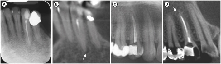
Figure 1 Images of PERI+CBCT and PAN+CBCT groups evaluated. Lower left canine in PERI (A) and CBCT coronal reconstruction (B). An upper right first premolar observed in cropped PAN (C) and CBCT sagittal reconstruction (D). Arrows indicate the apical radiolucencies detected in CBCT reconstructions.CBCT, cone-beam computed tomography; PAN, panoramic radiograph; PERI, periapical radiograph.

Figure 2 An apical radiolucency in a multi-rooted tooth that was not detected in PAN (A), but detected in the CBCT sagittal reconstruction, indicated by the arrow (B). The arrows are indicating an apical radiolucency detected on both PAN (C) and CBCT sagittal reconstruction (D) of a single-root tooth.CBCT, cone-beam computed tomography; PAN, panoramic radiograph.
Reference
-
1. Estrela C, Bueno MR, Azevedo BC, Azevedo JR, Pécora JD. A new periapical index based on cone beam computed tomography. J Endod. 2008; 34:1325–1331. PMID: 18928840.
Article2. Estrela C, Bueno MR, Leles CR, Azevedo B, Azevedo JR. Accuracy of cone beam computed tomography and panoramic and periapical radiography for detection of apical periodontitis. J Endod. 2008; 34:273–279. PMID: 18291274.
Article3. de Freitas JV, Baratto-Filho F, Coelho BS, Tomazinho FSF, Crozeta BM, de Sousa Neto MD, Gabardo MCL. Efficacy of different cone-beam computed tomographic protocols in the identification of mesiobuccal canals of maxillary first molars: a tomographic and ex vivo study. J Endod. 2017; 43:810–815. PMID: 28292600.
Article4. Patel S. New dimensions in endodontic imaging: part 2. Cone beam computed tomography. Int Endod J. 2009; 42:463–475. PMID: 19298576.
Article5. Moura MS, Guedes OA, De Alencar AH, Azevedo BC, Estrela C. Influence of length of root canal obturation on apical periodontitis detected by periapical radiography and cone beam computed tomography. J Endod. 2009; 35:805–809. PMID: 19482175.
Article6. Durack C, Patel S. Cone beam computed tomography in endodontics. Braz Dent J. 2012; 23:179–191. PMID: 22814684.
Article7. Patel S, Brown J, Pimentel T, Kelly RD, Abella F, Durack C. Cone beam computed tomography in endodontics - a review of the literature. Int Endod J. 2019; 52:1138–1152. PMID: 30868610.
Article8. Pope O, Sathorn C, Parashos P. A comparative investigation of cone-beam computed tomography and periapical radiography in the diagnosis of a healthy periapex. J Endod. 2014; 40:360–365. PMID: 24565653.
Article9. Nardi C, Calistri L, Pradella S, Desideri I, Lorini C, Colagrande S. Accuracy of orthopantomography for apical periodontitis without endodontic treatment. J Endod. 2017; 43:1640–1646. PMID: 28807372.
Article10. Nardi C, Calistri L, Grazzini G, Desideri I, Lorini C, Occhipinti M, Mungai F, Colagrande S. Is panoramic radiography an accurate imaging technique for the detection of endodontically treated asymptomatic apical periodontitis? J Endod. 2018; 44:1500–1508. PMID: 30154006.
Article11. Esposito S, Cardaropoli M, Cotti E. A suggested technique for the application of the cone beam computed tomography periapical index. Dentomaxillofac Radiol. 2011; 40:506–512. PMID: 22065800.
Article12. Kapila R, Harada N, Araki K, Sano T, Goto TK. Evaluation of juxta-apical radiolucency in cone beam CT images. Dentomaxillofac Radiol. 2014; 43:20130402. PMID: 24694213.
Article13. Patel S, Wilson R, Dawood A, Foschi F, Mannocci F. The detection of periapical pathosis using digital periapical radiography and cone beam computed tomography - part 2: a 1-year post-treatment follow-up. Int Endod J. 2012; 45:711–723. PMID: 22775142.
Article14. Davies A, Mannocci F, Mitchell P, Andiappan M, Patel S. The detection of periapical pathoses in root filled teeth using single and parallax periapical radiographs versus cone beam computed tomography - a clinical study. Int Endod J. 2015; 48:582–592. PMID: 25074727.
Article15. López FU, Kopper PM, Cucco C, Della Bona A, de Figueiredo JA, Vier-Pelisser FV. Accuracy of cone-beam computed tomography and periapical radiography in apical periodontitis diagnosis. J Endod. 2014; 40:2057–2060. PMID: 25306306.
Article16. Kanagasingam S, Lim CX, Yong CP, Mannocci F, Patel S. Diagnostic accuracy of periapical radiography and cone beam computed tomography in detecting apical periodontitis using histopathological findings as a reference standard. Int Endod J. 2017; 50:417–426. PMID: 27063209.
Article17. Abella F, Patel S, Durán-Sindreu F, Mercadé M, Bueno R, Roig M. An evaluation of the periapical status of teeth with necrotic pulps using periapical radiography and cone-beam computed tomography. Int Endod J. 2014; 47:387–396. PMID: 23889592.
Article18. Kanagasingam S, Hussaini HM, Soo I, Baharin S, Ashar A, Patel S. Accuracy of single and parallax film and digital periapical radiographs in diagnosing apical periodontitis - a cadaver study. Int Endod J. 2017; 50:427–436. PMID: 27063356.
Article19. Kruse C, Spin-Neto R, Wenzel A, Kirkevang LL. Cone beam computed tomography and periapical lesions: a systematic review analysing studies on diagnostic efficacy by a hierarchical model. Int Endod J. 2015; 48:815–828. PMID: 25283541.
Article
- Full Text Links
-
- Actions
-
Cited
- CITED
-
- Close
- Share
-
- Similar articles
-
- Use of preoperative cone-beam computed tomography to aid in establishment of endodontic working length: A systematic review and meta-analysis
- Cone-beam computed tomography versus digital periapical radiography in the detection of artificially created periapical lesions: A pilot study of the diagnostic accuracy of endodontists using both techniques
- Correlation between sagittal condylar guidance angles obtained using radiographic and protrusive occlusal record methods
- Radiographic evaluation of dentigerous cyst with cone beam CT
- Endodontic treatment of a C-shaped mandibular second premolar with four root canals and three apical foramina: a case report
