Correlation between sagittal condylar guidance angles obtained using radiographic and protrusive occlusal record methods
- Affiliations
-
- 1Department of Prosthodontics, Oral Science Research Center, College of Dentistry, Yonsei University, Seoul, Republic of Korea. jee917@yuhs.ac
- KMID: 2398078
- DOI: http://doi.org/10.4047/jap.2017.9.4.302
Abstract
- PURPOSE
This study compared the SCGAs measured in three types of radiographic images (panoramic, CBCT panoramic-section, and CBCT cross-section images) with values measured using the protrusive occlusal record.
MATERIALS AND METHODS
SCGAs were measured in 20 patients on a semi-adjustable articulator using the protrusive interocclusal record. Panoramic and CBCT images were obtained. SCGAs were measured on CBCT images in panoramic and cross sections. In all of the radiographic images, SCGAs were measured using the Frankfort horizontal reference line and the mean curvature line. The most-superior and most-inferior points of the curvatures were identified to determine the mean curvature line. Each measurement was performed twice by two operators independently. The data were analyzed by the t-test, Pearson's correlation test, and Cronbach's α using SPSS (α=.05).
RESULTS
The mean right and left SCGAs were as follows: protrusive occlusal record (30.1 and 30.2 degrees, respectively), panoramic (38.9 and 38.7 degrees), CBCT panoramic sections (35.4 and 36.8 degrees), and CBCT cross sections (35.3 and 36.1 degrees). The SCGAs differed significantly among the groups. The Pearson coefficients for the correlations with the protrusive occlusal record measurements on the left and right sides were as follows: panoramic (0.834 and 0.791, respectively), CBCT panoramic-section (0.918 and 0.837), and CBCT cross-section (0.918 and 0.845) images.
CONCLUSION
Strong correlations were found between SCGAs obtained using radiographic images and the protrusive occlusal record.
MeSH Terms
Figure
-

Fig. 1 Aluwax record placed on an articulator at 6 mm of protrusion. (A) Protrusive occlusal record, (B) The Protrusive occlusal record on an articulator.

Fig. 2 Measuring the SCGA in a radiographic tracing image. Line A: Frankfort line (Or - P), Line B: Most-superior and most-inferior points of the curvature, Or: Orbitale, P: Porion, Con: Condyle, Angle C: Angle made by the intersection of lines A and B.
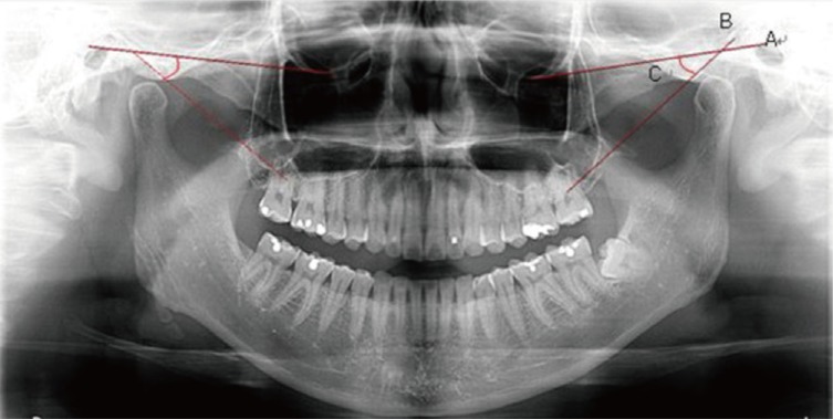
Fig. 3 Measuring the SCGA in panoramic images. Line A: Frankfort line (Or - P), Line B: Most-superior and most-inferior points of the curvature, Angle C: Angle made by the intersection of lines A and B.

Fig. 4 Determining the CBCT panoramic section in an axial view.
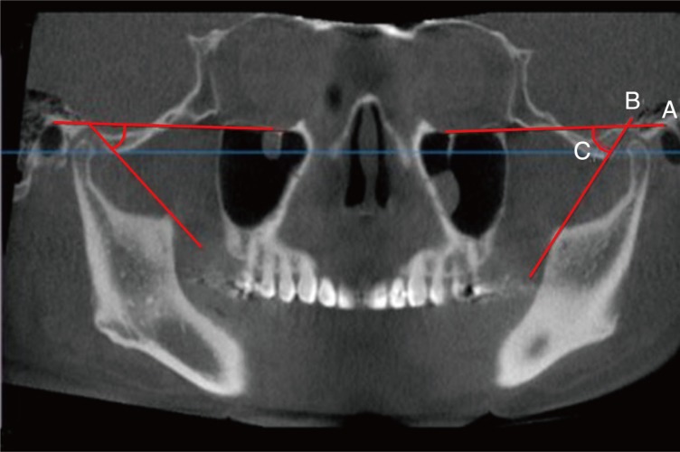
Fig. 5 Measuring the SCGA in a CBCT panoramic section image. Line A: Frankfort line (Or - P), Line B: Most-superior and most-inferior points of the curvature, Angle C: Angle made by the intersection of lines A and B.

Fig. 6 Determining the CBCT cross section in an axial view.
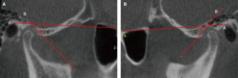
Fig. 7 Measuring the SCGA in a CBCT cross section image. (A) CBCT cross - section image (right), (B) CBCT cross - section image (left). Line A: Frankfort line (Or - P), Line B: Most-superior and most-inferior points of the curvature, Angle C: Angle made by the intersection of lines A and B.
Reference
-
1. Braly BV. Occlusal analysis and treatment planning for restorative dentistry. J Prosthet Dent. 1972; 27:168–171. PMID: 4500510.
Article2. Gilboa I, Cardash HS, Kaffe I, Gross MD. Condylar guidance: correlation between articular morphology and panoramic radiographic images in dry human skulls. J Prosthet Dent. 2008; 99:477–482. PMID: 18514670.
Article3. Prasad KD, Shetty M, Chandy BK. Evaluation of condylar inclination of dentulous subjects determined by axiograph and to compare with manual programming of articulators using protrusive interocclusal record. Contemp Clin Dent. 2015; 6:371–374. PMID: 26321837.
Article4. Shreshta P, Jain V, Bhalla A, Pruthi G. A comparative study to measure the condylar guidance by the radiographic and clinical methods. J Adv Prosthodont. 2012; 4:153–157. PMID: 22977723.
Article5. Utz KH, Müller F, Lückerath W, Fuss E, Koeck B. Accuracy of check-bite registration and centric condylar position. J Oral Rehabil. 2002; 29:458–466. PMID: 12028494.
Article6. Prasad KD, Shah N, Hegde C. A clinico-radiographic analysis of sagittal condylar guidance determined by protrusive interocclusal registration and panoramic radiographic images in humans. Contemp Clin Dent. 2012; 3:383–387. PMID: 23633793.
Article7. Christensen C. The problem of the bite. Dent Cosmos. 1905; 47:1184–1905.8. Tannamala PK, Pulagam M, Pottem SR, Swapna B. Condylar guidance: correlation between protrusive interocclusal record and panoramic radiographic image: a pilot study. J Prosthodont. 2012; 21:181–184. PMID: 22339685.
Article9. Scarfe WC, Levin MD, Gane D, Farman AG. Use of cone beam computed tomography in endodontics. Int J Dent. 2009; 2009:634567. PMID: 20379362.
Article10. Hatcher DC, Dial C, Mayorga C. Cone beam CT for pre-surgical assessment of implant sites. J Calif Dent Assoc. 2003; 31:825–833. PMID: 14696834.11. Christensen LV, Slabbert JC. The concept of the sagittal condylar guidance: biological fact or fallacy? J Oral Rehabil. 1978; 5:1–7. PMID: 272438.
Article12. Godavarthi AS, Sajjan MC, Raju AV, Rajeshkumar P, Premalatha A, Chava N. Correlation of condylar guidance determined by panoramic radiographs to one determined by conventional methods. J Int Oral Health. 2015; 7:123–128. PMID: 26464554.13. Millstein PL, Kronman JH, Clark RE. Determination of the accuracy of wax interocclusal registrations. J Prosthet Dent. 1971; 25:189–196. PMID: 5276189.
Article14. Assif D, Himel R, Grajower Y. A new electromechanical device to measure the accuracy of interocclusal records. J Prosthet Dent. 1988; 59:672–676. PMID: 3165452.
Article15. Freilich MA, Altieri JV, Wahle JJ. Principles for selecting interocclusal records for articulation of dentate and partially dentate casts. J Prosthet Dent. 1992; 68:361–367. PMID: 1501191.
Article16. Gross M, Nemcovsky C, Friedlander LD. Comparative study of condylar settings of three semiadjustable articulators. Int J Prosthodont. 1990; 3:135–141. PMID: 2133380.17. Posselt U, Franzen G. Registration of the condyle path inclination by intraoral wax records: Variations in three instruments. J Prosthet Dent. 1960; 10:441–454.
Article18. Craddock FW. The accuracy and practical value of records of condyle path inclination. J Am Dent Assoc. 1949; 38:697–710. PMID: 18150040.
Article19. Olthoff L, Meijer I, de Ruiter W, Bosman F, van der Zel J. Effect of virtual articulator settings on occlusal morphology of CAD/CAM restorations. Int J Comput Dent. 2007; 10:171–185. PMID: 17899892.
- Full Text Links
-
- Actions
-
Cited
- CITED
-
- Close
- Share
-
- Similar articles
-
- A study on the difference of the sagittal condylar guidance by semi-adjustable articulator and axiograph
- A study on the interrelationship of the condylar path, anterior occlusion and craniofacial morphology
- A comparative study to measure the condylar guidance by the radiographic and clinical methods
- Correlation between Cephalometric Reference Planes for Clinical Application to Articulators
- Comparison of condylar guidance using ARCUSdigma 2 and Checkbite
