A comparative study to measure the condylar guidance by the radiographic and clinical methods
- Affiliations
-
- 1Department of Prosthodontics, All India Institute of Medical Sciences, New Delhi, India. gunjan_prostho@yahoo.co.in
- KMID: 2176410
- DOI: http://doi.org/10.4047/jap.2012.4.3.153
Abstract
- PURPOSE
The study was conducted to compare the radiographic and clinical methods of measuring the horizontal condylar guidance (HCG) values.
MATERIALS AND METHODS
The condylar guidance was measured using the radiographic (CT scan) and three clinical methods i.e. the wax protrusive records, Lucia jig record and intraoral central bearing device in 12 patients aged between 20-40 years irrespective of sex. The records were taken and transferred on the semi-adjustable articulator to record the HCG values. The CT scan was taken for 3D reconstruction of the mid facial region. Frankfort horizontal plane (FHP) and a line extending from the superior anterior most point on the glenoid fossa to the most convex point on the apex of articular eminence (AE) was marked on the CT scan. An angle between these two lines was measured on both right and left sides to obtain condylar inclination angle. Three interocclusal protrusive wax and jig records were taken and transferred to the semi adjustable articulator. Three readings were recorded on each side. Similarly the records were taken and transferred to the same articulator using the intra oral central bearing device to record the readings.
RESULTS
The statistical analysis showed insignificant differences in the HCG values between the right and left sides [(P=.589 (CT), P=.928 (wax), P=.625 (jig), P=.886 (tracer)]. The clinical methods provided low Pearsons correlation values [(R = 0.423 (wax), R = 0.354 (jig), R = 0.265 (tracer)] for the right as well as the left sides when compared with the CT values. Among the clinical methods, jig and wax method showed strong level of association which is statistically significant while the intra-oral tracer showed weak association with the other two methods.
CONCLUSION
The right and left HCG values were almost similar. The CT scan showed higher HCG values than the clinical methods and among the clinical methods, values obtained from all the methods were comparable.
MeSH Terms
Figure
-

Fig. 1 Radiographic method to measure horizontal condylar guidance.
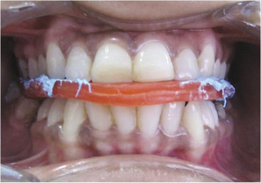
Fig. 2 Interocclusal protrusive wax record method.
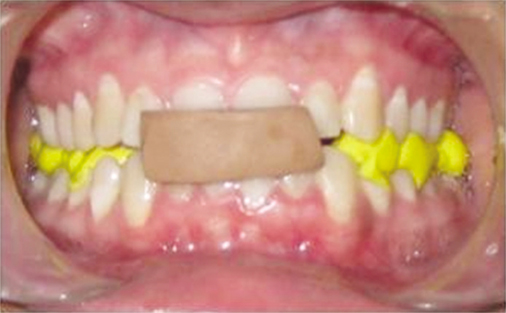
Fig. 3 Anterior jig method.
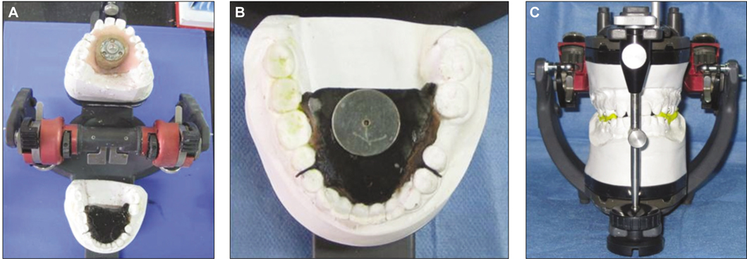
Fig. 4 A: Intraoral tracers attached to the maxillary and mandibular cast, B: Arrow point tracing, C: Protrusive record transferred to the articulator.

Fig. 5 Comparison of the CGA values obtained from different methods (X-axis-Method, Y- axis- Condylar guidance angles, CT-Computed Tomography, CGA- Condylar guidance angle, J-Jig, T-Tracer, W-Wax, R-Right, L-Left, -CT, -Clinical methods).

Fig. 6 Histogram showing comparison between right and left CGA obtained from different methods (X-axis-Method, Y- axis- Condylar guidance angles).
Cited by 1 articles
-
Correlation between sagittal condylar guidance angles obtained using radiographic and protrusive occlusal record methods
Oh-Kyun Kwon, Seung-Won Yang, Jee-Hwan Kim
J Adv Prosthodont. 2017;9(4):302-307. doi: 10.4047/jap.2017.9.4.302.
Reference
-
1. Zamacona JM, Otaduy E, Aranda E. Study of the sagittal condylar path in edentulous patients. J Prosthet Dent. 1992. 68:314–317.2. The glossary of prosthodontic terms. J Prosthet Dent. 2005. 94:10–92.3. Pelletier LB, Campbell SD. Comparison of condylar control settings using three methods: a bench study. J Prosthet Dent. 1991. 66:193–200.4. Posselt U. Physiology of occlusion and rehabilitation. 1968. 2nd ed. Oxford: Blackwell Science;121–132.5. Mohl ND, Zarb GA, Carlsson GE, Rugh JD. A textbook of occlusion. 1988. Chicago: Quintessence;139–140.6. Zarb GA, Bolender CL, Eckert SE, Fenton AH, Jacob RF, Mericske-Stern R. Prosthodontic treatment for edentulous patients: complete dentures and implant- supported prostheses. 2004. 12th ed. St. Louis: Mosby;294.7. Rosenstiel SF, Land MF, Fujimoto J. Contemporary fixed prosthodontics. 2006. 4th ed. St. Louis: Mosby;71.8. Hickey JC, Zarb GA, Bolender CL. Boucher's prosthodontic treatment for edentulous patients. 1985. 9th ed. St. Louis: CV Mosby;415.9. Gross M, Nemcovsky C, Friedlander LD. Comparative study of condylar settings of three semiadjustable articulators. Int J Prosthodont. 1990. 3:135–141.10. Gross M, Nemcovsky C, Tabibian Y, Gazit E. The effect of three different recording materials on the reproducibility of condylar guidance registrations in three semi-adjustable articulators. J Oral Rehabil. 1998. 25:204–208.11. dos Santos Júnior J, Nelson SJ, Nummikoski P. Geometric analysis of occlusal plane orientation using simulated ear-rod facebow transfer. J Prosthodont. 1996. 5:172–181.12. Christensen LV, Slabbert JC. The concept of the sagittal condylar guidance: biological fact or fallacy? J Oral Rehabil. 1978. 5:1–7.13. Gilboa I, Cardash HS, Kaffe I, Gross MD. Condylar guidance: correlation between articular morphology and panoramic radiographic images in dry human skulls. J Prosthet Dent. 2008. 99:477–482.14. el-Gheriani AS, Winstanley RB. Graphic tracings of condylar paths and measurements of condylar angles. J Prosthet Dent. 1989. 61:77–87.15. Brewka RE. Pantographic evaluation of cephalometric hinge axis. Am J Orthod. 1981. 79:1–19.16. Posselt U, Skytting B. Registration of the condyle path inclination: variations using the Gysi technique. J Prosthet Dent. 1960. 10:243–247.17. Donegan SJ, Christensen LV. Sagittal condylar guidance as determined by protrusion records and wear facets of teeth. Int J Prosthodont. 1991. 4:469–472.18. Price RB, Kolling JN, Clayton JA. Effects of changes in articulator settings on generated occlusal tracings. Part I: Condylar inclination and progressive side shift settings. J Prosthet Dent. 1991. 65:237–243.19. Ratzmann A, Mundt T, Schwahn C, Langforth G, Hutzen D, Gedrange T, Kordass B. Comparative clinical investigation of horizontal condylar inclination using the JMA electronic recording system and a protrusive wax record for setting articulators. Int J Comput Dent. 2007. 10:265–284.
- Full Text Links
-
- Actions
-
Cited
- CITED
-
- Close
- Share
-
- Similar articles
-
- Application of ARCUS digma I, II systems for full mouth reconstruction: a case report
- Non-Surgical Treatment of Mandibular Condylar Fracture with Functional Appliance: Clinical and Radiographic Analysis of 1 Case
- Correlation between sagittal condylar guidance angles obtained using radiographic and protrusive occlusal record methods
- A study on the interrelationship of the condylar path, anterior occlusion and craniofacial morphology
- Comparison of condylar position in transcranial radiography and polytomography from Polytome-U
