Management of diabetic foot ulcers: a narrative review
- Affiliations
-
- 1Department of Orthopaedic Surgery, Armed Forces Yangju Hospital, Yangju, Korea
- 2Department of Orthopaedic Surgery, Hallym University Chuncheon Sacred Heart Hospital, College of Medicine, Hallym University, Chuncheon, Korea
- 3Department of Orthopaedic Surgery, Soonchunhyang University Seoul Hospital, Seoul, Korea
- KMID: 2547356
- DOI: http://doi.org/10.12701/jyms.2023.00682
Abstract
- Diabetic foot ulcers (DFUs) are among the most serious complications of diabetes and are a source of reduced quality of life and financial burden for the people involved. For effective DFU management, an evidence-based treatment strategy that considers the patient's clinical context and wound condition is required. This treatment strategy should include conventional practices (surgical debridement, antibiotics, vascular assessment, offloading, and amputation) coordinated by interdisciplinary DFU experts. In addition, several adjuvant therapies can be considered for nonhealing wounds. In this narrative review, we aim to highlight the current trends in DFU management and review the up-to-date guidelines.
Keyword
Figure
-
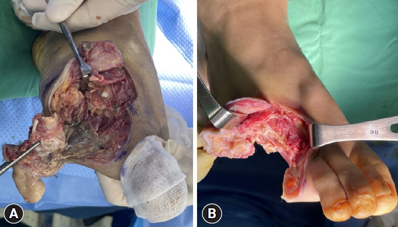
Fig. 1. Clinical images show differences in wound bed condition (A) before and (B) after surgical debridement.
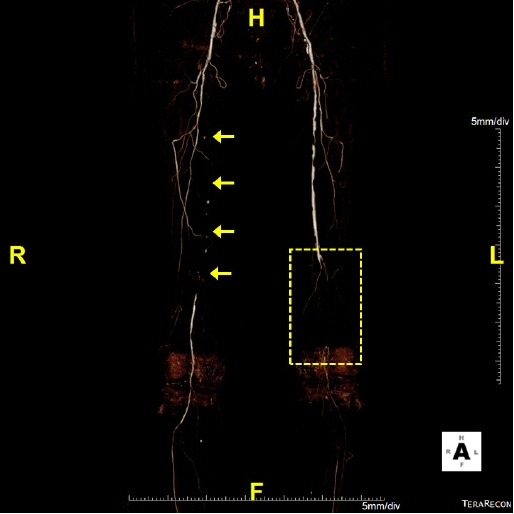
Fig. 2. Computed tomography angiography of a patient with ischemic diabetic foot ulcer. The right proximal to middle superficial femoral artery shows total occlusion (arrows) and the ankle-brachial index (ABI) is 0.38. Left distal superficial femoral and popliteal arteries reveal total occlusion and the ABI is not measurable due to poor vascular status (dotted box).

Fig. 3. (A) Total contact cast. (B) Removable ankle-high offloading device.
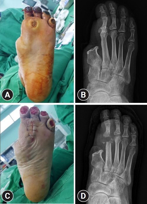
Fig. 4. A 62-year-old male with a history of first-ray amputation. (A) Chronic forefoot plantar ulcer due to repetitive loading is detected and (B) plain radiograph shows osteomyelitis on the second metatarsal head. (C, D) A second metatarsal head resection was performed for offloading.
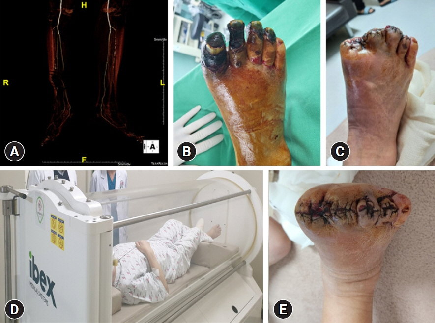
Fig. 5. A 73-year-old male with a 3-month history of diabetic foot ulcer. (A) Computed tomography angiography shows multifocal mild to moderate stenosis at both the superficial femoral artery and popliteal arteries. (B) Ischemic necrosis accompanied by infectious edema is detected on the first to fourth toes. (C) Toe amputation has been performed. (D) The patient has undergone adjuvant hyperbaric therapy both preoperatively for wound demarcation and postoperatively for wound healing. (E) The amputation wound has healed with improved infection and swelling.
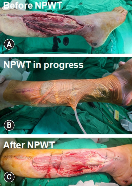
Fig. 6. Serial images describing negative pressure wound therapy (NPWT). (A) Before NPWT, the diabetic wound was debrided and (B) NPWT was applied. (C) After NPWT, the wound was ready to close with flap surgery.
Cited by 1 articles
-
Unveiling the challenges of diabetic foot infections: diagnosis, pathogenesis, treatment, and rehabilitation
Chul Hyun Park
J Yeungnam Med Sci. 2023;40(4):319-320. doi: 10.12701/jyms.2023.01011.
Reference
-
References
1. van Netten JJ, Bus SA, Apelqvist J, Chen P, Chuter V, Fitridge R, et al. Definitions and criteria for diabetes-related foot disease (IWGDF 2023 update). Diabetes Metab Res Rev. 2023; e3654.
Article2. Armstrong DG, Boulton AJ, Bus SA. Diabetic foot ulcers and their recurrence. N Engl J Med. 2017; 376:2367–75.
Article3. Musuuza J, Sutherland BL, Kurter S, Balasubramanian P, Bartels CM, Brennan MB. A systematic review of multidisciplinary teams to reduce major amputations for patients with diabetic foot ulcers. J Vasc Surg. 2020; 71:1433–46.
Article4. Naves CC. The diabetic foot: a historical overview and gaps in current treatment. Adv Wound Care (New Rochelle). 2016; 5:191–7.
Article5. Lavery LA, Davis KE, Berriman SJ, Braun L, Nichols A, Kim PJ, et al. WHS guidelines update: diabetic foot ulcer treatment guidelines. Wound Repair Regen. 2016; 24:112–26.
Article6. Lipsky BA, Berendt AR, Cornia PB, Pile JC, Peters EJ, Armstrong DG, et al. 2012 Infectious Diseases Society of America clinical practice guideline for the diagnosis and treatment of diabetic foot infections. Clin Infect Dis. 2012; 54:e132–73.7. Schaper NC, van Netten JJ, Apelqvist J, Bus SA, Fitridge R, Game F, et al. Practical guidelines on the prevention and management of diabetes-related foot disease (IWGDF 2023 update). Diabetes Metab Res Rev. 2023; e3657.
Article8. Braun L, Kim PJ, Margolis D, Peters EJ, Lavery LA; Wound Healing Society. What’s new in the literature: an update of new research since the original WHS diabetic foot ulcer guidelines in 2006. Wound Repair Regen. 2014; 22:594–604.
Article9. Everett E, Mathioudakis N. Update on management of diabetic foot ulcers. Ann N Y Acad Sci. 2018; 1411:153–65.
Article10. Tan JS, File TM Jr. Diagnosis and treatment of diabetic foot infections. Baillieres Best Pract Res Clin Rheumatol. 1999; 13:149–61.
Article11. Vas PR, Edmonds M, Kavarthapu V, Rashid H, Ahluwalia R, Pankhurst C, et al. The diabetic foot attack: “tis too late to retreat!”. Int J Low Extrem Wounds. 2018; 17:7–13.
Article12. Kim J, Chun DI, Kim S, Yang HJ, Kim JH, Cho JH, et al. Trends in lower limb amputation in patients with diabetic foot based on vascular intervention of peripheral arterial disease in Korea: a population-based nationwide study. J Korean Med Sci. 2019; 34:e178.
Article13. Kolossváry E, Farkas K, Colgan MP, Edmonds M, Fitzgerald HP, Fox M, et al. “No more amputations”: a complex scientific problem and a challenge for effective preventive strategy implementation on vascular field. Int Angiol. 2017; 36:107–15.
Article14. Mills JL Sr, Conte MS, Armstrong DG, Pomposelli FB, Schanzer A, Sidawy AN, et al. The Society for Vascular Surgery Lower Extremity Threatened Limb Classification System: risk stratification based on wound, ischemia, and foot infection (WIfI). J Vasc Surg. 2014; 59:220–34.
Article15. Yavuz M. Plantar shear stress: is it the H pylori of diabetic foot ulcers? Clin Biomech (Bristol, Avon). 2022; 92:105581.
Article16. Yavuz M, Ersen A, Hartos J, Schwarz B, Garrett AG, Lavery LA, et al. Plantar shear stress in individuals with a history of diabetic foot ulcer: an emerging predictive marker for foot ulceration. Diabetes Care. 2017; 40:e14–5.
Article17. van Deursen R. Mechanical loading and off-loading of the plantar surface of the diabetic foot. Clin Infect Dis. 2004; 39(Suppl 2):S87–91.
Article18. de Oliveira AL, Moore Z. Treatment of the diabetic foot by offloading: a systematic review. J Wound Care. 2015; 24:560–70. 560, 562-70.
Article19. Bus SA, Armstrong DG, Crews RT, Gooday C, Jarl G, Kirketerp-Moller K, et al. Guidelines on offloading foot ulcers in persons with diabetes (IWGDF 2023 update). Diabetes Metab Res Rev. 2023; e3647.
Article20. Bus SA. The role of pressure offloading on diabetic foot ulcer healing and prevention of recurrence. Plast Reconstr Surg. 2016; 138(3 Suppl):179S–187S.
Article21. Chun DI, Kim S, Kim J, Yang HJ, Kim JH, Cho JH, et al. Epidemiology and burden of diabetic foot ulcer and peripheral arterial disease in Korea. J Clin Med. 2019; 8:748.
Article22. Chun DI, Kim J, Kang EM, An CY, Min TH, Kim S, et al. Does amputation negatively influence the incidence of depression in diabetic foot patients?: a population-based nationwide study. Appl Sci. 2022; 12:1653.
Article23. Smith DG, Ehde DM, Legro MW, Reiber GE, del Aguila M, Boone DA. Phantom limb, residual limb, and back pain after lower extremity amputations. Clin Orthop Relat Res. 1999; (361):29–38.
Article24. Koob TJ, Rennert R, Zabek N, Massee M, Lim JJ, Temenoff JS, et al. Biological properties of dehydrated human amnion/chorion composite graft: implications for chronic wound healing. Int Wound J. 2013; 10:493–500.
Article25. Zelen CM. An evaluation of dehydrated human amniotic membrane allografts in patients with DFUs. J Wound Care. 2013; 22:347–351. 347-8, 350-1.
Article26. Tettelbach W, Cazzell S, Sigal F, Caporusso JM, Agnew PS, Hanft J, et al. A multicentre prospective randomised controlled comparative parallel study of dehydrated human umbilical cord (EpiCord) allograft for the treatment of diabetic foot ulcers. Int Wound J. 2019; 16:122–30.
Article27. Sledge I, Maislin D, Bernarducci D, Snyder R, Serena TE. Use of a dual-layer amniotic membrane in the treatment of diabetic foot ulcers: an observational study. J Wound Care. 2020; 29(Suppl 9):S8–12.
Article28. Liu Y, Min D, Bolton T, Nubé V, Twigg SM, Yue DK, et al. Increased matrix metalloproteinase-9 predicts poor wound healing in diabetic foot ulcers. Diabetes Care. 2009; 32:117–9.
Article29. Ren Y, Gu G, Yao M, Driver VR. Role of matrix metalloproteinases in chronic wound healing: diagnostic and therapeutic implications. Chin Med J (Engl). 2014; 127:1572–81.30. Kulahin N, Kiselyov V, Kochoyan A, Kristensen O, Kastrup JS, Berezin V, et al. Dimerization effect of sucrose octasulfate on rat FGF1. Acta Crystallogr Sect F Struct Biol Cryst Commun. 2008; 64(Pt 6):448–52.
Article31. Edmonds M, Lázaro-Martínez JL, Alfayate-García JM, Martini J, Petit JM, Rayman G, et al. Sucrose octasulfate dressing versus control dressing in patients with neuroischaemic diabetic foot ulcers (Explorer): an international, multicentre, double-blind, randomised, controlled trial. Lancet Diabetes Endocrinol. 2018; 6:186–96.
Article32. Löndahl M, Tarnow L, Karlsmark T, Lundquist R, Nielsen AM, Michelsen M, et al. Use of an autologous leucocyte and platelet-rich fibrin patch on hard-to-heal DFUs: a pilot study. J Wound Care. 2015; 24:172–8. 172-4, 176-8.
Article33. Game F, Jeffcoate W, Tarnow L, Jacobsen JL, Whitham DJ, Harrison EF, et al. LeucoPatch system for the management of hard-to-heal diabetic foot ulcers in the UK, Denmark, and Sweden: an observer-masked, randomised controlled trial. Lancet Diabetes Endocrinol. 2018; 6:870–8.34. Löndahl M, Katzman P, Nilsson A, Hammarlund C. Hyperbaric oxygen therapy facilitates healing of chronic foot ulcers in patients with diabetes. Diabetes Care. 2010; 33:998–1003.
Article35. Wenhui L, Changgeng F, Lei X, Baozhong Y, Guobin L, Weijing F. Hyperbaric oxygen therapy for chronic diabetic foot ulcers: an overview of systematic reviews. Diabetes Res Clin Pract. 2021; 176:108862.
Article36. Kurze C, Farn CJ, Siow J. The interdisciplinary approach: preventive and therapeutic strategies for diabetic foot ulcers. Foot Ankle Clin. 2022; 27:529–43.37. Korzon-Burakowska A, Dziemidok P. Diabetic foot: the need for comprehensive multidisciplinary approach. Ann Agric Environ Med. 2011; 18:314–7.38. Rubio JA, Aragón-Sánchez J, Jiménez S, Guadalix G, Albarracín A, Salido C, et al. Reducing major lower extremity amputations after the introduction of a multidisciplinary team for the diabetic foot. Int J Low Extrem Wounds. 2014; 13:22–6.
Article39. Wang C, Mai L, Yang C, Liu D, Sun K, Song W, et al. Reducing major lower extremity amputations after the introduction of a multidisciplinary team in patient with diabetes foot ulcer. BMC Endocr Disord. 2016; 16:38.
Article40. Buggy A, Moore Z. The impact of the multidisciplinary team in the management of individuals with diabetic foot ulcers: a systematic review. J Wound Care. 2017; 26:324–39.
Article
